Medivisor India Treatment Logo
  • Home
  • About Us
    • About Us
    • Our Services
    • Our Team
    • Our Medical Advisors
    • Our Safety Measures
  • Treatment
    • Loading treatments...
  • Hospitals
    • Loading hospitals...
  • Patient Guide
    • Treatment Cost
    • Treatment Process
    • Visa Process
    • Travel Guide
    • FAQs
    • Why Medivisor
  • Gallery
    • Success Stories
    • Patient Activities
    • Social Activities
    • News Coverage
    • Blog
  • Become a Partner

+91 83407 80250

Medivisor Logo

Medivisor India Treatment is a trusted New Delhi–based medical travel partner connecting global patients to advanced, affordable, high-quality healthcare in India.

  • Medivisor House 359, Sector 1, Vaishali, Ghaziabad, (Delhi/NCR) India
  • +91 8340 780 250
  • info@medivisorhealth.com

About Us

  • About Us
  • Our Services
  • Our Team
  • Our Medical Advisors
  • Our Safety Measures
  • Join Our Network
  • Contact Us

India Treatment

  • Hospitals
  • Treatments
  • Treatment Cost
  • Treatment Process
  • Visa Process
  • Travel Guide
  • FAQs
  • Why Medivisor

Gallery

  • Patient Testimonials
  • Patient Activities
  • News Coverage
  • Blog

Get in Touch

© 2026 Medivisor India Treatment. All rights reserved.

Vascular Surgery hospital

Vascular Surgery

  1. Home
  2. Departments
  3. Vascular Surgery

Vascular Surgery Department

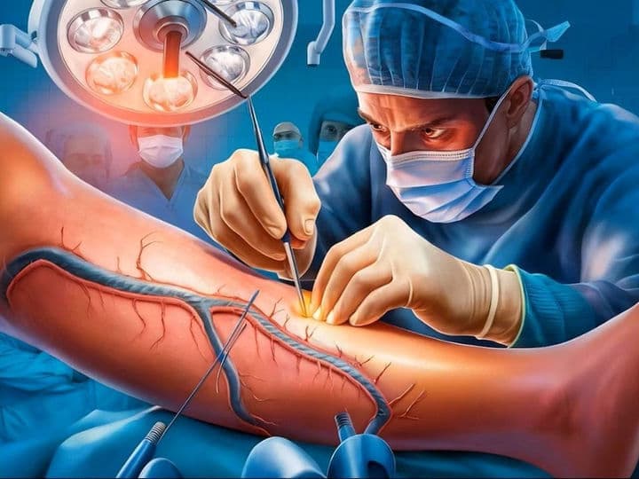
The Vascular Surgery Department is the hospital’s specialized "circulation hub," dedicated to the vast and intricate network of arteries and veins. While cardiac surgeons focus on the heart, vascular surgeons manage every blood vessel from the neck to the toes. For international patients, this wing represents the pinnacle of modern "plumbing" science, utilizing both traditional open surgery and high-tech "endovascular" interventions to ensure every organ and limb receives the life-sustaining blood flow it requires.

A Specialized Team of Circulation Experts

Vascular care is a multidisciplinary field where surgical mastery meets interventional medicine:

  • Vascular & Endovascular Surgeons: Primary experts trained in both traditional "open" vessel repair and "keyhole" stent placements.

  • Podiatrists (Limb Salvage Specialists): Experts focused on diabetic foot care and the healing of ulcers caused by poor circulation.

  • Vascular Technologists: Specialized sonographers who use Doppler ultrasound to map blockages, clots, and leaky valves in real-time.

  • Wound Care Specialists: Clinical nurses expert in treating chronic sores and skin changes resulting from venous insufficiency.

Advanced Infrastructure and Endovascular Technology

The department utilizes high-resolution imaging and miniaturized hardware to restore blood flow with minimal trauma:

  • The Hybrid Cath Lab: A futuristic surgical suite that allows for minimally invasive stent procedures with the capability to switch to open surgery instantly if needed.

  • Stent-Graft Technology: Fabric-covered mesh "sleeves" used to reinforce aneurysms (vessel bulges) from the inside, preventing life-threatening ruptures.

  • High-Pressure Angioplasty & Atherectomy: Precision tools that use balloons or rotating "shaving" devices to clear plaque and hard calcium from clogged arteries.

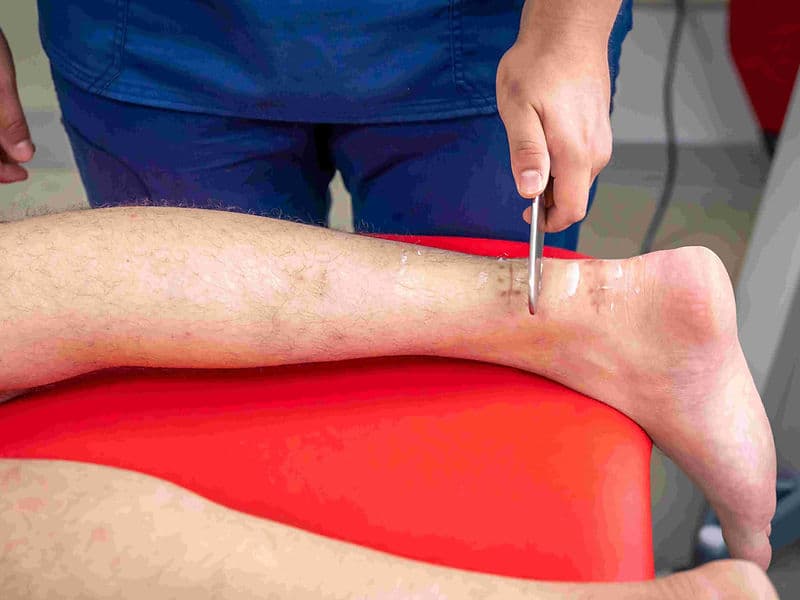
  • Laser & Radiofrequency Fibers: Modern "incisionless" tools that seal diseased varicose veins from the inside, redirecting blood to healthier pathways.

  • Vena Cava Filters: Microscopic metal "umbrellas" designed to catch traveling blood clots before they can reach the lungs.

Specialized Functional and Diagnostic Units

To ensure a total recovery, the department houses several dedicated diagnostic and treatment hubs:

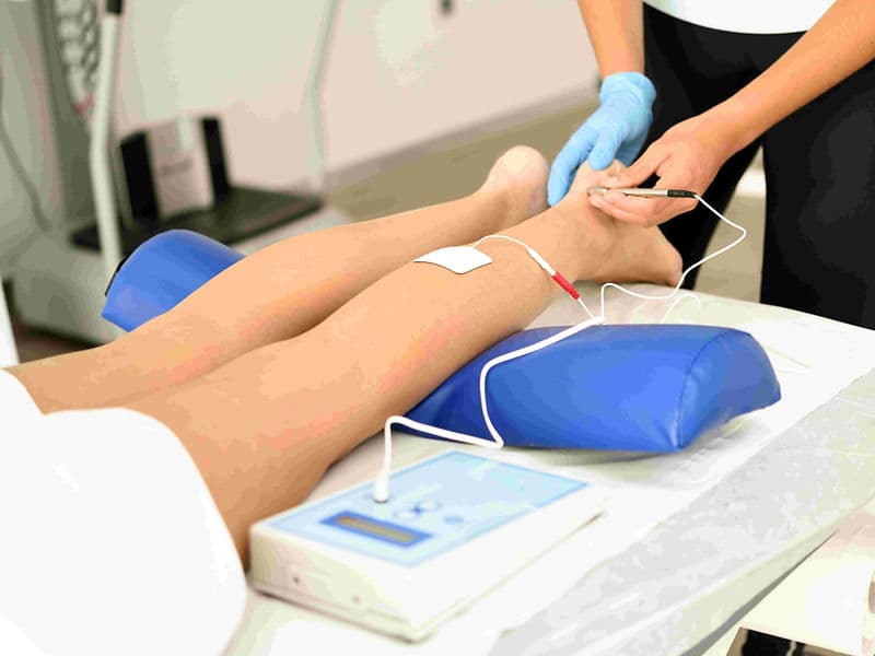
  • Non-Invasive Vascular Lab: A screening center using the Ankle-Brachial Index (ABI) to compare blood pressure and identify hidden blockages.

  • The Dialysis Access Clinic: A specialized unit where surgeons create vital "fistulas" for patients requiring long-term kidney dialysis.

  • Varicose Vein & Daycare Suite: A specialized area for walk-in, walk-out laser treatments for bulging or painful leg veins.

  • Limb Salvage Center: A high-intensity wing focused on preventing amputations through aggressive circulation restoration in patients with severe peripheral artery disease (PAD).

A Focus on Preservation and Stroke Prevention

The department’s philosophy centers on restoring natural function and preventing emergency events:

  • Limb Preservation Excellence: Advanced re-opening techniques that have reduced the need for major amputations by over 50% in recent years.

  • Aneurysm Protection: Repairing aortic bulges through tiny groin punctures rather than large abdominal incisions, significantly lowering patient risk.

  • Carotid Artery Management: Precision "cleaning" of the neck arteries to significantly reduce the risk of major strokes in high-risk patients.

  • Emergency Clot Retrieval: Utilizing the legendary Fogarty Catheter to manually retrieve fresh blood clots, saving limbs within the critical "golden hour."

  • International Patient Support: A dedicated lounge for global families to coordinate 3D CT Angiograms, surgical bookings, and long-term travel plans.

May we help you?

Enter digits only (6-15 digits). We'll add your country code.

Vascular Surgery Treatments

Arteriovenous (AV) Fistula Surgery

Arteriovenous (AV) Fistula Surgery

An Arteriovenous (AV) Fistula is a surgically created connection between an artery and a vein, designed to provide a reliable, long-term access point for hemodialysis. It remains the "gold standard" for vascular access because it is constructed entirely from the patient's own biological tissue. Compared to synthetic grafts or temporary catheters, an AV fistula offers a significantly lower risk of infection, fewer instances of clotting, and the longest functional lifespan, making it the foundation of high-quality renal care.

When You Should Consider An AV Fistula

  • End-Stage Renal Disease (ESRD): When kidney function has declined to the point where regular hemodialysis is necessary to filter waste from the blood.

  • Long-Term Dialysis Planning: For patients expected to be on dialysis for several years, as the fistula provides the most durable access.

  • Vessel Health: When a patient has healthy, adequately sized veins and arteries in the arm that can support the increased blood flow.

  • Infection Prevention: For individuals who are highly susceptible to infections or have had complications with synthetic grafts or central venous catheters.

  • Home Hemodialysis: It is often the preferred access for patients performing their own treatments at home due to its reliability and safety profile.

Methods Of An AV Fistula

  • Radiocephalic Fistula: Created at the wrist by joining the radial artery and cephalic vein; usually the first choice to preserve upper arm veins.

  • Brachiocephalic Fistula: Created at the elbow; often used when wrist veins are too small or have been damaged by previous medical procedures.

  • Brachiobasilic Transposition: A more complex surgery where a deep vein in the upper arm is moved closer to the skin to make it reachable for needles.

  • Endovascular (Percutaneous) Fistula: A modern, "no-scalpel" approach using radiofrequency energy to join vessels through a tiny needle stick, leaving no surgical scar.

  • Gracz Fistula: A specific type of elbow connection involving the perforating vein, often used when other elbow options are limited.

How Is Performed

  • Vessel Mapping: Before surgery, an ultrasound is used to find the healthiest artery and vein to ensure the highest chance of success.

  • Anesthesia: The procedure is performed under local anesthesia with light sedation, or a regional "block" that numbs the entire arm.

  • The Connection: The surgeon makes a small incision and carefully stitches the side of the vein to the side (or end) of the artery.

  • Immediate Flow Check: Once the connection is made, the surgeon can usually feel a "thrill" (vibration), indicating arterial blood is successfully entering the vein.

  • Incision Closure: The skin is closed with small sutures or surgical glue, and a light protective dressing is applied.

  • Endovascular Alternative: If using a "no-scalpel" system, the vessels are fused using a specialized catheter under X-ray or ultrasound guidance.

Pre-Procedure Preparation

  • Vein Preservation: The chosen arm must be "protected"—meaning no blood draws, IVs, or blood pressure checks should be performed on that arm once a fistula is planned.

  • Early Planning: Specialists recommend creating the fistula 3 to 6 months before you expect to start dialysis to ensure it is ready for use on day one.

  • Diagnostic Imaging: Undergoing a formal "vessel map" ultrasound to confirm the diameter and depth of the target vessels.

  • Fasting (NPO): Depending on the type of sedation used, you may be asked to fast for 8 hours prior to the procedure.

Tests Before An AV Fistula

  • Duplex Ultrasound: To measure the size of the arteries and veins and check for any existing blockages or clots.

  • Allen's Test: A manual clinical test to ensure the hand has adequate blood supply from both the radial and ulnar arteries.

  • Blood Panels: Routine labs to check for anemia, electrolyte balance, and clotting factors before the minor surgical intervention.

  • Cardiac Evaluation: In some cases, to ensure the heart can handle the increased workload created by the new "shunt" in the circulation.

Life After An AV Fistula

  • The "Ripening" Phase: A fistula needs 6 to 12 weeks to mature. During this time, the vein thickens and toughens so it can safely handle dialysis needles.

  • Fistula Exercises: Patients are often taught "stress ball" or "grip" exercises to strengthen the arm and encourage the vein to enlarge.

  • Daily Monitoring: Patients are taught to touch their fistula daily to feel for a constant vibration (the thrill) and listen for the whooshing sound (the bruit).

  • Arm Protection: 1. Never let anyone take blood pressure on the fistula arm. 2. Never let anyone draw blood or start an IV in that arm. 3. Avoid wearing tight jewelry or restrictive sleeves.

  • Activity: Once the initial surgical wound heals, patients can return to normal activities, though they should avoid sleeping with the fistula arm tucked under their body.

Benefits Of An AV Fistula

  • Superior Durability: Once matured, a fistula can last for many years, often outperforming all other types of dialysis access.

  • Lower Infection Rates: Since no foreign material is implanted, the risk of life-threatening bloodstream infections is significantly reduced.

  • High Blood Flow: It provides the robust, high-volume blood flow necessary for the dialysis machine to clean the blood effectively.

  • Better Health Outcomes: Studies consistently show that patients with a functioning AV fistula have better overall survival rates on dialysis.

  • Natural Healing: Because it is made of your own tissue, the site heals itself after each dialysis session, reducing the need for long-term maintenance.

AVM (Arteriovenous Malformation) Surgery

AVM (Arteriovenous Malformation) Surgery

Arteriovenous Malformation (AVM) Surgery, also known as surgical resection, is an intricate procedure to remove a tangled mass of abnormal blood vessels that bypasses the normal capillary system. This "nidus" of vessels is often found in the brain or spinal cord and can be life-threatening if it ruptures and causes a hemorrhage. The primary goal of surgery is to completely remove the malformation to eliminate the risk of bleeding while preserving the surrounding healthy neural tissue.

When You Should Consider AVM Surgery

  • Prior Hemorrhage: If the AVM has already bled, the risk of a second, more dangerous rupture increases significantly.

  • Seizure Management: When the AVM irritates the surrounding brain tissue, leading to chronic or severe seizures that are difficult to control with medication.

  • Progressive Neurological Deficits: If the malformation is "stealing" blood from healthy brain tissue, causing worsening weakness, numbness, or vision changes.

  • AVM Size and Location: For AVMs located in accessible areas of the brain where surgical removal carries a lower risk than the lifelong risk of rupture.

  • Severe Headaches: In cases where the high-pressure blood flow within the AVM causes chronic, debilitating migraines or localized head pain.

Methods Of AVM Surgery

  • Microsurgical Resection: The primary surgical method using a high-powered operating microscope to meticulously separate the AVM from healthy brain tissue.

  • Stereotactic Radiosurgery (Gamma Knife): A non-invasive alternative using targeted radiation to slowly shrink and close the vessels over 1 to 3 years; often used for deep or small AVMs.

  • Endovascular Embolization: A catheter-based technique where "glue" or coils are injected to block blood flow; often used as a precursor to make the main surgery safer.

  • Staged Resection: Breaking the removal into multiple smaller surgeries to allow the brain’s blood flow patterns to adapt gradually.

  • Image-Guided Navigation: Using specialized "GPS-like" computer systems to map the exact boundaries of the AVM in real-time during the operation.

How Is Performed

  • Accessing the Site: A craniotomy (opening the skull) or laminectomy (opening the spine) is performed to provide direct access to the site of the malformation.

  • Microdissection: Using a high-powered operating microscope, the surgeon carefully identifies and isolates the feeding arteries that supply the AVM.

  • Sealing Feeders: The surgeon uses specialized tiny surgical clips to seal off the high-pressure feeding arteries one by one.

  • En Bloc Removal: Once the blood supply is cut off, the entire tangled mass (the nidus) is delicately separated from healthy brain or spinal tissue and removed as a single piece.

  • Preserving Drainage: The draining veins are typically left intact until the very end of the procedure to prevent the AVM from swelling and rupturing during dissection.

  • Intraoperative Confirmation: ICG videoangiography (a fluorescent dye test) is often used to ensure no hidden shunts or fragments of the AVM remain before closing.

Pre-Procedure Preparation

  • Imaging & Planning: High-resolution Cerebral Angiography, MRI, and CT scans are mandatory to map the complex "feeding" and "draining" patterns of the vessels.

  • Pre-Surgical Embolization: Many patients undergo a separate catheter procedure days before surgery to "plug" parts of the AVM and reduce the risk of intraoperative bleeding.

  • Multidisciplinary Review: The case is typically reviewed by a team of neurosurgeons and interventional radiologists to assess the risk to critical (eloquent) brain areas.

  • Medication Adjustment: Patients may be started on anti-seizure medications or steroids to reduce brain swelling before the intervention.

  • Fasting (NPO): No food or drink for 8–12 hours prior to the procedure to ensure safety under general anesthesia.

Tests Before AVM Surgery

  • Cerebral Angiography: The gold standard test for visualizing the exact blood flow architecture and identifying any associated aneurysms.

  • Functional MRI (fMRI): Used to map the AVM’s proximity to critical brain functions like speech, movement, or memory.

  • CT Angiography (CTA): Provides a rapid, 3D view of the AVM in relation to the skull and bony structures.

  • Baseline Neurological Exam: A comprehensive assessment of strength, coordination, and cognitive function to serve as a benchmark for post-operative recovery.

Life After AVM Surgery

  • Hospital Stay: Typically 4 to 7 days, with at least the first 24 hours spent in a Neuro-ICU for continuous neurological monitoring.

  • Immediate Recovery: It is normal to experience fatigue, severe headaches, and swelling or bruising around the incision site or eyes for the first week.

  • Activity Restrictions: Strenuous activity, heavy lifting, and contact sports are strictly prohibited for 4 to 6 weeks to allow the skull and brain to heal.

  • Rehabilitation: Depending on the AVM's location, many patients require physical, occupational, or speech therapy to regain or optimize function.

  • Surveillance: Follow-up imaging (Angiography or MRI) is typically performed at 1 month and then periodically for several years to ensure no recurrence.

Benefits Of AVM Surgery

  • Eliminates Rupture Risk: If the AVM is completely removed, the patient is considered cured, and the lifelong threat of a brain hemorrhage is eliminated.

  • Seizure Control: Removal often leads to a significant reduction or total elimination of seizures caused by the malformation.

  • Permanent Solution: Unlike radiation, which takes years to work, surgery provides an immediate result once the resection is complete.

  • Restores Normal Circulation: By removing the "shunt," blood flow is redirected back to the healthy brain tissue that was previously deprived of oxygen.

  • Peace of Mind: Provides long-term security for patients, knowing the abnormal vessel mass is no longer present in their nervous system.

Carotid Endarterectomy

Carotid Endarterectomy

Carotid Endarterectomy (CEA) is a major surgical procedure used to remove fatty deposits (plaque) from the carotid artery in the neck. This is the primary surgical method for stroke prevention when the artery is significantly narrowed (stenosis). By cleaning out the artery, the surgery restores healthy blood flow to the brain and removes the source of potential blood clots.

When You Should Consider Carotid Endarterectomy

  • Significant Stenosis: When the carotid artery is blocked by more than 70%, even if you have not experienced symptoms.

  • Symptomatic Narrowing: When the artery is blocked by more than 50% and you have already experienced a stroke or a Transient Ischemic Attack (TIA/mini-stroke).

  • TIA (Mini-Stroke): Warning signs such as sudden numbness, facial drooping, or speech difficulty that resolve within 24 hours but indicate high stroke risk.

  • Amaurosis Fugax: Temporary loss of vision in one eye, often described as a "shade being pulled down," caused by a small piece of plaque blocking a retinal artery.

  • Failed Medical Management: When plaque continues to build up despite the use of blood thinners and high-dose cholesterol medications.

Methods of Carotid Endarterectomy

  • Traditional CEA: The standard method where the artery is opened vertically to peel out the plaque and then closed with a patch to widen the vessel.

  • Eversion CEA: A technique where the internal carotid artery is cut at its base, turned inside out to remove the plaque, and then reattached to the main artery.

  • CEA under Local/Regional Anesthesia: Performing the surgery while the patient is awake to allow the surgeon to monitor neurological function (speech and grip) in real-time.

  • CEA with Shunting: Using a temporary plastic tube to reroute blood to the brain while the artery is being cleaned, ensuring continuous oxygen delivery.

  • Patch Angioplasty: The use of a synthetic (Dacron) or biological (vein) patch during closure to prevent the artery from narrowing again.

How Carotid Endarterectomy Is Performed

  • Exposure: A vertical incision (approx. 7–10 cm) is made along the side of the neck, following a natural skin crease to minimize scarring.

  • Control: The surgeon identifies the common, internal, and external carotid arteries and places temporary surgical clamps to pause blood flow to the treatment site.

  • Plaque Removal: A vertical cut is made in the artery, and the surgeon meticulously "peels" out the yellow, waxy plaque from the inner lining of the vessel.

  • Widening: To ensure the artery remains wide and open, a patch made of synthetic material or a vein harvested from the leg is stitched over the incision.

  • Flushing and Restoration: The surgeon carefully flushes the artery to remove any debris before removing the clamps and restoring full blood flow to the brain.

  • Closure: A small drain may be left in the neck for 24 hours to prevent fluid buildup, and the skin is closed with fine sutures or surgical glue.

Pre-Procedure Preparation

  • Vascular Mapping: Diagnosis is confirmed via Carotid Ultrasound, CT Angiography (CTA), or MR Angiography (MRA) to determine the exact location and "hardness" of the plaque.

  • Cardiac Clearance: Because carotid disease often coexists with heart disease, a cardiology evaluation is often required to ensure the heart is stable for surgery.

  • Medication Review: Patients are typically instructed to continue taking aspirin but may need to adjust other blood thinners under surgical guidance.

  • Fasting (NPO): No food or drink for 8–12 hours before the procedure to ensure safety during anesthesia.

Tests Before Carotid Endarterectomy

  • Carotid Duplex Ultrasound: A non-invasive test that uses sound waves to measure the speed of blood flow and the degree of narrowing.

  • CT Angiography (CTA): Provides a detailed 3D map of the neck arteries and identifies calcified (hard) vs. soft plaque.

  • Electrocardiogram (EKG): To assess heart rhythm and check for underlying coronary artery disease before the stress of surgery.

  • Neurological Assessment: A baseline exam of speech, vision, and motor strength to allow for accurate post-operative comparisons.

Life After Carotid Endarterectomy

  • Hospital Stay: Typically 24 to 48 hours; intensive blood pressure monitoring is critical during the first 12 hours to prevent stress on the repair.

  • Immediate Recovery: Soreness and numbness around the neck incision are common and can last for several weeks or months.

  • Activity Resumption: Most patients return to normal light activities within 1 to 2 weeks. Driving is usually restricted for one week until full neck rotation is comfortable.

  • Lifelong Medication: Daily Aspirin and Statins (cholesterol medication) are mandatory to prevent new plaque from forming in the repaired artery.

  • Long-Term Surveillance: A follow-up Carotid Ultrasound is typically performed at 1 month, 6 months, and then annually to ensure the artery remains open.

Benefits of Carotid Endarterectomy

  • Significant Stroke Prevention: Reduces the risk of a future stroke on the operated side by over 60–80% compared to medication alone.

  • Restores Cerebral Blood Flow: Increases the volume of oxygenated blood reaching the brain, which can improve cognitive clarity and reduce "mini-stroke" symptoms.

  • Long-Term Durability: The artery remains open in approximately 95% of cases over a 10-year period.

  • Minimal Scarring: The use of skin-crease incisions and fine suturing techniques ensures a subtle cosmetic result once fully healed.

Embolectomy / Thrombectomy

Embolectomy / Thrombectomy

Embolectomy and Thrombectomy are emergency surgical procedures used to remove a physical blockage from a blood vessel to restore blood flow. While these terms are often used interchangeably, an embolectomy specifically removes an embolus (a clot that traveled from elsewhere in the body), whereas a thrombectomy removes a thrombus (a clot that formed locally within the vessel). These are critical interventions used to prevent permanent tissue death, organ failure, or limb loss.

When You Should Consider Embolectomy or Thrombectomy

  • Acute Limb Ischemia: A sudden loss of blood flow to a leg or arm, characterized by the "6 Ps": Pain, Pallor (pale skin), Pulselessness, Paresthesia (numbness), Paralysis, and Perishing Cold.

  • Ischemic Stroke: When a large blood vessel in the brain is blocked, leading to sudden facial drooping, arm weakness, or speech difficulties.

  • Massive Pulmonary Embolism (PE): A large clot lodged in the lung arteries that causes severe shortness of breath, low blood pressure, and strain on the heart.

  • Failed Thrombolysis: When "clot-busting" medications (like tPA) are either unsafe for the patient or have failed to dissolve a large, stubborn blockage.

  • Organ Ischemia: Sudden blockage of the arteries supplying the kidneys or intestines (Mesenteric Ischemia), which is a surgical emergency.

Methods of Clot Removal

  • Surgical (Open) Embolectomy: The traditional method where the vessel is opened manually to remove the blockage under direct vision.

  • Mechanical (Endovascular) Thrombectomy: A minimally invasive approach using specialized catheters to retrieve or dissolve the clot from within the vessel.

  • Aspiration Thrombectomy: Using high-powered suction catheters to "vacuum" the clot out of the artery or vein.

  • Stent-Retriever Thrombectomy: A mesh-like device is expanded into the clot, snagging it so it can be safely pulled out of the body; this is the gold standard for many stroke treatments.

  • Fogarty Balloon Catheterization: A classic surgical technique where a balloon is passed beyond the clot, inflated, and withdrawn to sweep the blockage out.

How Embolectomy and Thrombectomy Are Performed

  • Rapid Access: For a surgical embolectomy, an incision is made directly over the affected artery (often in the groin). For mechanical procedures, a small puncture is made in the groin or wrist.

  • Navigation: Using real-time X-ray guidance (fluoroscopy), the surgeon steers a catheter to the exact location of the blockage.

  • Clot Extraction:
    Open: The surgeon opens the vessel, inserts a Fogarty catheter past the clot, inflates the balloon, and pulls the blockage out through the incision.
    Mechanical: A stent-retriever or suction device is deployed to capture and remove the thrombus through the catheter.

  • Flow Assessment: Contrast dye is injected to perform an angiogram, ensuring that blood flow is fully restored to all downstream branches.

  • Vessel Repair: In open surgery, the artery is meticulously sewn shut. In endovascular cases, the catheter is removed, and the small puncture site is closed with a pressure device or a special "plug."

Pre-Procedure Preparation

  • Emergency Mapping: Rapid diagnosis via CT Angiogram (CTA) or Duplex Ultrasound is mandatory to locate the exact position and size of the clot.

  • Immediate Anticoagulation: Patients are typically started on high-dose Heparin immediately to prevent the existing clot from growing while they wait for surgery.

  • Vital Stabilization: Managing blood pressure and oxygen levels to keep the "starved" tissue alive as long as possible before the procedure.

  • Fasting (NPO): While these are emergencies, patients are kept from eating or drinking as soon as the diagnosis is suspected to prepare for potential general anesthesia.

Tests Before Embolectomy or Thrombectomy

  • CT Angiography (CTA): The most common test to provide a high-definition 3D map of the blocked vessels.

  • Duplex Ultrasound: A quick, bedside tool used to visualize clots in the limbs or neck.

  • Blood Panels: To check clotting times (PT/INR), kidney function (for contrast safety), and "markers" of muscle damage (CPK).

  • Electrocardiogram (EKG): To determine if a heart rhythm issue, such as Atrial Fibrillation, was the source of the wandering clot (embolus).

Life After Embolectomy or Thrombectomy

  • Hospital Stay: Typically 3 to 7 days, often starting in a specialized Intensive Care Unit (ICU) for close monitoring.

  • Frequent Checks: Nurses will perform hourly pulse checks, skin temperature assessments, and neurological exams to ensure the vessel remains open.

  • Fasciotomy Care: In severe cases of limb swelling (Compartment Syndrome), patients may have surgical incisions in their muscles that require specialized wound care.

  • Lifelong Anticoagulation: Most patients will require long-term blood thinners (like Eliquis or Warfarin) to prevent new clots from forming.

  • Rehabilitation: Physical therapy is often necessary to recover muscle strength or coordination lost during the time the tissue was without blood flow.

Benefits of Embolectomy and Thrombectomy

  • Life and Limb Salvage: Effectively prevents the need for amputation or the death of vital organ tissue.

  • Rapid Recovery of Function: In stroke cases, successful thrombectomy can lead to the immediate return of speech or movement.

  • Prevents Heart Strain: Removing a massive pulmonary embolism immediately reduces the workload on the heart, preventing right-sided heart failure.

  • High Technical Success: Modern mechanical devices allow surgeons to reach and remove clots in very small or deep vessels that were previously unreachable.

Laser Varicose Vein Treatment

Laser Varicose Vein Treatment

Laser Varicose Vein Treatment, scientifically known as Endovenous Laser Ablation (EVLA) or EVLT, is a minimally invasive procedure used to seal shut diseased veins. Unlike traditional "vein stripping," this approach uses targeted laser heat to collapse the vein from the inside, naturally rerouting blood flow to healthier veins. It is considered the modern gold standard for treating the underlying cause of painful, bulging varicose veins.

When You Should Consider Laser Varicose Vein Treatment

  • Chronic Venous Insufficiency: When leaky valves in the leg veins cause blood to pool, leading to leg heaviness, aching, and swelling.

  • Bulging Varicose Veins: Large, twisted veins on the surface of the legs that are often painful or cause skin irritation.

  • Venous Ulcers: Open sores near the ankles caused by long-term high pressure in the leg veins.

  • Phlebitis: Recurrent inflammation or painful "clots" in the superficial veins.

  • Skin Changes: Brownish discoloration (hyperpigmentation) or thickening of the skin near the ankles, which indicates advanced vein disease.

Methods of Laser Varicose Vein Treatment

  • Endovenous Laser Ablation (EVLA): The primary method using a specialized laser fiber to deliver heat energy directly to the vein wall.

  • Radiofrequency Ablation (RFA): A similar technique that uses high-frequency electrical energy instead of light to generate heat and close the vein.

  • Ultrasound-Guided Sclerotherapy: Often used as a secondary treatment to close smaller "branch" veins that remain after the main vein is sealed.

  • VenaSeal (Medical Adhesive): A non-thermal alternative that uses a specialized medical "glue" to seal the vein without the need for heat or tumescent numbing.

  • Clarivein (MOCA): A mechanical-chemical approach that uses a rotating wire and a liquid sclerosant to close the vein without heat.

How Laser Varicose Vein Treatment Is Performed

  • Mapping: Under ultrasound guidance, the surgeon identifies the exact "leaky" segment of the Great Saphenous Vein or Small Saphenous Vein.

  • Access: A tiny needle is used to create a small "stab" entry point, usually near the knee or ankle.

  • Fiber Placement: A thin laser fiber is threaded through a catheter and positioned at the top of the diseased vein, near the groin or behind the knee.

  • Tumescent Anesthesia: A large volume of numbing fluid (lidocaine and saline) is injected around the vein. This "cushion" numbs the area and protects the skin and nerves from the laser's heat.

  • Laser Activation: As the surgeon slowly withdraws the fiber, the laser emits intense light energy that collapses and seals the vein wall.

  • Completion: The fiber is removed, and since the entry point is so small, no stitches are required—only a simple adhesive bandage.

[Image showing the steps of EVLT from catheter insertion to vein closure]

Pre-Procedure Preparation

  • Venous Doppler Ultrasound: A mandatory "mapping" scan to identify the location of leaky valves and measure the diameter of the veins.

  • Compression Fitting: Patients should be measured for medical-grade compression stockings to be worn immediately after the procedure.

  • Attire: Wear loose-fitting clothing or shorts to the clinic to accommodate the bandages and stockings.

  • Medication: Most patients can continue their normal medications, as the procedure is performed under local rather than general anesthesia.

Tests Before Laser Varicose Vein Treatment

  • Duplex Ultrasound: The primary tool used to confirm "reflux" (blood flowing the wrong way) and plan the surgical path.

  • Visual Assessment: To document the presence of edema (swelling), skin changes, or ulcers for insurance and clinical staging.

  • Ankle-Brachial Index (ABI): Occasionally performed to ensure the arterial circulation in the legs is healthy before applying high-pressure compression.

Life After Laser Varicose Vein Treatment

  • Immediate Mobilization: This is a "walk-in, walk-out" procedure; you are required to walk for 15–20 minutes immediately after the session.

  • Compression Therapy: High-pressure stockings must be worn 24/7 for the first 3–7 days, and then during the day for another 1–2 weeks to ensure the vein remains closed.

  • Activity: Normal daily activities and walking can resume immediately. However, heavy weightlifting and hot baths should be avoided for 2 weeks.

  • Healing Sensations: It is normal to feel a "tightness" or a pulling sensation along the inner thigh for 5–10 days as the vein naturally turns into scar tissue.

  • Follow-up Scan: An ultrasound is typically performed within the first week to confirm the vein is successfully occluded and to rule out any deep vein clots.

Benefits of Laser Varicose Vein Treatment

  • Extremely High Success Rate: Over 95% of treated veins remain permanently closed and are eventually absorbed by the body.

  • No Surgical Incisions: Eliminates the need for large cuts, preventing scarring and significantly reducing the risk of infection.

  • Minimal Downtime: Most patients return to work the following day with very little discomfort.

  • Symptom Relief: Provides rapid relief from the "heavy leg" sensation, aching, and nighttime cramping associated with venous disease.

  • Cosmetic Improvement: Bulgy surface veins often shrink or disappear once the high-pressure "root cause" is sealed shut.

Peripheral Bypass Surgery (Leg arteries)

Peripheral Bypass Surgery (Leg arteries)

Peripheral Bypass Surgery (also known as Lower Extremity Bypass) is a major vascular procedure used to reroute blood flow around a blocked artery in the leg. It is the primary surgical treatment for advanced Peripheral Artery Disease (PAD) to restore circulation, relieve severe pain, and prevent tissue death or amputation. By creating a new pathway for blood, the surgery ensures that oxygen and nutrients reach the lower leg and foot.

When You Should Consider Peripheral Bypass Surgery

  • Critical Limb Ischemia: Severe leg pain that occurs even at rest, often waking you up at night.

  • Non-Healing Ulcers: Sores or wounds on the toes, feet, or legs that do not heal despite standard wound care.

  • Gangrene: Visible tissue death in the foot or toes due to a total lack of blood supply.

  • Failed Conservative Care: When walking exercise programs, smoking cessation, and medications have failed to improve symptoms.

  • Ineligible for Stenting: When the blockage is too long, too hard (calcified), or located in an area where a stent would easily fail or kink.

Methods of Peripheral Bypass

  • Autologous Vein Bypass (Gold Standard): Using the patient's own healthy vein (usually the Great Saphenous Vein) to create the new bridge. This has the highest long-term success rate.

  • Synthetic Graft Bypass: Utilizing a medical-grade plastic tube (such as PTFE or Dacron) if the patient's natural veins are too small or diseased.

  • In-Situ Bypass: Leaving the patient's vein in its natural place but stripping the internal valves and connecting it to the blocked artery above and below.

  • Reversed Vein Bypass: Harvesting the vein, turning it around so the valves don't block blood flow, and stitching it into the new position.

  • Composite Graft: Using a combination of a natural vein and a synthetic tube for very long bypasses that extend from the groin to the ankle.

How Peripheral Bypass Surgery Is Performed

  • Mapping: The surgeon uses pre-operative imaging to identify the exact "inflow" (healthy artery above) and "outflow" (healthy artery below) for the graft.

  • Incisions: Two main incisions are made—one in the groin to access the femoral artery and another near the knee or ankle to access the target artery.

  • Graft Preparation: The surgeon either harvests the patient's saphenous vein or prepares the synthetic graft for implantation.

  • Tunneling: The graft is carefully "tunneled" through the tissues, either under the skin or deep beneath the muscles, to bypass the clogged arterial segment.

  • Anastomosis: Using extremely fine sutures and magnification, the surgeon stitches the graft into the healthy sections of the artery at both ends.

  • Flow Verification: A completion angiogram (dye test) or Doppler ultrasound is performed in the operating room to ensure blood is pulsing through the new bypass without leaks or kinks.

Pre-Procedure Preparation

  • Vascular Mapping: A CT Angiogram (CTA) or MR Angiogram (MRA) is mandatory to provide a detailed "road map" of the blockages.

  • Vein Ultrasound: A specialized ultrasound to check if the leg veins are large and healthy enough to be used as a graft.

  • Smoking Cessation: Patients must stop smoking for at least 4 weeks prior; nicotine causes the new graft to clog almost immediately and prevents wound healing.

  • Medication Review: Coordination of blood thinners and diabetic medications to ensure the body is ready for a long surgical procedure.

  • Fasting (NPO): No food or drink for 8–12 hours before the procedure to ensure safety under general or spinal anesthesia.

Tests Before Peripheral Bypass Surgery

  • CT or MR Angiography: To visualize the exact length and location of the arterial blockages.

  • Duplex Ultrasound: To evaluate the quality of the "donor" veins and the speed of blood flow in the remaining healthy arteries.

  • Ankle-Brachial Index (ABI): A baseline measurement of the blood pressure in the legs compared to the arms to assess the severity of the PAD.

  • Cardiac Clearance: Because PAD often coexists with heart disease, an EKG or stress test is often required to ensure the heart can handle the surgery.

Life After Peripheral Bypass Surgery

  • Hospital Stay: Typically 3 to 7 days; nurses will check the pulses in your foot every hour for the first 24 hours to ensure the graft is open.

  • Early Mobilization: You will be encouraged to stand and take short walks within 24–48 hours to prevent blood clots and pneumonia.

  • Leg Elevation: Keeping the leg elevated when sitting is critical for the first 4 weeks to manage the significant swelling that follows the return of blood flow.

  • Lifelong Medication: Daily aspirin and usually a second blood thinner (like Clopidogrel) are required indefinitely to keep the graft from clotting.

  • Surveillance: Regular Duplex Ultrasound scans are required every 3–6 months for the first two years to monitor the bypass and catch any narrowing early.

Benefits of Peripheral Bypass Surgery

  • Limb Salvage: Effectively prevents the need for amputation in patients with critical limb ischemia and gangrene.

  • Dramatic Pain Relief: Restoring blood flow immediately eliminates the severe "rest pain" caused by a lack of oxygen to the tissues.

  • Heals Chronic Wounds: Provides the necessary circulation for long-standing ulcers and sores to finally heal.

  • Restores Mobility: Allows patients to walk significantly further without the cramping and weakness associated with PAD.

  • Long-Term Durability: When performed with a natural vein, the bypass can remain open and functional for many years, significantly improving quality of life.

May we help you?

Enter digits only (6-15 digits). We'll add your country code.

Get Treated

Join thousands of patients who trust us with their health. Get started today and experience world-class medical care at your fingertips.

Trusted by 2,500+ patients worldwide 🌍