Bankart Repair (Shoulder instability)
Bankart Repair
Bankart Repair is a surgical procedure used to treat recurrent shoulder dislocation by reattaching the torn labrum—the cuff of cartilage that lines the shoulder socket—to the bone. This injury typically occurs during an anterior shoulder dislocation, where the humerus (ball) pops out of the glenoid (socket) and rips the labrum away from the front-bottom of the joint.
When You Should Consider Bankart Repair
Chronic Instability: The shoulder feels "loose" or has dislocated multiple times, hindering daily activities.
Recurrent Subluxation: Frequent episodes where the joint partially slips out of place and snaps back.
Younger, Active Patients: Individuals who participate in sports or physically demanding jobs that require a stable shoulder.
Failure of Physical Therapy: Persistent instability despite 3–6 months of targeted strengthening of the rotator cuff.
Confirmed Bankart Lesion: A specific tear at the bottom-front of the socket identified via diagnostic imaging.
Methods of Bankart Repair
Arthroscopic Bankart Repair: The most common minimally invasive approach, using tiny incisions, a camera, and suture anchors.
Open Bankart Repair: A traditional surgical approach involving a larger incision, sometimes preferred for patients with very high-impact needs or specific bone defects.
Arthroscopic Capsular Shift: A technique performed alongside the repair to "tuck" or tighten a loose joint capsule.
Thermal Capsulorrhaphy: A historical technique using heat to shrink the capsule, though largely replaced by mechanical tightening (suturing).
How Bankart Repair Is Performed
Joint Debridement: The surgeon cleans the edge of the glenoid (socket) to create a "bleeding bone" surface, which is essential for the cartilage to knit back to the bone.
Anchor Placement: Small, screw-like suture anchors (made of biocomposite or fiber) are drilled into the rim of the bone socket.
Labral Reattachment: High-strength sutures from the anchors are looped through the torn labrum.
Cinching the Joint: The threads are tied down, pulling the labrum firmly against the bone to restore the deep "cup" shape of the socket.
Capsular Tightening: The surgeon may "pleat" the joint capsule (capsulorrhaphy) to reduce overall joint laxity and further stabilize the shoulder.
Pre-Procedure Preparation
Diagnostic confirmation via an MRI Arthrogram, where dye is injected into the joint to highlight the Bankart lesion.
Assessment for bone loss; if the socket is significantly worn down, a different procedure (such as a Latarjet) may be recommended.
Fasting (NPO) for 8–12 hours prior to the procedure.
Coordination of an Interscalene Nerve Block to provide localized numbness and pain relief for the first day after surgery.
Tests Before Bankart Repair
MRI Arthrogram: The primary imaging tool used to visualize the specific detachment of the labrum from the glenoid.
CT Scan: Used if the surgeon suspects "Bony Bankart" (where a piece of bone broke off with the labrum) or other socket defects.
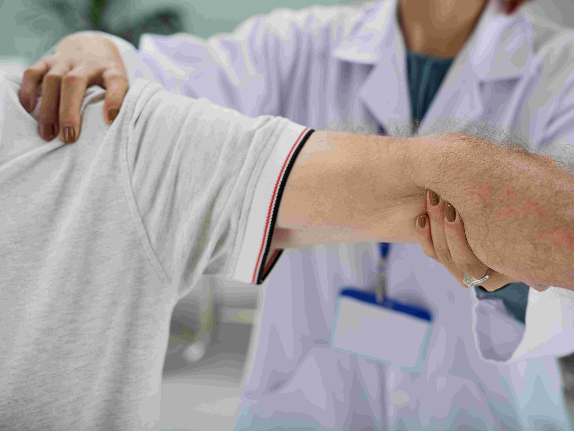
Apprehension and Relocation Tests: Physical exams where the surgeon moves the arm to reproduce the sensation of instability.
Blood Panels: Routine testing to ensure the patient is fit for general anesthesia and outpatient surgery.
Life After Bankart Repair
This is almost always an outpatient procedure, allowing patients to return home the same day.
A shoulder sling must be worn 24/7 for 4 to 6 weeks to protect the repair while the tissue knits to the bone.
Initial physical therapy (weeks 1–6) focuses on "passive" motion only; "external rotation" (turning the hand outward) is strictly forbidden to avoid tearing the new stitches.
Active strengthening of the rotator cuff and shoulder blade muscles begins around week 6 to 8.
Return to non-contact sports typically occurs at 3 to 4 months, while contact sports (football, rugby) require 6 to 9 months of rehabilitation.
Benefits of Bankart Repair
Excellent success rates for preventing future dislocations and restoring confidence in the joint.
Restores the natural anatomy of the shoulder socket, providing a more stable "cradle" for the humerus.
Minimally invasive techniques lead to smaller scars and less post-operative pain than open surgery.
Significantly reduces the long-term risk of developing shoulder arthritis caused by repeated dislocations.