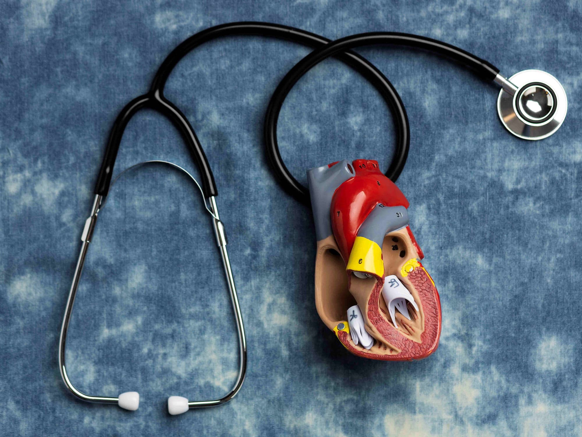
Cardiac Tumour Removal Surgery
Cardiac Tumour Removal Surgery
Cardiac Tumour Removal Surgery is a specialized procedure to excise abnormal growths from within or on the heart. While the majority of primary heart tumours (75–80%) are benign (non-cancerous), such as myxomas, they can still be life-threatening. These growths can obstruct blood flow, damage heart valves, or lead to strokes if pieces of the tumour break off and travel to the brain. Advanced imaging and robotic techniques allow for highly precise removal of these rare growths.
When You Should Consider Cardiac Tumour Removal
Benign Myxomas: The most common heart tumour, typically found in the left atrium, which requires removal to prevent blood flow obstruction.
Papillary Fibroelastomas: Small growths on heart valves that carry a high risk of causing a stroke or heart attack.
Symptoms of Obstruction: If a tumour causes dizziness, fainting, or sudden shortness of breath by blocking a heart valve.
Embolic Events: If pieces of a suspected tumour have already broken off and caused "mini-strokes" (TIAs) or blood clots in the limbs.
Malignant Sarcomas: Rare, aggressive cancers that require surgery to relieve symptoms or as part of a multi-stage treatment plan involving chemotherapy.
Surgical Approaches
Open-Heart Surgery (Median Sternotomy): The traditional approach providing the most direct view, necessary for large, complex, or malignant tumours.
Minimally Invasive Surgery: Uses small incisions (3–5 cm) between the ribs, often assisted by a 3D endoscope to reduce pain and scarring.
Robotically Assisted Surgery: A standard for precision, where surgeons use robotic arms to remove tumours in tight spaces within the heart.
Cardiopulmonary Bypass (CPB): Using a heart-lung machine to maintain circulation while the heart is stopped for the safe excision of the growth.
Reconstructive Surgery: Using a patch (synthetic or from the patient’s own pericardium) to repair any holes left in the heart wall after the tumour is removed.
How Is Performed
Access: The surgeon reaches the heart via a sternotomy or a minimally invasive port-access between the ribs.
Bypass: The patient is connected to the heart-lung machine, allowing the surgeon to open the heart chambers in a bloodless environment.
Excision: The tumour is meticulously removed, usually along with a small "margin" of healthy tissue to ensure no cells are left behind to regrow.
Repair: If the tumour was attached to a valve, the surgeon performs a valve repair or replacement during the same session.
Verification: The heart is closed and restarted, and an intraoperative echocardiogram is performed to ensure the tumour is gone and the valves are functioning perfectly.
Pre-Procedure Preparation
Fasting: Required for at least 8–12 hours before surgery, as the procedure is performed under general anesthesia.
Blood Work: Extensive blood work and cross-matching for blood transfusions, which are common in complex cardiac resections.
Dental Clearance: To ensure no bacteria from the mouth could infect the surgical site or any repair patches.
Medication Adjustment: Stopping certain medications, particularly blood thinners, several days before the operation.
Logistics: Arranging for a hospital stay of roughly one week and a support person for the multi-week recovery at home.
Tests Before Cardiac Tumour Removal
Echocardiogram (TTE/TEE): The primary tool used to identify the tumour's size, mobility, and attachment point.
Cardiac MRI: Provides high-definition 3D tissue characterization to help distinguish between benign and malignant growths.
Cardiac CT Scan: Used to evaluate the tumour’s relationship with the coronary arteries and the chest wall.
Coronary Angiogram: Performed in older patients to check for blockages that may need to be bypassed during the same surgery.
PET Scan: Occasionally used if a malignant tumour is suspected, to check if the cancer has spread elsewhere in the body.
Life After Cardiac Tumour Removal
ICU Stay: Patients spend 1–2 days in the Intensive Care Unit for constant monitoring of heart rhythm and oxygen levels.
Hospital Discharge: Most patients go home after 5 to 10 days, depending on whether the approach was open or minimally invasive.
Activity Restrictions: No heavy lifting (over 4 kg) for 6 to 12 weeks to allow the breastbone or rib incisions to heal fully.
Cardiac Rehabilitation: Supervised exercise is strongly recommended to rebuild physical strength and cardiovascular endurance.
Long-term Monitoring: Annual echocardiograms are usually required for several years to ensure the tumour does not recur.
Benefits of Cardiac Tumour Removal
Cure for Benign Growths: For tumours like myxomas, surgery is often completely curative with excellent long-term results.
Stroke Prevention: Removing highly mobile tumours significantly reduces the risk of life-altering strokes or organ damage.
Restores Blood Flow: Eliminates heart failure symptoms caused by tumours obstructing the heart valves.
Specialized Outcomes: In-hospital mortality is relatively low (approximately 3%) for such a specialized and complex procedure.
Symptom Relief: Most patients experience an immediate improvement in energy levels and a reduction in fainting or palpitations.