Corpectomy (Vertebral Body Removal)
Corpectomy (Vertebrectomy)
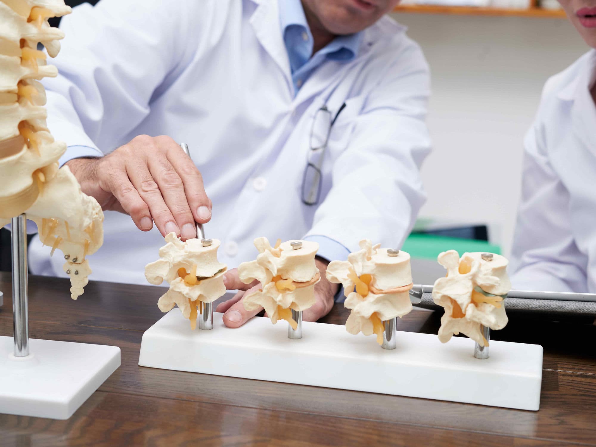
A Corpectomy, also known as a vertebrectomy, is a major spinal surgery involving the removal of all or part of a vertebral body to relieve significant pressure on the spinal cord and nerves. Unlike a discectomy, which only removes disc material, a corpectomy is used when disease or damage extends into the bone of the vertebra itself.
When You Should Consider Corpectomy
Surgeons typically recommend this procedure for severe conditions that cannot be treated with less invasive methods. Indications include:
Severe Spinal Stenosis: Confluent narrowing that extends behind the vertebral body.
Vertebral Tumors: Primary or metastatic tumors that destroy the bone and compress the spinal cord.
Spinal Fractures: Traumatic burst fractures where bone fragments are pushed into the spinal canal.
Bone Infections: Conditions like osteomyelitis or tuberculosis that cause vertebral collapse.
Cervical Myelopathy: Compression of the spinal cord in the neck causing loss of coordination or bladder control.
Methods of Corpectomy
Anterior Cervical Corpectomy: Performed through the front of the neck to access the cervical spine.
Side-Access Lumbar Corpectomy: Approached from the side of the body for issues in the lower back.
Reconstruction with Strut Grafts: Using bone from the patient (autograft) or a donor (allograft) to fill the gap.
Reconstruction with Expandable Cages: Using titanium or synthetic mesh cages packed with bone graft for structural support.
How Corpectomy Is Performed
Surgical Access: The surgeon makes an incision, most commonly through the front or side, depending on the location of the affected vertebra.
Vertebral Removal: The surgeon removes the damaged vertebral body along with the discs directly above and below it.
Reconstruction: To fill the resulting gap, the "anterior column" is rebuilt using a graft or a specialized expandable cage.
Stabilization: Metal plates and screws are attached to the remaining vertebrae to hold the reconstruction in place while the bones fuse.
Pre-Procedure Preparation
Fasting: Patients must fast for 8–12 hours prior to the procedure.
Medical Clearances: Extensive blood tests, ECG, and chest X-rays are required to assess fitness for major surgery.
Medication Review: Guidance from the cardiology or surgical team on adjusting medications that may affect bleeding or healing.
Recovery Planning: Arranging for significant post-operative support and home modifications for the initial recovery phase.
Tests Before Corpectomy
MRI Scan: Essential for visualizing spinal cord compression and soft tissue involvement.
CT Scan: Provides detailed mapping of the bony structures and the extent of vertebral damage.
X-rays: Used to evaluate overall spinal alignment and stability.
Cardiac Catheterization or Stress Test: May be required for older patients or those with high-risk factors to measure heart health before major surgery.
Life After Corpectomy (Recovery)
Hospital Stay: Typically requires 1 to 3 days, though complex lumbar cases may stay longer.
Initial Restrictions: Patients often wear a cervical collar or back brace for 4 to 8 weeks to protect the fusion site.
Activity: Desk work and light daily activities can often be resumed within 3 to 6 weeks.
Long-term Healing: Complete bony fusion between the graft and the vertebrae typically takes 6 months to 1 year.
Benefits of Corpectomy
Spinal Cord Protection: Stops the progression of neurological damage and protects the lungs and body from further disability.
Structural Stability: Restores the integrity of the spinal column following trauma or tumor-related destruction.
Long-term Cure: Provides a definitive treatment for complex bone-related nerve compression with high success rates.
Functional Improvement: Significant improvement in coordination, strength, and overall physical stamina.