Cross-linking (for Keratoconus)
Corneal Collagen Cross-linking (CXL)
Corneal Collagen Cross-linking (CXL) is a specialized medical procedure used to treat Keratoconus, a progressive condition where the cornea thins and bulges into a cone shape. Unlike LASIK or SMILE, which are designed to correct vision, the primary goal of CXL is to strengthen the corneal tissue to stop the disease from worsening and prevent future vision loss.
When You Should Consider CXL
Progressive Keratoconus: When repeat eye exams show that the cornea is continuing to thin or the "cone" shape is becoming more pronounced.
Post-LASIK Ectasia: A rare complication where the cornea becomes unstable and thins following refractive surgery.
Pellucid Marginal Degeneration: A similar corneal thinning condition that affects the lower part of the cornea.
Fluctuating Vision: When your eyeglass or contact lens prescription is changing rapidly due to corneal instability.
Early Diagnosis: It is highly effective when performed early to stabilize the cornea before significant vision loss occurs.
How Is Performed
Numbing: Anesthetic eye drops are applied so the patient remains comfortable and feels no pain during the process.
Epithelium Management:
Epi-off Method: The thin outer layer (epithelium) is gently removed to allow the medication to saturate the deeper layers more effectively.
Epi-on (Trans-epithelial): The outer layer is left intact, which may reduce post-operative discomfort.Riboflavin Saturation: Vitamin B2 (riboflavin) drops are applied to the eye every few minutes for about 30 minutes until the cornea is fully saturated.
UV Light Exposure: The eye is exposed to a controlled amount of Ultraviolet A (UVA) light for several minutes.
The Chemical Reaction: The interaction between the Riboflavin and UV light creates new "cross-links" (chemical bonds) between the collagen fibers, making the cornea stiffer and more stable.
Bandage Lens: A clear, soft contact lens is placed on the eye to protect the surface while it heals.
Duration: The entire treatment typically takes 30 to 60 minutes.
Pre-Procedure Preparation
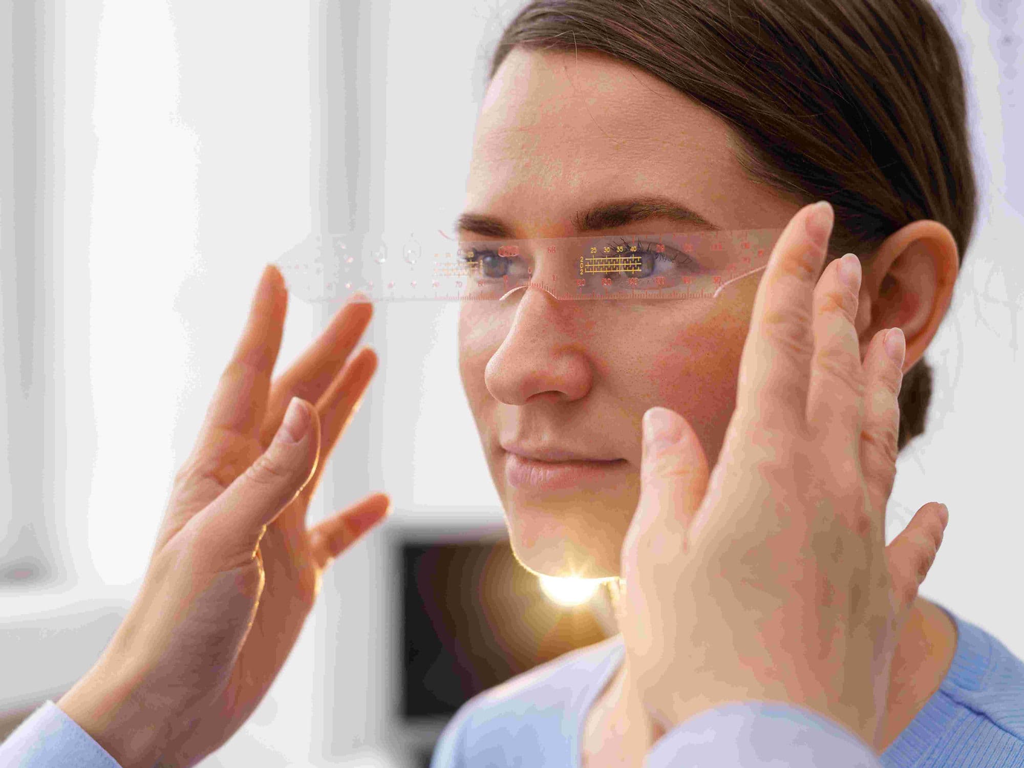
Corneal Mapping: Detailed scans (topography) are taken to document the current shape and thickness of the cornea as a baseline.
Contact Lens Holiday: Patients must stop wearing hard or gas-permeable contact lenses for several weeks before the procedure to allow the cornea to settle.
Medication Review: Discussing any history of slow healing or eye infections with your specialist.
Transportation: Arranging for a ride home, as the eye may be sensitive to light and vision may be blurry immediately after the UV exposure.
Tests Before CXL
Pachymetry: To measure the thinnest point of the cornea; a minimum thickness is often required to safely perform the UV light stage.
Corneal Topography/Tomography: To identify the "apex" of the cone and track the rate of disease progression.
Slit-Lamp Examination: To check for any pre-existing corneal scarring that might affect the treatment outcome.
Visual Acuity: Establishing the current level of corrected and uncorrected vision.
Life After CXL
Initial Discomfort: Especially in "Epi-off" cases, the eye may feel sore, gritty, or highly light-sensitive for the first 3–5 days.
Vision Fluctuations: It is normal for vision to be slightly "foggy" or worse immediately after the procedure before it stabilizes over several weeks.
Medication Regimen: Patients must use prescribed antibiotic and steroid drops for several weeks to prevent infection and manage inflammation.
Healing Timeline: While the surface heals within a week, the full strengthening effect of the cross-linking takes 3 to 6 months to reach maximum stability.
Follow-up Care: Regular checkups are essential to monitor the "haze" (temporary cloudiness) and ensure the epithelium has regrown properly.
Why Specialized Treatment Is Highly Effective
Prevents Disease Progression: CXL has a success rate of over 90% in stopping Keratoconus from getting worse.
Avoids Major Surgery: By stabilizing the cornea early, the vast majority of patients can avoid the need for a full corneal transplant later in life.
Long-Term Stability: The new chemical bonds created between collagen fibers provide a permanent increase in corneal rigidity.
Preserves Vision: While it does not eliminate the need for glasses, it "locks" the vision in place and prevents further deterioration.
Minimally Invasive: It is an outpatient procedure that does not require stitches or a long hospital stay.