External Beam Radiation Therapy
Bone and Soft Tissue Tumor Surgery
Surgery for bone and soft tissue tumors (primarily sarcomas) is a highly specialized field where the goal is to remove the cancer completely while preserving as much physical function and appearance as possible. Because these tumors often grow near major nerves, blood vessels, and joints, the surgery requires meticulous planning. The standard of care involves using 3D-printed models and computer-assisted navigation to achieve precise "clear margins" while sparing the limb.
When You Should Consider Sarcoma Surgery
Primary Bone Cancer: For malignancies such as osteosarcoma, Ewing sarcoma, or chondrosarcoma.
Soft Tissue Sarcoma: When a cancerous mass is identified in the muscle, fat, nerves, or connective tissues (e.g., liposarcoma or synovial sarcoma).
Benign but Aggressive Tumors: For non-cancerous growths like Giant Cell Tumors (GCT) that can destroy local bone if not removed.
Metastatic Bone Disease: When cancer from another organ (like the lung or breast) has spread to a bone and threatens to cause a fracture.
Recurrent Tumors: When a previously treated tumor returns in the same anatomical compartment.
Types of Surgical Margins
The "margin" is the area of healthy tissue removed along with the tumor. Surgeons use specific classifications to define how much tissue to take:
Intralesional (Curettage): The tumor is scraped out from the inside. This is generally used only for benign (non-cancerous) bone tumors.
Marginal Excision: The tumor is removed exactly at its edge (pseudocapsule). This is often used for benign soft tissue tumors like lipomas.
Wide Excision: The tumor is removed with a continuous "cuff" of healthy tissue surrounding it. This is the standard of care for malignant tumors (sarcomas) to ensure no microscopic cells are left behind.
Radical Resection: Removal of the entire anatomical compartment (the whole bone or muscle group) containing the tumor.
Advanced Reconstruction Techniques
Once a tumor is removed, the resulting gap must be rebuilt to restore strength and mobility:
Biological Reconstruction: Uses the body's own ability to heal.
Allograft: Uses donated bone from a bone bank to act as a scaffold.
Autograft: Uses the patient's own bone, such as the fibula (calf bone), often moved with its blood vessels intact.Mechanical Reconstruction: Uses artificial megaprostheses (large metal implants) to replace joints or long sections of bone. These allow for immediate weight-bearing.
Distraction Osteogenesis: Using devices like the Ilizarov fixator to slowly "grow" new bone to fill a gap.
Specialized Procedures for Children
Because children's bones are still growing, surgery requires unique solutions to prevent leg-length discrepancies:
Expandable Prostheses: Metal implants that can be lengthened non-invasively using magnets as the child grows to keep the legs equal in length.
Rotationplasty: A specialized procedure where the middle of the leg is removed, and the lower leg is rotated 180° and reattached to the thigh. The ankle then functions as a knee joint.
Growth Plate Sparing: Advanced 3D navigation allows surgeons to remove tumors while saving the natural growth plates whenever possible.
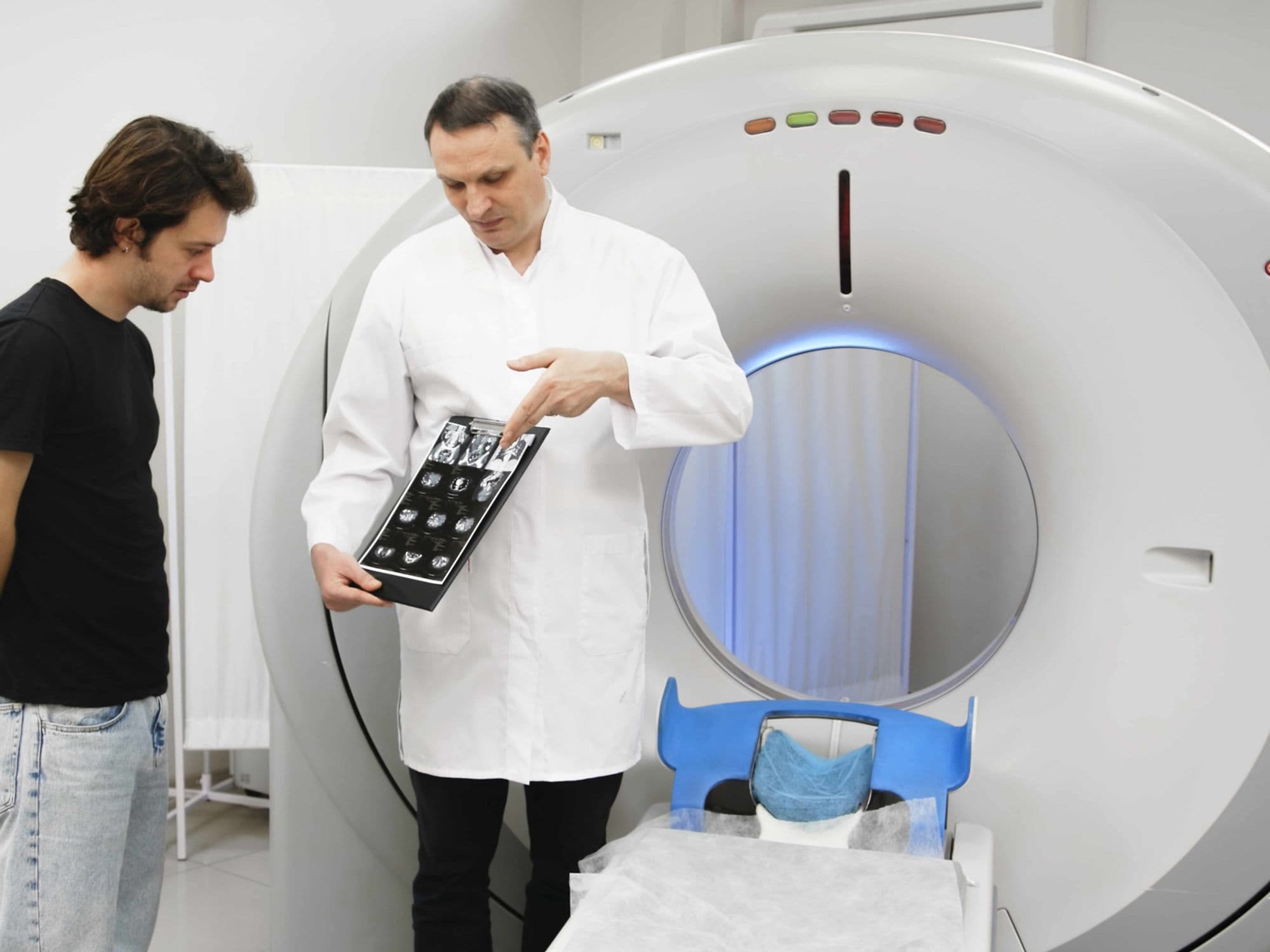
[Image showing an expandable "growing" prosthesis for a pediatric patient]
How Is Performed
Anesthesia: Performed under general anesthesia. Advanced nerve blocks are often used to provide long-term pain relief to the limb.
Computer-Assisted Navigation: Surgeons use "GPS for surgery" to follow a pre-planned 3D map, ensuring they cut exactly where the cancer ends and healthy bone begins.
Multidisciplinary Collaboration: If the tumor involves major blood vessels or leaves a large skin defect, vascular and plastic surgeons work simultaneously to perform bypasses or skin flaps.
3D-Printed Cutting Guides: Custom-made templates are placed on the bone during surgery to guide the saw blade with sub-millimeter precision.
Intraoperative Imaging: Using O-arm or C-arm technology to verify the placement of implants and the completeness of the resection before the patient leaves the OR.
Pre-Procedure Preparation
Tumor Board Review: Your case is reviewed by a team of radiologists, pathologists, and oncologists to determine the best sequence of treatment.
3D Virtual Planning: Surgeons use MRI/CT scans to create a virtual 3D model of your limb to practice the surgery before the actual procedure.
Physical Therapy Baseline: Establishing a baseline for your limb's strength and range of motion to guide your post-operative recovery.
Nutritional Optimization: Ensuring high protein intake to support the extensive bone and tissue healing required.
"Pre-hab" Exercise: Strengthening the healthy limbs to prepare for the period of restricted weight-bearing on the operated side.
Tests Before Bone and Soft Tissue Tumor Surgery
High-Resolution MRI: The most critical test for visualizing the tumor's relationship to muscles, nerves, and blood vessels.
Systemic CT Scan: To rule out "skip lesions" or spread to the lungs, which is common with certain sarcomas.
PET-CT Scan: To identify any other areas of metabolic activity that might indicate the cancer has moved elsewhere.
Core Needle Biopsy: To confirm the exact grade and type of the sarcoma, which dictates how wide the surgical margins must be.
Angiography: To map out the blood supply of the limb, especially if a vascularized bone graft (autograft) is planned.
Life After Sarcoma Surgery (Recovery & Risks)
Hospital Stay: Typically 5 to 10 days depending on the complexity of the reconstruction and the level of pain management needed.
Rehabilitation: This is the most critical phase. Physical therapy usually begins within 24–48 hours and can continue for 6 to 12 months.
Weight-Bearing Rules: Depending on the type of bone graft or prosthesis, you may need to use crutches or a walker for 3 to 6 months while the bone heals.
Mechanical Wear: Over many years, metal megaprostheses can wear out or loosen, potentially requiring a revision surgery.
Infection Monitoring: Large implants and pre-operative chemotherapy can increase the risk of infection, requiring long-term monitoring.
Why Specialized Treatment Is Highly Effective
Limb Salvage Success: Over 90% of sarcoma patients can have their limbs saved rather than amputated, with no loss in survival rates.
Precise Margin Control: Advanced 3D navigation has significantly lowered the risk of local recurrence by ensuring no microscopic cells are missed.
Functional Recovery: Modern megaprostheses and biological grafts allow many patients to return to walking, swimming, and an active lifestyle.
Growth Management: Expandable technology ensures that children can reach their full height without the need for multiple major open surgeries.
Integrated Care: When surgery is combined with modern immunotherapy and targeted radiation, the long-term cure rates for sarcomas are higher than ever before.