IVC Filter Placement
Inferior Vena Cava (IVC) Filter Placement
Inferior Vena Cava (IVC) Filter Placement is a minimally invasive procedure to insert a small, cage-like metal device into the body's largest vein (the IVC). Its purpose is to trap blood clots traveling from the legs or pelvis before they can reach the heart and lungs, thereby preventing a life-threatening Pulmonary Embolism (PE). While blood thinners remain the standard treatment, this transcatheter technique has expanded significantly for patients who cannot safely take anticoagulants.
When You Should Consider IVC Filter Placement
Active bleeding (e.g., gastrointestinal or brain bleed) that prevents the use of blood thinners.
New blood clots forming or traveling to the lungs despite proper blood-thinning medication.
Recent major surgery or massive trauma where anticoagulation is not an option.
High-risk prophylaxis for patients undergoing specialized high-risk surgeries.
Evidence of significant heart overload or potential for massive pulmonary embolism.
Methods of IVC Filter Placement
Transcatheter Placement: Minimally invasive method using a neck or groin catheter to "plug" the vein with a filter.
Retrievable (Optional) Filters: Devices designed to be removed once the immediate risk of blood clots has passed.
Permanent Filters: Intended for patients with a lifelong risk of clots who can never safely take blood thinners.
Below-Renal Deployment: Placing the filter just below the kidney veins to avoid interfering with renal blood flow.
Device Occlusion: Deployment of "soft" low-profile metal devices to block clots without major surgery.
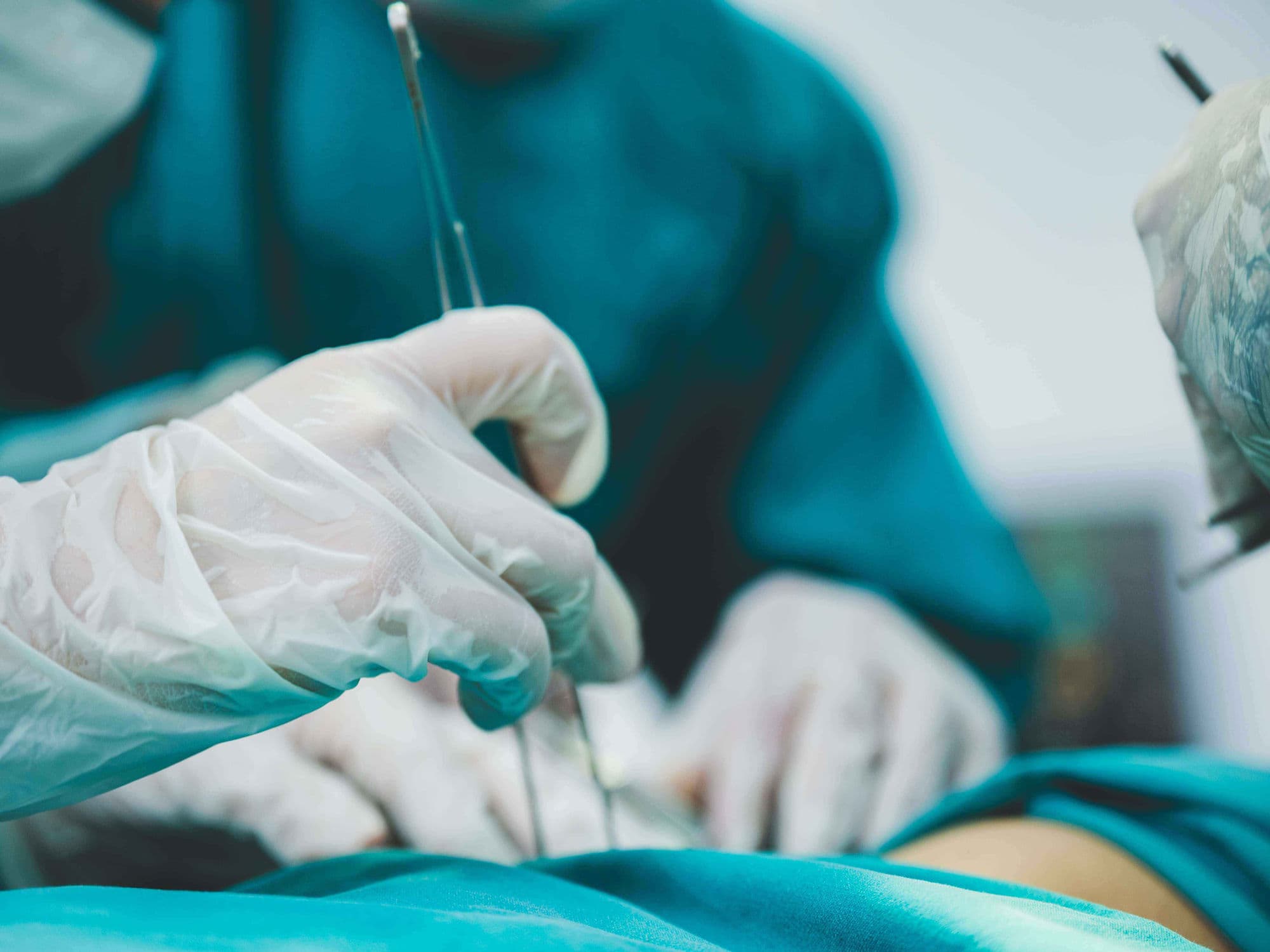
How IVC Filter Placement Is Performed
Catheter Access: A thin tube is guided through the internal jugular vein (neck) or femoral vein (groin) to the heart.
Imaging Guidance: Real-time X-ray (fluoroscopy) and contrast dye ensure the filter is perfectly positioned before finishing.
Filter Deployment: A collapsed occluder-like device is expanded across the vein to trap clots permanently or temporarily.
Release: Once positioned, the filter attaches to the vein walls using small hooks or radial pressure.
Monitoring: Doctors verify the filter is securely anchored below the renal veins before removing the delivery system.
Pre-Procedure Preparation
Fasting for 8-12 hours before the catheterization procedure.
Blood tests, ECG, and chest X-rays to assess overall health and kidney function.
Adjusting current medications as directed by the cardiology or radiology team.
Discussing any allergies, particularly to the metal in the device or contrast dye.
Arranging for post-operative care and a support person for the recovery period.
Tests Before IVC Filter Placement
Duplex Ultrasound to determine the size and location of existing blood clots.
Cardiac Catheterization to measure lung pressures and map the venous anatomy.
Cardiac MRI or CT scan for detailed 3D mapping of the inferior vena cava.
ECG to monitor the heart's electrical rhythm and check for strain.
Pulse oximetry to evaluate oxygen saturation levels in the blood.
Life After IVC Filter Placement
Short hospital stay, usually 1-2 days for device closure, often as an outpatient procedure.
Avoid strenuous activity and heavy lifting for a few days post-procedure.
Most patients return to normal daily activities within 24 to 48 hours.
Regular follow-up visits with a cardiologist to monitor the repair site and discuss retrieval.
Immediate protection against life-threatening pulmonary embolism and improved peace of mind.
Benefits of IVC Filter Placement
Restores safety by trapping dangerous clots before they reach the heart and lungs.
Protects the lungs from permanent damage caused by massive pulmonary emboli.
Provides a vital alternative for patients who cannot tolerate traditional blood-thinning medications.
Reduces the risk of sudden cardiac events and enlargement of the heart's chambers.
Provides a long-term or temporary solution with very high technical success rates.