Laparoscopic Hernia Repair
Laparoscopic Hernia Repair
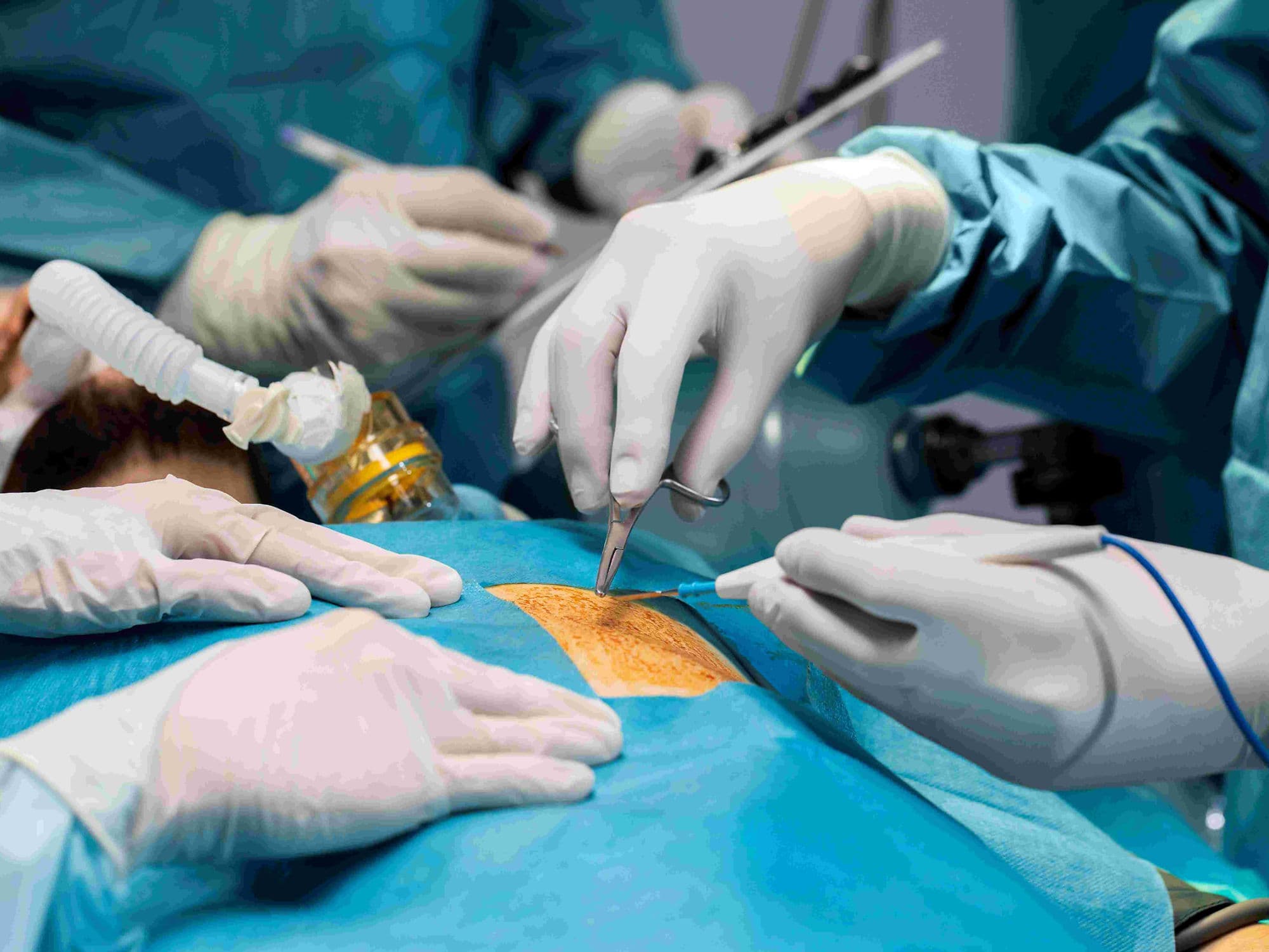
Laparoscopic Hernia Repair, also known as minimally invasive or keyhole surgery, uses specialized tools and a camera to repair a weakness in the abdominal wall from the inside out. It is most commonly used for inguinal (groin), umbilical (navel), and ventral hernias.
When You Should Consider Laparoscopic Hernia Repair
A visible bulge in the groin or abdomen that may become more prominent when standing or coughing.
Pain or pressure at the hernia site during physical activity or lifting.
Inguinal hernias that occur on both sides (bilateral) of the groin.
Recurrent hernias where a previous open surgical repair has failed.
Desire for a faster return to work and daily activities with minimal scarring.
Methods of Laparoscopic Hernia Repair
TAPP (Transabdominal Preperitoneal): The surgeon enters the peritoneal cavity where the organs are located to reach and repair the hernia.
TEP (Totally Extraperitoneal): The surgeon operates in the space between the muscle and the abdominal lining, avoiding the organ cavity entirely.
IPOM (Intraperitoneal Onlay Mesh): Primarily used for ventral hernias, where a specialized mesh is placed on the inside of the abdominal wall.
Robotic-Assisted Laparoscopy: Using a robotic interface for enhanced precision and 3D visualization during the repair.
How Laparoscopic Hernia Repair Is Performed
Insufflation: The abdominal cavity is inflated with Carbon Dioxide (CO2) gas to create a dome-shaped workspace.
Port Insertion: Three to four small incisions (0.5–1 cm) are made to allow the insertion of a laparoscope and long, thin instruments.
Reduction: Protruding tissue, such as fat or a loop of intestine, is carefully pulled back into the abdominal cavity from the inside.
Mesh Reinforcement: A lightweight, flexible synthetic mesh is unfurled over the defect to reinforce the weakened wall.
Fixation: The mesh is secured in place using surgical tacks, staples, or specialized surgical glue to prevent shifting.
Pre-Procedure Preparation
Fasting (NPO) for 6–8 hours prior to the surgery to ensure safety during anesthesia.
Pausing blood-thinning medications several days in advance as coordinated with the surgical team.
Medical clearance to ensure the patient can tolerate general anesthesia and abdominal CO2 inflation.
Discussing the specific approach (TAPP vs. TEP) based on the hernia's location and surgical history.
Tests Before Laparoscopic Hernia Repair
Physical Examination: The primary method to determine if the hernia is "reducible" or "incarcerated."
Ultrasound or CT Scan: Imaging used to confirm the diagnosis and map the size of the abdominal wall defect.
Electrocardiogram (ECG): Often required for patients over a certain age to ensure heart health for general anesthesia.
Blood Panels: To check for infection markers and ensure proper kidney and liver function.
Life After Laparoscopic Hernia Repair
Most patients return home the same day as the procedure (day-care surgery).
Walking is encouraged immediately following surgery to prevent blood clots.
Desk-based work can typically be resumed within 3 to 7 days.
Strict lifting restrictions—usually no more than 5–10 kg—must be followed for 4–6 weeks.
Monitoring for temporary shoulder pain, which is a common side effect of the CO2 gas used during surgery.
Benefits of Laparoscopic Hernia Repair
Results in significantly smaller scars and a lower risk of wound infection compared to open surgery.
Offers a faster recovery timeline and a quicker return to professional and athletic activities.
Provides a superior view for the surgeon to identify and repair multiple hernia defects through the same incisions.
Utilizes a "tension-free" mesh technique that lowers the risk of the hernia returning.