Lobectomy (Cancer)
Lobectomy
A Lobectomy is the surgical removal of an entire lobe of an organ. While it can be performed on the liver, brain, or thyroid, it is most commonly the "gold standard" surgical treatment for early-stage Non-Small Cell Lung Cancer (NSCLC). In 2026, the procedure is frequently performed using robotic-assisted technology to ensure the most precise removal of the tumor and surrounding lymph nodes.
When You Should Consider a Lobectomy
Early-Stage Lung Cancer: For Stage I or II NSCLC where the tumor is confined to a single lobe of the lung.
Localized Tumors: When the malignancy is centrally located within a lobe, making a smaller "wedge" resection insufficient.
Curative Intent: When the goal is to remove the primary tumor along with its dedicated lymphatic drainage system.
Infectious Disease: Occasionally performed for severe, localized infections like tuberculosis or fungal balls that do not respond to medication.
Congenital Abnormalities: To remove a lobe that has not formed correctly or is causing recurrent health issues.
Surgical Approaches
RATS (Robotic-Assisted Thoracic Surgery): The 2026 preferred method for complex cases. The surgeon operates robotic arms from a console, offering high-definition 3D visualization and greater dexterity for removing deep lymph nodes.
VATS (Video-Assisted Thoracoscopic Surgery): A minimally invasive technique using 2–4 small incisions (1–3 cm). A camera (thoracoscope) guides the surgeon, resulting in less pain and a faster recovery than open surgery.
Thoracotomy (Open Surgery): A traditional 15–20 cm incision made between the ribs. This provides a direct view and is used for larger tumors or those near major blood vessels.
Sleeve Lobectomy: A specialized approach where a piece of the main bronchus is also removed and "re-sleeved" to save the rest of the lung tissue.
How a Lobectomy Is Performed
Anesthesia: Performed under general anesthesia using a "double-lumen" tube, which allows the surgeon to deflate the lung being operated on while the other lung continues to breathe.
Anatomic Dissection: The surgeon carefully separates and identifies the specific pulmonary artery, pulmonary vein, and bronchus belonging to the affected lobe.
Precision Stapling: These major structures are sealed and cut using advanced surgical staplers to prevent bleeding and air leaks.
Lymphadenectomy: Surgeons remove nearby mediastinal lymph nodes to check for microscopic cancer spread, which determines the need for "mop-up" chemotherapy.
Chest Tube Placement: A tube is inserted into the pleural space to drain air and fluid, allowing the remaining lung lobes to expand and fill the chest cavity.
Inflation Test: Before closing, the remaining lung is reinflated under water to check for bubbles, ensuring the surgical site is airtight.
Pre-Surgery Preparation
Pulmonary Function Tests (PFTs): Essential tests (Spirometry) to ensure your remaining lung lobes can support your breathing needs after the surgery.
Smoking Cessation: You must stop smoking for at least 4 weeks prior to surgery to reduce the risk of post-operative pneumonia and air leaks.
Cardiac Clearance: Undergoing an EKG or stress test to ensure your heart can handle the circulatory changes during a lung resection.
Nutritional Loading: Adhering to a high-protein diet to provide the body with the resources needed for the pleura (lung lining) to heal quickly.
Incentive Spirometry Training: Learning how to use a breathing exercise device before the surgery so you can effectively clear your lungs during recovery.
Pre-Surgery Tests
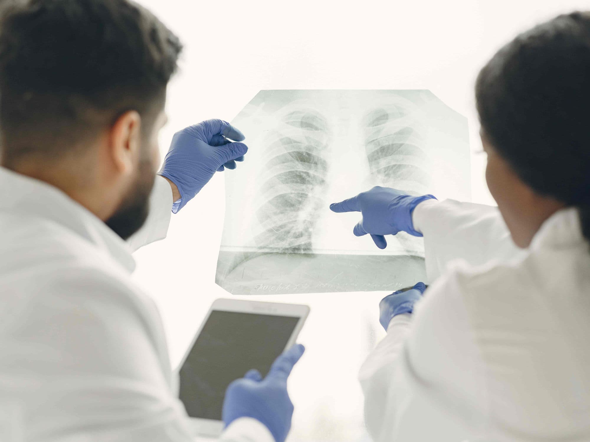
High-Resolution CT Scan: To map the tumor’s exact location in relation to the complex branching of the pulmonary vessels.
PET-CT Scan: To confirm that the cancer has not spread to other organs, ensuring that a lobectomy is the most effective curative path.
Quantitative V/Q Scan: In borderline cases, this determines exactly how much "work" the lobe to be removed is currently doing.
EBUS (Endobronchial Ultrasound): A specialized biopsy of the lymph nodes near the windpipe to confirm the cancer's stage before the main surgery.
Baseline Blood Work: Comprehensive panels (CBC/CMP) to check for anemia or kidney issues that could affect healing.
Life After a Lobectomy (Recovery & Risks)
Hospital Stay: Usually 3–4 days for VATS/Robotic surgery and 5–7 days for an open thoracotomy.
Chest Tube Removal: The tube is usually removed on day 2 or 3 once the drainage stops and the "air leak" is gone.
Air Leak Management: The most common complication; most small leaks heal within a few days while the chest tube remains in place.
Atrial Fibrillation (AFib): A temporary irregular heart rhythm (10–20% of cases) caused by inflammation near the heart; it is typically managed with medication.
Full Activity: Most patients return to light daily tasks within 2 weeks and full physical activity within 6 to 8 weeks.
Why Specialized Treatment Is Highly Effective
The "Gold Standard": Lobectomy provides the lowest rate of local cancer recurrence compared to smaller, "sub-lobar" resections.
Compensatory Growth: If the remaining lobes are healthy, they will typically expand and "re-model" to fill the space, resulting in minimal long-term shortness of breath.
Robotic Accuracy: 2026 data shows that robotic lobectomy leads to a more thorough lymph node harvest, providing the most accurate cancer staging possible.
Enhanced Recovery (ERAS): Specialized thoracic protocols allow for earlier walking and eating, which significantly reduces the risk of blood clots.
Multidisciplinary Success: When paired with modern 2026 immunotherapy, a lobectomy provides the strongest foundation for long-term lung cancer survival.