Lung & Thoracic Cancer Surgery
Lung and Thoracic Cancer Surgery
Lung and Thoracic Cancer Surgery involves the surgical removal of tumours from the lungs, chest wall, or the mediastinum (the space between the lungs). The primary goal is to achieve an "R0 resection," meaning the entire tumour is removed with clear, cancer-free margins. Clinical standards favor minimally invasive approaches like VATS and RATS to preserve respiratory function and accelerate recovery.
When You Should Consider Thoracic Surgery
Early-Stage NSCLC: For Non-Small Cell Lung Cancer (Stage I or II) where surgery offers the highest chance of a permanent cure.
Solitary Pulmonary Nodules: When a suspicious "spot" on the lung is growing or has high-risk features on a PET-CT.
Mediastinal Tumours: Malignancies located in the center of the chest, such as thymomas or germ cell tumours.
Metastatic "Oligometastases": When cancer from another organ (like the kidney or colon) has spread only to a limited area of the lung.
Chest Wall Involvement: When a lung tumour has invaded the ribs, requiring a combined resection and reconstruction.
Types of Lung Resections
Wedge Resection: Removal of a small, pie-shaped piece of lung; reserved for very small peripheral tumours or patients with limited lung capacity.
Segmentectomy: Removal of a specific functional segment. This 2026 standard preserves more healthy tissue than a lobectomy for early-stage "ground-glass" opacities.
Lobectomy: The "gold standard" for most lung cancers. One of the five lobes (three right, two left) is removed entirely to capture all local lymph drainage.
Pneumonectomy: Removal of an entire lung; only performed for centrally located tumours involving the main bronchus.
Sleeve Resection: A lung-sparing alternative to pneumonectomy where a section of the bronchus is removed and the healthy ends are sewn back together.
Surgical Approaches
RATS (Robotic-Assisted Thoracic Surgery): The 2026 preferred method for complex dissections. It provides 3D visualization and extreme precision for removing lymph nodes in the narrow mediastinum.
VATS (Video-Assisted Thoracoscopic Surgery): A minimally invasive approach using 2–3 small incisions (1–3 cm). It results in significantly less pain and faster return to activity.
Thoracotomy (Open Surgery): A larger incision on the side of the chest where ribs are spread; necessary for very large tumours or those involving major heart vessels.
Mediastinoscopy: A small incision at the base of the neck used to biopsy lymph nodes and confirm the cancer hasn't spread before a major resection.
Pleurodesis: A procedure for fluid buildup (effusion) where a sterile agent is used to make the lung stick to the chest wall, preventing fluid return.
How Thoracic Surgery Is Performed
Anaesthesia: Performed under general anaesthesia, typically using a "double-lumen" tube to deflate the lung being operated on.
Nodal Staging: Regardless of resection type, surgeons perform a mandatory lymphadenectomy to check for microscopic spread.
Airlock Testing: Before closing, the lung is reinflated under water to check for bubbles, ensuring the surgical site is airtight.
Chest Tube Placement: One or two tubes are placed in the pleural space to drain air and fluid, allowing the lung to remain fully expanded during healing.
Pathologic Staging: The removed tissue is analyzed to determine if "adjuvant" chemotherapy or immunotherapy is needed post-surgery.
Pre-Surgery Preparation
PFT/Spirometry: Completing a Pulmonary Function Test to ensure the remaining lung tissue can support your breathing needs after surgery.
Smoking Cessation: Adhering to a strict "zero-tobacco" policy for at least 4 weeks prior to surgery to reduce the risk of post-operative pneumonia.
Incentive Spirometry: Training with a breathing exercise device to strengthen respiratory muscles before the procedure.
Cardiac Risk Stratification: Undergoing an EKG or Echo to ensure the heart can handle the circulatory changes of thoracic surgery.
Nutritional Optimization: A high-protein diet to ensure the pleura (lung lining) heals quickly and prevents prolonged air leaks.
Pre-Surgery Tests
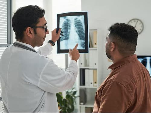
High-Resolution CT (Chest): To map the tumour's exact location in relation to the pulmonary arteries and veins.
PET-CT Scan: To rule out any metabolic activity in other parts of the body, ensuring the surgery remains a curative option.
Quantitative V/Q Scan: In borderline cases, this determines exactly how much "work" each lobe of your lung is doing.
EBUS (Endobronchial Ultrasound): A specialized internal ultrasound used to biopsy lymph nodes near the windpipe before the main surgery.
Liquid Biopsy: 2026 protocols may include a blood test to check for circulating tumour DNA (ctDNA) as a baseline for recovery.
Life After Lung Surgery (Recovery & Risks)
Chest Tube Management: Tubes are usually removed within 2–4 days once the "air leak" has stopped and drainage is minimal.
AFib Monitoring: Irregular heart rhythms occur in 10–20% of patients due to inflammation near the heart; this is typically temporary.
Early Mobilization: You will be encouraged to sit up and walk within 24 hours to prevent blood clots and help the lung expand.
Subcutaneous Emphysema: A "crackling" sensation under the skin if air traps there; it is harmless and usually resolves on its own.
Long-Term Breathlessness: Most patients return to normal activity in 4–8 weeks, though heavy aerobic exercise may feel different depending on the amount of lung removed.
Why Specialized Treatment Is Highly Effective
Robotic Precision: RATS allows for more thorough lymph node removal than traditional surgery, leading to more accurate staging and treatment.
Lung-Sparing Techniques: 2026 advancements in segmentectomy and sleeve resections allow for cancer removal while saving as much healthy lung as possible.
Enhanced Recovery (ERAS): Specialized thoracic protocols significantly reduce the need for heavy narcotics, allowing for faster mental and physical recovery.
Curative Foundation: Surgery remains the single most effective way to eliminate early-stage lung cancer and prevent future spread.