Permanent Pacemaker Implantation
Permanent Pacemaker Implantation (PPI)
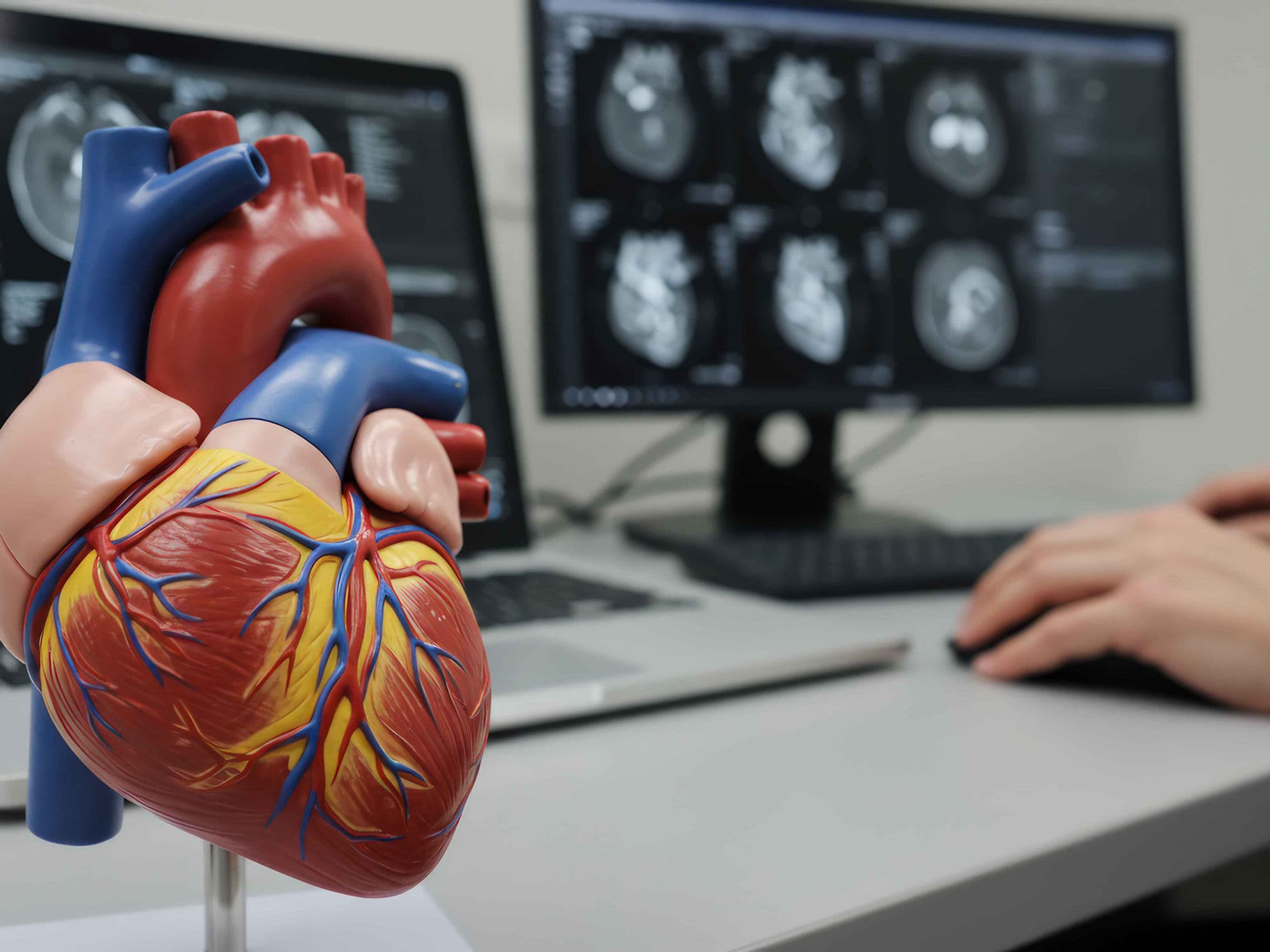
Permanent Pacemaker Implantation (PPI) is a routine, minimally invasive procedure to place a small electronic device in the chest to regulate a slow or irregular heartbeat (arrhythmia). The device monitors the heart's rhythm and sends electrical impulses to stimulate a contraction only when the heart's natural rate drops below a pre-set threshold. While traditional lead-based systems are common, newer leadless "capsule" devices have expanded treatment options by eliminating the need for chest incisions and wires.
When You Should Consider PPI
Chronic slow heart rate (bradycardia) causing fatigue or dizziness.
Irregular heart rhythms that do not respond to medication.
Evidence of heart block or significant heart overload detected during testing.
Heart failure symptoms where coordinated pumping is required (Biventricular pacing).
Risk of sudden fainting or cardiac events due to electrical conduction issues.
Core Components and Types
Pulse Generator: A small metal box containing a battery and a computer to monitor and pace the heart.
Leads (Electrodes): Thin, insulated wires that "sense" heart activity and "pace" the muscle by delivering signals.
Single-Chamber Pacemaker: Uses one lead in either the right atrium or right ventricle.
Dual-Chamber Pacemaker: Uses two leads to coordinate contractions between the upper and lower chambers.
Biventricular (CRT): Uses three leads to ensure the left and right ventricles pump in sync for heart failure patients.
How PPI Is Performed
Incision & Pocket Creation: A 2-inch incision is made below the collarbone to create a small "pocket" for the generator.
Vascular Access: A large vein is punctured to provide a path to the heart for the leads.
Lead Placement: Using real-time X-ray (fluoroscopy), leads are guided through the vein and secured to the heart wall.
Leadless Deployment: In leadless cases, a capsule device is implanted directly via a catheter in the groin.
Testing & Connection: The doctor verifies the leads trigger a heartbeat before plugging them into the generator and closing the site.
Pre-Procedure Preparation
Fasting for 8-12 hours before the surgery or catheterization.
Blood tests, ECG, and chest X-rays to assess overall cardiac health.
Adjusting current medications as directed by the cardiology team.
Discussing any allergies, particularly to materials in the device or contrast dye.
Arranging for post-operative care and a support person for the recovery period.
Tests Before PPI
Echocardiogram (TTE) to determine the heart's structure and pumping function.
Cardiac Catheterization to evaluate heart pressures if necessary.
ECG to monitor the heart's electrical rhythm and confirm the type of arrhythmia.
Pulse oximetry to evaluate oxygen saturation levels in the blood.
Chest X-ray to provide a baseline for the heart and lungs before implantation.
Life After PPI
Short hospital stay, usually involving one night for observation and a follow-up chest X-ray.
Avoid strenuous activity and lifting more than 4.5 kg for 4 to 6 weeks post-surgery.
Restrictions on raising the arm on the pacemaker side above shoulder level initially to prevent lead displacement.
Regular follow-up visits every 3 to 12 months to monitor the device and repair site.
Immediate improvement in heart rate regulation, breathing, and energy levels.
Benefits of PPI
Restores a normal heart rate and prevents symptoms of a slow or irregular heartbeat.
Protects the heart from long-term damage and enlargement caused by conduction issues.
Reduces the risk of fainting and other life-threatening cardiac events.
Provides a long-term solution with battery lives typically ranging from 5 to 15 years.
Allows for remote monitoring, ensuring constant cardiac safety and oversight.