Phacoemulsifacation
Phacoemulsification (Cataract Surgery)
Phacoemulsification, commonly referred to as "phaco," is the modern gold standard for cataract surgery. This procedure uses ultrasonic energy to fragment a clouded natural lens (cataract), allowing it to be removed through a microscopic, often stitchless incision. It is then replaced with a permanent artificial intraocular lens (IOL) to restore clear vision.
When You Should Consider Phacoemulsification
Cloudy or Blurry Vision: When daily activities like reading or driving become difficult due to a "foggy" lens.
Glare and Halos: Increased sensitivity to light, especially during night driving.
Fading Colors: When vibrant colors appear yellowed, dull, or faded.
Frequent Prescription Changes: Rapid changes in eyeglass or contact lens strength.
Double Vision: Experiencing multiple images in a single eye.
How Is Performed
Anesthesia: Numbing eye drops (topical anesthesia) or a local injection are used so the patient remains awake but feels no pain.
The Incision: A microscopic, self-sealing incision (typically 2.2 to 2.8 mm) is made at the edge of the cornea.
Capsulorhexis: A precise circular opening is created in the thin membrane (capsule) that surrounds the lens.
Phacoemulsification: A specialized probe emitting ultrasonic waves vibrates at high frequencies to fragment the cataract into tiny pieces.
Aspiration: The fragmented pieces are gently suctioned out of the eye using the same high-tech probe.
IOL Implantation: A foldable artificial lens is inserted through the tiny incision; it unfolds naturally inside the lens capsule to restore focus.
Closure: Because of the precise shape of the incision, stitches are usually unnecessary.
Pre-Procedure Preparation
Eye Measurement (Biometry): Precise ultrasound or laser measurements are taken to determine the correct power of the artificial lens (IOL).
Medication Audit: Patients may be asked to start antibiotic or anti-inflammatory drops a few days before the procedure.
Fasting: Following specific instructions regarding food and drink intake on the morning of the surgery.
Transportation: Arranging for a family member or friend to drive you home, as vision will be blurry immediately after the procedure.
Tests Before Cataract Surgery
Visual Acuity Test: To measure exactly how much the cataract is affecting your sight.
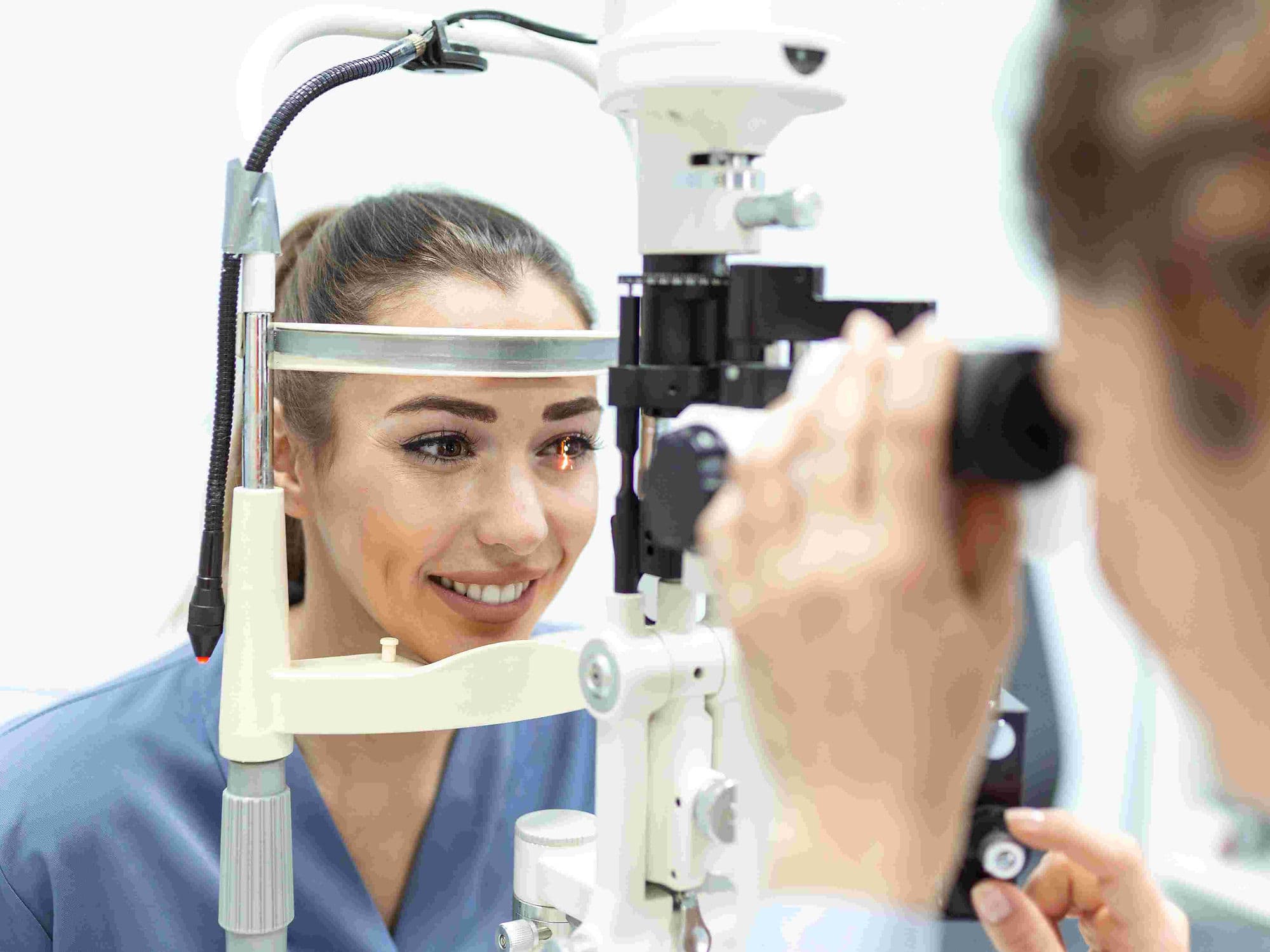
Slit-Lamp Examination: A detailed microscopic look at the front structures of the eye, including the lens.
Retinal Exam: Checking the back of the eye to ensure the retina is healthy and capable of good vision after surgery.
Keratometry: Measuring the curvature of the cornea to help select the most appropriate artificial lens.
Life After Phacoemulsification
Initial Vision: Vision may be blurry for the first 24–48 hours but typically improves rapidly as the eye heals.
Medication: Patients must use prescribed antibiotic and anti-inflammatory eye drops for several weeks to prevent infection.
Eye Protection: A plastic shield is often worn while sleeping for the first week to prevent accidental rubbing or pressure.
Activity Restrictions: Avoid heavy lifting, bending over, or getting water/soap in the eye for at least the first 2–3 weeks.
Follow-up: Regular checkups ensure the IOL is perfectly positioned and the eye pressure remains stable.
Why Specialized Treatment Is Highly Effective
Rapid Recovery: Most patients return to light daily activities within one to two days.
Micro-Incision Precision: Smaller incisions significantly reduce the risk of surgically induced astigmatism.
Customized Vision: Modern IOLs can correct pre-existing nearsightedness, farsightedness, or even presbyopia (reading vision).
Minimal Complications: Advanced technology has lowered the risks of infection, bleeding, and inflammation compared to older methods.
Permanent Solution: Once the clouded lens is removed and replaced, a cataract cannot grow back on the artificial lens.