Septum Resection
Hysteroscopic Septum Resection (Septoplasty)
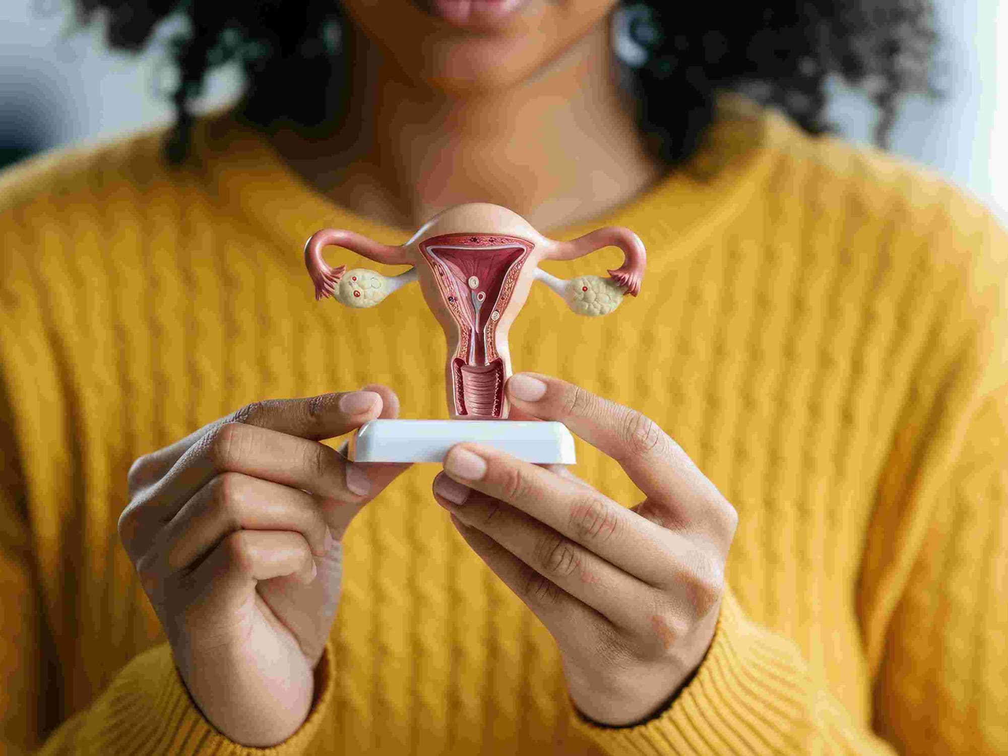
Hysteroscopic Septum Resection, or Septoplasty, is a specialized surgical procedure used to correct a uterine septum—a congenital condition where a wedge-shaped longitudinal wall of tissue divides the uterine cavity. This tissue is typically fibrous and has a poor blood supply, which can interfere with the healthy development of a pregnancy. The procedure aims to unify the cavity into a single, functional space.
When You Should Consider a Septum Resection
Recurrent Miscarriage: A uterine septum is one of the most common correctable causes of repeated pregnancy loss, as an embryo may implant on the septum where there is insufficient blood flow to support growth.
Infertility: While a septum does not always prevent conception, it can interfere with the proper implantation of a fertilized egg into a healthy part of the uterine lining.
Risk of Preterm Labor: A divided cavity limits the physical space available for a growing fetus, significantly increasing the risk of early delivery or malpresentation (such as a breech position).
Congenital Abnormality: When a 3D ultrasound or MRI confirms the presence of a partial or complete septum that distorts the internal triangular shape of the uterus.
How Is Performed
Access: This is a "scarless" outpatient procedure performed entirely through the vagina and cervix using a hysteroscope (a thin camera). No abdominal incisions are required.
Anesthesia: The surgery is typically performed under general or spinal anesthesia and takes approximately 20 to 45 minutes.
Distension: The uterus is expanded with a saline solution to provide the surgeon with a clear, magnified view of the dividing septum and the surrounding cavity walls.
Resection: The surgeon uses micro-scissors, a laser, or a specialized electrosurgical loop (resectoscope) to precisely cut through the midline of the fibrous tissue. Because the septum lacks major blood vessels, there is usually very little bleeding during the procedure.
Completion: The resection continues until the uterine cavity reaches a normal, unified triangular shape, ensuring there is no obstruction between the two sides of the uterus.
Pre-Procedure Preparation
Advanced Imaging: A 3D ultrasound or pelvic MRI is essential to differentiate a septate uterus from a bicornuate (heart-shaped) uterus, which requires a different surgical approach.
Timing Your Procedure: Ideally scheduled during the first week after your period ends, when the uterine lining is at its thinnest for the best surgical visualization.
Pregnancy Test: A mandatory check to ensure the procedure is safe to perform.
Cervical Ripening: You may be given medication to take a few hours before surgery to help soften the cervix, allowing the hysteroscope to pass more easily.
Fasting: Following "nothing by mouth" instructions for 6–8 hours prior to your scheduled anesthesia.
Tests Before Hysteroscopic Septum Resection
3D Pelvic Ultrasound: The gold standard for measuring the depth and thickness of the septum and the outer contour of the uterus.
Hysterosalpingogram (HSG): An X-ray that uses dye to show the internal "V" or "Y" shape of the divided cavity.
Diagnostic Hysteroscopy: Often performed just before the resection to confirm the surgical plan and check the health of the fallopian tube openings.
ECG: A standard heart check to ensure you are healthy enough for the administration of anesthesia.
Life After Hysteroscopic Septum Resection
Recovery Time: Most patients return to their normal daily routine within 1 to 2 days.
Immediate Symptoms: Mild cramping and light vaginal spotting or a "watery" discharge are normal for 2 to 5 days as the uterus heals.
Hormonal Support: Some surgeons prescribe estrogen therapy for a few weeks post-op to encourage the healthy uterine lining to grow over the area where the septum was removed.
Follow-up Imaging: A "second-look" diagnostic hysteroscopy or a follow-up 3D ultrasound is often performed 4 to 8 weeks later to confirm the cavity is fully open.
Pregnancy Timing: Most doctors recommend waiting 2 to 3 menstrual cycles before attempting to conceive to ensure the uterine lining has completely regenerated and is ready for implantation.
Why Specialized Treatment Is Highly Effective
Significant Improvement in Outcomes: Successful resection can reduce miscarriage rates from as high as 80–90% down to 10–15% in women with this condition.
Incision-Free Recovery: Using the natural opening of the cervix means no external scars and a much faster return to work and family life.
Micro-Precision Tools: The use of specialized micro-scissors or lasers allows for the removal of the septum without damaging the sensitive, vascular parts of the uterine wall.
Unified Uterine Space: Restoring the natural triangular shape of the cavity provides the maximum possible space for a baby to grow to full term.
Definitive Correction: Unlike many other fertility issues, a uterine septum is a structural problem that can be permanently corrected with a single, short surgical procedure.