Slip Disc (Lumbar Discectomy)
Lumbar Discectomy
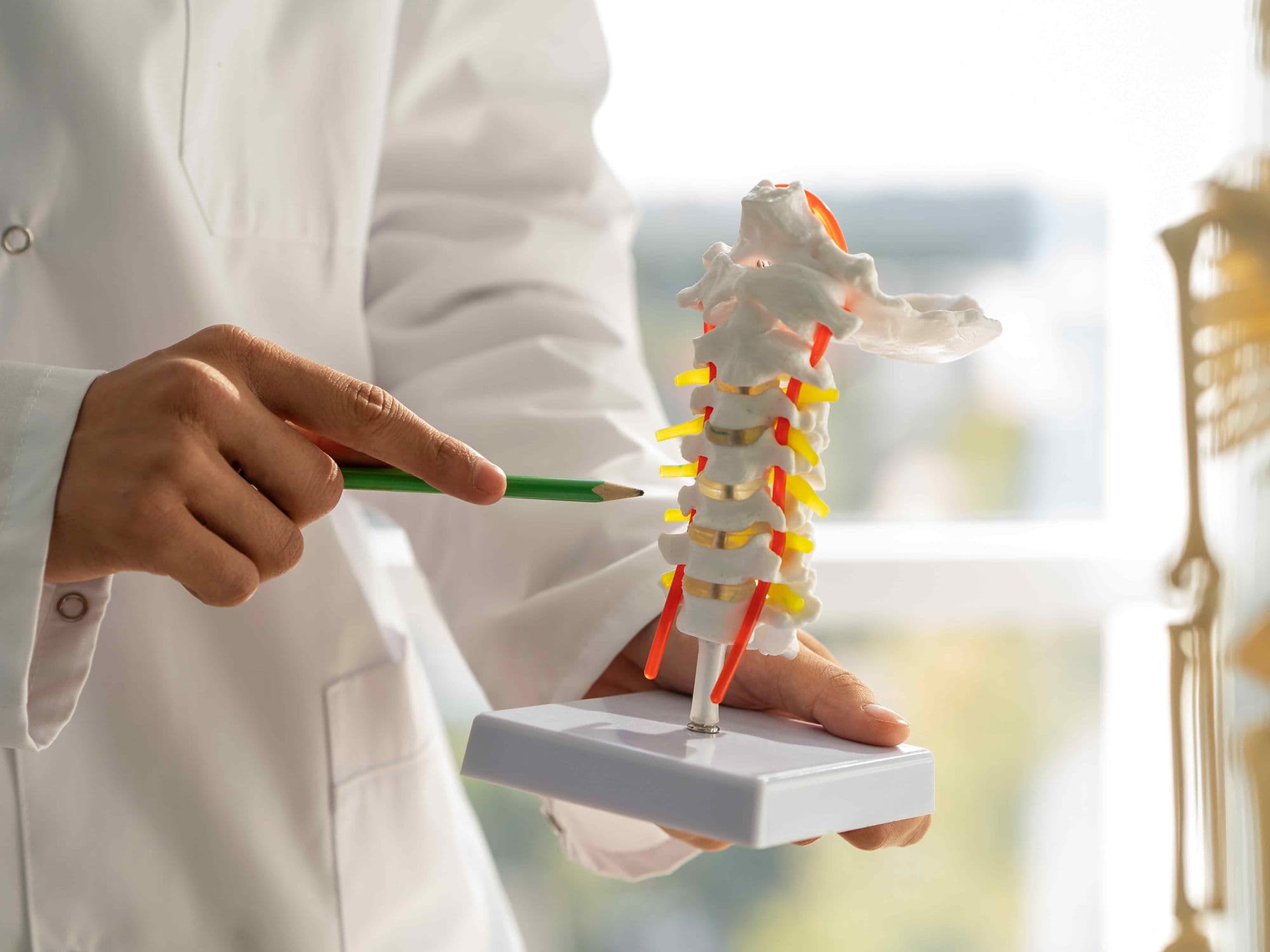
Lumbar Discectomy is a surgical procedure to remove the fragmented or protruding portion of a herniated disc (slip disc) that is compressing a spinal nerve. It is most commonly performed in the lower back (lumbar spine) to relieve radiating leg pain, known as sciatica, by decompressing the affected nerve root.
When You Should Consider Lumbar Discectomy
Failed Conservative Treatment: When 6–12 weeks of physical therapy, NSAIDs, or steroid injections fail to provide relief.
Radiculopathy (Sciatica): Severe, radiating pain, numbness, or weakness that travels down the leg and into the foot.
Neurological Deficit: Progressive muscle weakness or a "foot drop" caused by sustained nerve compression.
Cauda Equina Syndrome: An emergency condition involving loss of bowel or bladder control or "saddle anesthesia" (numbness in the groin).
Significant Functional Impairment: When back and leg pain prevents the performance of basic daily activities or work.
Methods of Lumbar Discectomy
Microdiscectomy (Gold Standard): Using a high-powered operating microscope to minimize the incision size and improve visualization of the nerve.
Endoscopic Discectomy: An ultra-minimally invasive technique using a tiny camera and specialized tools inserted through a small tube.
Laminotomy/Laminectomy: Removing a small portion of the vertebral bone (lamina) to create a window to access the spinal canal.
Tubular Retractor Discectomy: Using a series of dilating tubes to part the muscles rather than cutting them, reducing post-operative soreness.
Sequestrectomy: Removing only the free-floating disc fragment without entering the main disc space, which may reduce the risk of future collapse.
How Lumbar Discectomy Is Performed
Positioning: The patient is placed face down on a specialized surgical frame that opens the spaces between the vertebrae.
Incision: A small 2–3 cm midline incision is made in the lower back directly over the level of the herniation (most commonly L4-L5 or L5-S1).
Exposure: The surgeon moves the spinal muscles aside and removes a small amount of ligament and bone to view the spinal canal.
Nerve Protection: The compressed nerve root is gently retracted to one side to expose the herniated disc material underneath.
Fragment Removal: The surgeon identifies the "jelly-like" protrusion and removes it. The healthy portion of the disc is left intact to serve as a shock absorber.
Closure: The muscles return to their original position, and the skin is closed with dissolvable stitches and surgical glue.
Pre-Procedure Preparation
Confirmation of the herniation level via MRI to ensure the surgical site matches the patient's clinical symptoms.
Smoking cessation is mandatory for several weeks prior, as nicotine restricts blood flow to the spine and significantly hinders healing.
Fasting (NPO) for 8–12 hours before the procedure to ensure safety during general anesthesia.
Discussion of the "BLT" (Bending, Lifting, Twisting) restrictions that will be required immediately following the surgery.
Tests Before Lumbar Discectomy
Lumbar MRI: The primary diagnostic tool used to visualize the disc herniation and its relationship to the nerve roots.
X-ray (Flexion/Extension): Performed to ensure there is no underlying spinal instability or "slipped" vertebrae (spondylolisthesis).
Electromyography (EMG): Occasionally used to confirm which specific nerve is being damaged and to assess the severity of the nerve injury.
Blood Panels: Routine screens to ensure the patient is fit for anesthesia and has no signs of active infection.
Life After Lumbar Discectomy
Most procedures are performed as same-day (outpatient) surgeries or require only a single overnight stay.
Patients are encouraged to stand and walk within 4 hours of waking up to promote circulation and prevent stiffness.
The "BLT" Rule: For the first 6 weeks, you must strictly avoid Bending at the waist, Lifting anything over 2kg, and Twisting the spine.
Incisions must be kept dry for the first 3–5 days; stitches are usually dissolvable and do not require removal.
Physical therapy typically begins around the 6-week mark to strengthen the core and multifidus muscles that support the spine.
Benefits of Lumbar Discectomy
Over 90% of patients report immediate and dramatic relief from radiating leg pain (sciatica).
Minimally invasive techniques allow for smaller scars, less muscle damage, and a faster return to daily life.
Prevents permanent nerve damage by removing the source of chronic compression and inflammation.
Restores the ability to perform physical activities, work, and exercise without the limitation of debilitating leg pain.