Spinal Decompression Surgery
Spinal Decompression Surgery
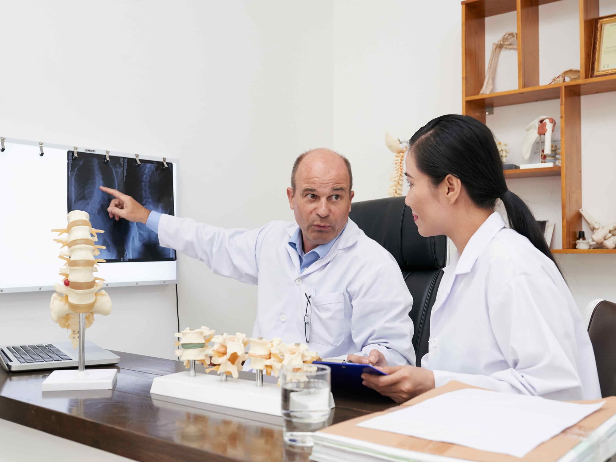
Spinal Decompression Surgery is a general term for various procedures performed to relieve pressure on the spinal cord or nerve roots. It is most commonly used to treat Spinal Stenosis (narrowing of the spinal canal) caused by bone spurs, thickened ligaments, or bulging discs, allowing the neural structures to function without compression.
When You Should Consider Spinal Decompression Surgery
Neurogenic Claudication: Leg pain, heaviness, or cramping that occurs when walking or standing and is relieved by sitting or leaning forward.
Radiculopathy: Shooting pain, numbness, or "pins and needles" that radiates into the arms or legs due to a pinched nerve.
Failed Conservative Care: When physical therapy, activity modification, and epidural steroid injections fail to improve quality of life after 3–6 months.
Progressive Weakness: Measurable loss of motor function, such as a weakened grip or a "foot drop," indicating severe nerve compromise.
Spinal Stenosis: Diagnostic confirmation of a narrowed spinal canal that correlates with the patient's physical limitations and pain patterns.
Methods of Spinal Decompression
Laminectomy: The "gold standard" procedure where the entire bony arch (lamina) at the back of the vertebra is removed to create significant room for the spinal cord.
Laminotomy: A less invasive approach where only a small portion of the lamina is removed, creating a "window" to access a specific pinched nerve.
Foraminotomy: Enlarging the "exit holes" (foramina) where the nerve roots leave the spinal canal to relieve localized compression.
Discectomy: Removing the specific portion of a herniated disc that is pressing directly against a spinal nerve.
Corpectomy: An extensive procedure where a portion of the vertebral body and adjacent discs are removed to decompress the spinal cord across a larger area.
How Spinal Decompression Surgery Is Performed
Positioning: The patient is placed face down (prone) on a specialized surgical frame that minimizes pressure on the abdomen and helps open the spinal spaces.
Incision: A midline incision is made over the affected area of the spine. The length of the incision depends on how many levels of the spine require decompression.
Muscle Retraction: The spinal muscles are gently moved aside to expose the bony elements of the vertebrae.
Bone and Ligament Removal: The surgeon carefully removes the bone spurs (osteophytes), thickened ligaments, or portions of the lamina that are encroaching on the spinal canal.
Nerve Inspection: The surgeon uses magnification to ensure the nerve roots are completely free and "floating" within the newly enlarged space.
Closure: The muscles are allowed to return to their natural position, and the incision is closed with sutures, staples, or surgical glue.
Pre-Procedure Preparation
Confirmation of the degree of narrowing via high-resolution MRI or CT Myelogram to plan the exact surgical levels.
Smoking cessation is mandatory for at least 4 weeks prior to surgery, as nicotine significantly hinders bone and tissue healing and increases the risk of infection.
Fasting (NPO) for 8–12 hours before the procedure to ensure safety under general anesthesia.
Evaluation of spinal stability via X-rays to determine if a fusion might be necessary in addition to the decompression.
Tests Before Spinal Decompression Surgery
Lumbar or Cervical MRI: The primary diagnostic tool used to visualize the soft tissues, nerves, and the extent of the canal narrowing.
X-rays (Flexion/Extension): Used to check for spinal instability, such as one vertebra sliding over another (spondylolisthesis).
CT Scan: Provides detailed images of the bony structures, which is helpful for mapping out dense bone spurs or ligament calcification.
Electrodiagnostic Studies (EMG/NCS): Performed to confirm that the symptoms are caused by spinal compression rather than peripheral nerve issues like diabetes or carpal tunnel.
Life After Spinal Decompression Surgery
Hospital stays vary from same-day discharge for simple procedures to 2–4 days for multi-level laminectomies.
Patients are encouraged to stand and walk within 4–6 hours of surgery to promote circulation and prevent complications like blood clots or pneumonia.
The "BLT" Restrictions: For the first 6 weeks, patients must strictly avoid Bending at the waist, Lifting anything over 3-5kg, and Twisting the spine.
Physical therapy typically begins 4–6 weeks post-operatively to strengthen the core and back muscles that support the spine.
While leg or arm pain often improves dramatically and quickly, the surgical site may remain sore for several weeks during the healing process.
Benefits of Spinal Decompression Surgery
Over 80% of patients experience a significant reduction in radiating limb pain and an improved ability to walk longer distances.
Effectively halts the progression of neurological damage, such as permanent numbness or muscle wasting.
Restores the ability to engage in daily activities, hobbies, and work that were previously limited by spinal stenosis symptoms.
Provides a durable, long-term solution for mechanical compression that does not respond to non-surgical interventions.