Strabismus Surgery
Strabismus Surgery (Strabeculectomy)
Strabeculectomy, commonly known as strabismus surgery, is a specialized procedure performed to correct the misalignment of the eyes (crossed eyes or "squint"). By 2026, the procedure is used not only for children but increasingly for adults to improve depth perception, eliminate double vision, and restore a natural appearance.
When You Should Consider Strabismus Surgery
To correct eye misalignment (crossed eyes or "squint") in both children and adults.
If you are experiencing persistent double vision (diplopia) due to ocular misalignment.
To restore or improve depth perception and binocular vision.
When seeking to restore a natural, symmetrical appearance to the eyes.
In complex cases where previous treatments or prism glasses have not achieved proper alignment.
Methods of Strabismus Surgery
Recession (Weakening): Detaching a muscle and reattaching it further back on the eye to relax its pull.
Resection (Strengthening): Removing a small section of a muscle to make it shorter, tighter, and stronger.
Plication: A modern, less traumatic alternative where the muscle is folded over itself to shorten it without cutting.
Adjustable Sutures: A 2026 advancement where knots are fine-tuned after surgery while the patient is awake to ensure perfect alignment.
Extraocular Manipulation: Focused entirely on the six small muscles outside the eyeball rather than internal eye structures.
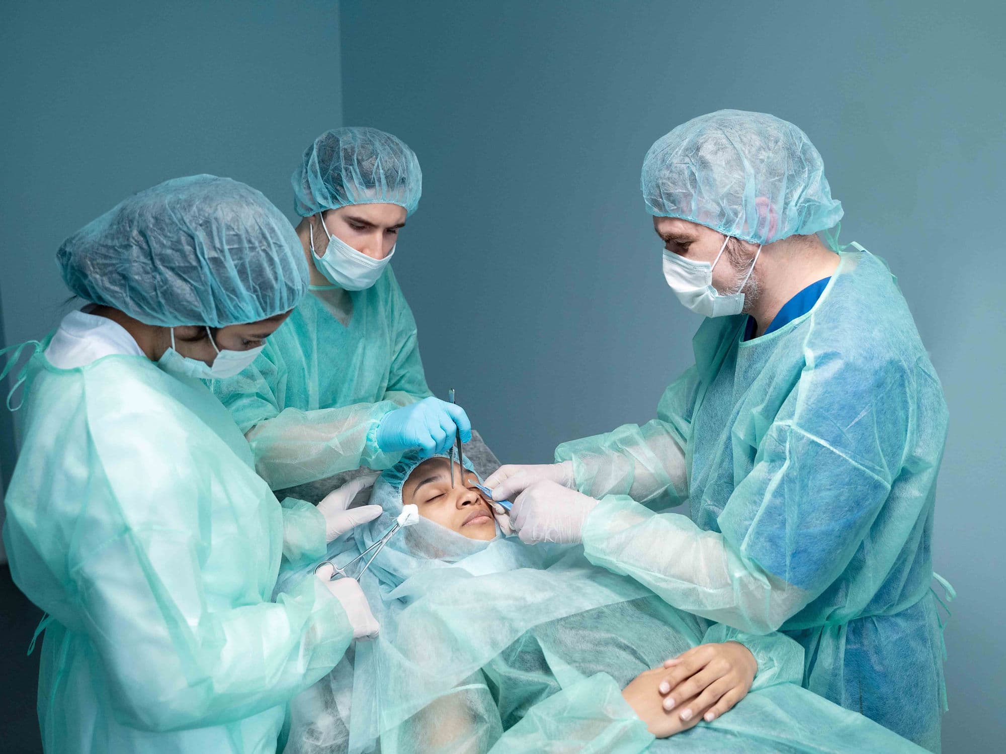
How Strabismus Surgery Is Performed
Anesthesia: Children receive general anesthesia, while adults may choose local anesthesia with heavy sedation.
Accessing Muscles: The surgeon makes a small incision in the conjunctiva to reach the extraocular muscles.
Alignment Correction: Muscles are either weakened, strengthened, or folded based on the specific direction of the squint.
Suture Adjustment: In cases using adjustable sutures, the alignment is verified and "fine-tuned" shortly after the patient wakes up.
Closure: The microscopic incisions are closed with dissolvable stitches that do not require removal.
Duration: The procedure typically takes between 30 to 90 minutes.
Pre-Procedure Preparation
Comprehensive eye muscle evaluation to determine which muscles are overactive or underactive.
Discussion of the goal: whether for functional improvement (double vision), depth perception, or cosmetic restoration.
Planning for a one-week downtime from work or school.
Preparing for the temporary "bloodshot" appearance of the eyes post-surgery.
Identifying whether the patient is a candidate for adjustable sutures to increase success rates.
Tests Before Strabismus Surgery
Prism Cover Test: To measure the exact degree of eye deviation in multiple directions of gaze.
Sensory Testing: To evaluate the brain's ability to use both eyes together (binocularity).
Visual Acuity Test: To check for underlying conditions like amblyopia (lazy eye).
Ocular Motility Exam: A detailed check of how well each individual eye muscle is functioning.
Life After Strabismus Surgery
Expect the white of the eye to appear very red for 2 to 4 weeks; this is a normal part of the healing process.
Use prescription drops as directed to manage inflammation and prevent infection.
Manage "scratchy" or "sandy" sensations in the eye during the first few days.
Avoid swimming and heavy lifting for 10 to 14 days to protect the muscle attachments.
Monitor for temporary double vision as the brain learns to merge images from the newly aligned eyes.
Benefits of Strabismus Surgery
Significantly increases the success rate for complex cases through the use of adjustable suture technology.
Enhances quality of life by eliminating the social and functional challenges of misaligned eyes.
Provides a non-invasive internal approach, as the surgery is performed entirely on the outside of the eyeball.
Restores the ability of the eyes to work together, improving coordination and depth perception.
Offers a rapid recovery with most patients returning to their normal routine within a single week.