Vaginal Hysterectomy (Open/Laparoscopic/Robotic)
Vaginal Hysterectomy
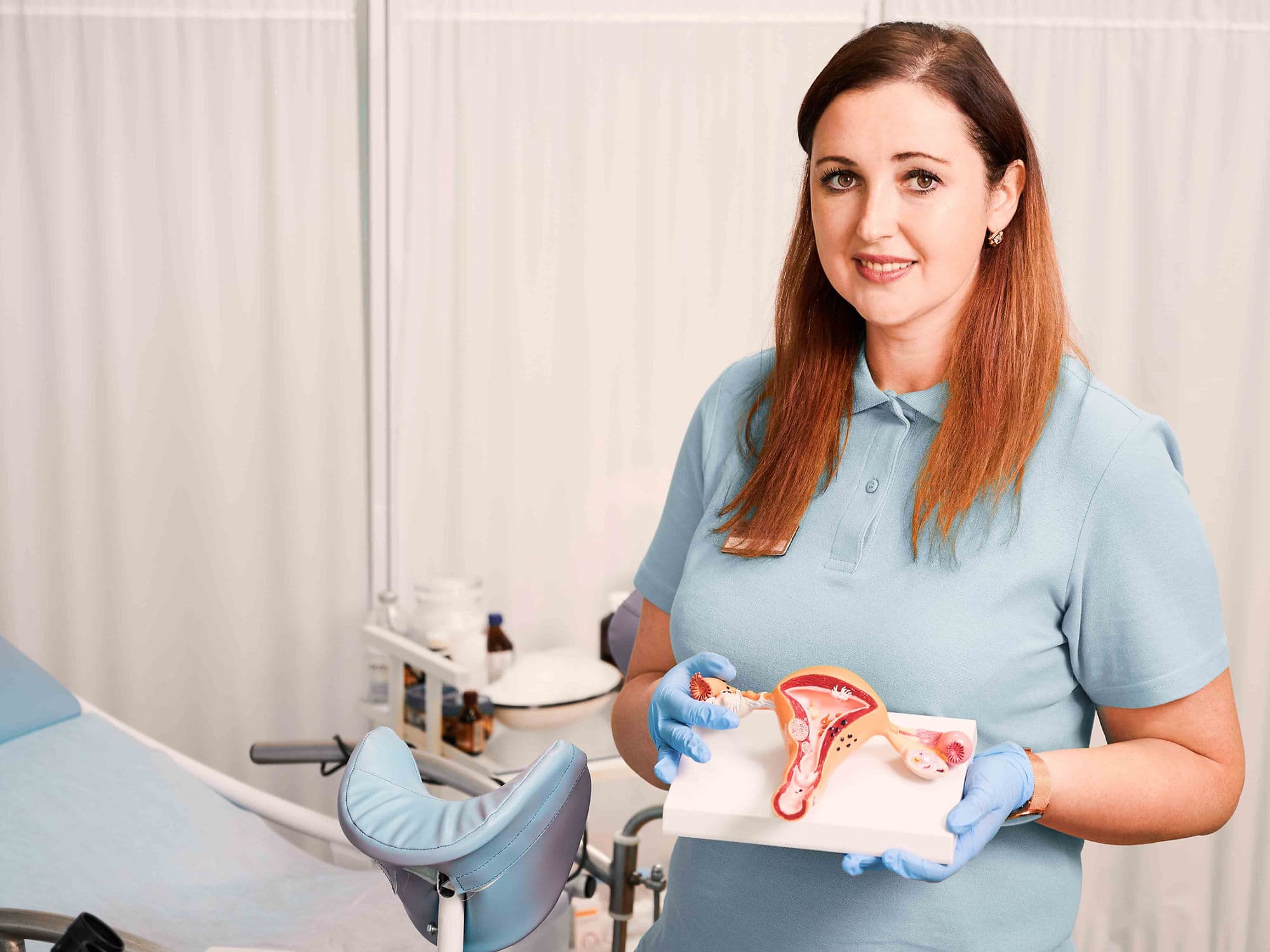
A vaginal hysterectomy is a surgical procedure to remove the uterus through the vaginal canal. Unlike an abdominal hysterectomy, this approach requires no external incisions on the abdomen, which typically results in a shorter hospital stay, lower costs, and a significantly faster recovery. It is a preferred method for treating various non-cancerous gynecological conditions.
When You Should Consider a Vaginal Hysterectomy
Uterine Fibroids: Benign growths in the uterine wall that cause heavy menstrual bleeding, pelvic pressure, or persistent pain.
Uterine Prolapse: When the pelvic floor muscles and ligaments weaken, causing the uterus to slip down into or even out of the vaginal canal.
Adenomyosis: A condition where the uterine lining grows into the muscular wall of the uterus, causing severe cramping and heavy periods.
Endometriosis: When tissue similar to the uterine lining grows outside the uterus, leading to chronic pain and scarring.
Abnormal Uterine Bleeding: When heavy or irregular bleeding cannot be managed with medication or less invasive procedures.
Surgical Approaches
Traditional Vaginal Hysterectomy: The entire procedure is performed through an incision inside the vagina. This is the least invasive method and leaves no visible scars.
Laparoscopically Assisted Vaginal Hysterectomy (LAVH): A thin camera (laparoscope) and micro-instruments are inserted through 3–4 tiny "keyhole" incisions in the belly to detach the upper uterus. The uterus is then removed through the vagina.
Robotic-Assisted Hysterectomy: Similar to the laparoscopic approach, but the surgeon uses a robotic console for superior 3D visualization and enhanced precision, which is ideal for complex cases like extensive endometriosis.
Vaginal Vault Creation: After the uterus is removed, the surgeon sews the top of the vagina closed, creating what is known as a "vaginal cuff."
How Is Performed
Anesthesia: The surgery is performed under general anesthesia or regional anesthesia (spinal/epidural) and typically takes 1 to 2 hours.
Access: The surgeon makes an incision inside the vagina to reach the uterus.
Detachment: Using specialized tools, the surgeon clamps and cuts the blood vessels and ligaments that support the uterus.
Removal: The uterus (and sometimes the fallopian tubes or ovaries) is withdrawn through the vaginal opening.
Closure: The internal incisions are closed with dissolvable stitches. Because there are no abdominal cuts in the traditional approach, there is no external scarring.
Pre-Procedure Preparation
Pelvic Examination: To assess the size of the uterus and ensure it can be safely removed through the vaginal canal.
Fasting: Adhering to strict "nothing by mouth" instructions for at least 8 hours prior to your general anesthesia.
Medication Audit: You may need to stop taking blood thinners, aspirin, or certain herbal supplements 7–10 days before the procedure.
Bowel Prep: In some cases, your surgeon may recommend a mild laxative or a specific diet the day before surgery.
Smoking Cessation: Stopping smoking at least 4 weeks before surgery is vital to promote healthy tissue healing and reduce the risk of infection.
Tests Before Vaginal Hysterectomy
Pelvic Ultrasound: To map the size and location of fibroids and check the health of the ovaries.
Endometrial Biopsy: To rule out any cancerous or precancerous cells within the uterine lining.
Pap Smear: To ensure there are no cervical abnormalities before the uterus and cervix are removed.
Blood Panels: Checking hemoglobin levels and blood type to prepare for the rare possibility of a transfusion.
ECG: A standard heart check to ensure you are healthy enough for the administration of anesthesia.
Life After Vaginal Hysterectomy
Hospital Stay: Most patients are discharged the same day or after one night of observation.
Physical Activity: Walking is encouraged immediately to prevent blood clots, but you must avoid heavy lifting (over 5 kg) and strenuous exercise for 4 to 6 weeks.
Vaginal Health: You must not use tampons or have sexual intercourse for 6 to 8 weeks to allow the vaginal cuff to heal completely.
Hormonal Changes: If the ovaries are left intact, you will not enter menopause prematurely, though you will no longer have periods. If ovaries are removed, menopause begins immediately.
Follow-up Care: A post-operative checkup is typically scheduled for 2 to 6 weeks after surgery to ensure the internal stitches are dissolving correctly.
Why Specialized Treatment Is Highly Effective
Faster Recovery: Patients generally return to normal activities much sooner than those undergoing traditional abdominal surgery.
No Visible Scarring: Because the entry point is internal, there are no cosmetic changes to the abdomen.
Reduced Complication Rates: Vaginal approaches are associated with lower risks of wound infection and less post-operative pain.
Cost-Efficient: Shorter hospital stays and quicker operating times often result in lower overall medical costs.
High Patient Satisfaction: Most women report a significant improvement in quality of life once the symptoms of fibroids or prolapse are resolved.