+91 83682 47758
info@medivisorhealth.com
Medivisor India Treatment Logo
  • Home
    • About Us
    • Our Services
    • Our Team
    • Our Safety Measures
    • Loading treatments...
    • Loading hospitals...
    • Loading doctors...
    • Treatment Cost
    • Treatment Process
    • Visa Process
    • Travel Guide
    • FAQs
    • Why Medivisor
    • Success Stories
    • Patient Activities
    • Social Activities
    • News Coverage
    • Blog
  • Become a Partner

Medivisor India Treatment is a trusted New Delhi-based medical travel company assisting international patients with advanced, affordable, and seamless treatment in India.

  • Medivisor House 359, Sector 1, Vaishali, Ghaziabad, (Delhi/NCR) India
  • +91 8368 247 758
  • info@medivisorhealth.com

About Us

  • About Us
  • Our Services
  • Our Team
  • Our Medical Advisors
  • Our Safety Measures
  • Join Our Network
  • Contact Us

India Treatment

  • Hospitals
  • Treatments
  • Treatment Cost
  • Treatment Process
  • Visa Process
  • Travel Guide
  • FAQs
  • Why Medivisor

Gallery

  • Patient Testimonials
  • Patient Activities
  • News Coverage
  • Blog

Get in Touch

© 2026 Medivisor India Treatment. All rights reserved.

Dr Vishal Soni

Gastroenterologist

Apollo Hospital International Ltd, Ahmedabad

Gastroenterologist

15+ years experience

Dr Vishal Soni, Gastroenterologist at Apollo Hospital International Ltd, Ahmedabad - Apollo Hospitals
  1. Home
  2. Doctors
  3. Dr Vishal Soni

About Dr Vishal Soni

Dr. Vishal Soni is a highly accomplished Robotic Gastro-Intestinal, Hernia (Abdominal Wall Reconstruction), and Obesity Surgeon currently practicing at Apollo Hospitals International Limited, Ahmedabad. He is proficient in English, Marathi, Hindi, and Gujarati, enabling him to communicate effectively with a diverse patient population. Renowned for his patient-centered approach and commitment to surgical excellence, he offers advanced care in complex gastrointestinal, hernia, and obesity cases.

Robotic, Hernia, and Bariatric Expertise

He is a Fellow of the American College of Surgeons (FACS), a prestigious recognition he received in 2021. Dr. Soni has completed multiple fellowships in minimally invasive and advanced techniques, including a Fellowship in Advanced Laparoscopic Surgery (FALS) with specializations in Robotics, Bariatric, and Hernia Surgery from the Indian Association of Gastro-Intestinal EndoSurgeons (IAGES) in 2022. He also holds a Fellowship of the Indian Association of Gastro-Intestinal EndoSurgeons (FIAGES), alongside a Fellowship and Diploma in Minimal Access Surgery (F.MAS & D.MAS) from the World Laparoscopy Hospital, Delhi.

Dr. Vishal Soni at a Glance

  • Specialized Robotic Gastro-Intestinal, Hernia (AWR), and Obesity Surgeon.

  • Practices at the premier Apollo Hospitals International Limited, Ahmedabad.

  • Awarded Fellowship of the prestigious American College of Surgeons (FACS) in 2021.

  • Completed advanced FALS credentials in Robotics, Bariatric, and Hernia Surgery in 2022.

  • Holds specialized minimal access surgery qualifications (FIAGES, F.MAS, and D.MAS).

  • Quadrilogual proficiency in English, Marathi, Hindi, and Gujarati to support personalized care.

M.S
F.M.A.S & D.MAS
FIAGES
FACS
FALS - Robotics, Bariatric & Hernia Surgery
Board Certified in Gastroenterologist

No awards & achievements available
Affiliated Hospitals

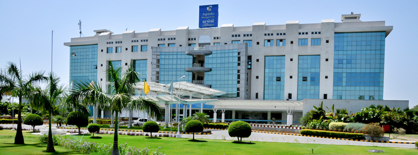
Apollo Hospital International Ltd, Ahmedabad

Multi Specialty

Ahmedabad, Gujarat

2003

Estd.

300+

Beds

100+

Doctors

May we help you?

Enter digits only (6-15 digits). We'll add your country code.

Related Treatments

Endoscopic Retrograde Cholangiopancreatography (ERCP)

Endoscopic Retrograde Cholangiopancreatography (ERCP) is a highly specialized procedure used primarily to treat disorders of the bile ducts and pancreatic ducts. It uniquely combines advanced endoscopy with real-time X-ray imaging (fluoroscopy). By 2026, ERCP has evolved from a diagnostic tool into a powerful therapeutic intervention, allowing physicians to clear obstructions, place stents, and repair leaks within the delicate ductal system of the liver and pancreas without the need for invasive abdominal surgery.

Why ERCP is Performed

  • Common Bile Duct Stones: Removing gallstones that have migrated out of the gallbladder and are blocking the main drainage duct.

  • Malignant Obstructions: Relieving blockages caused by tumors in the pancreas, bile ducts, or liver.

  • Biliary Strictures: Opening narrowed areas of the ducts caused by scar tissue or chronic inflammation.

  • Post-Surgical Bile Leaks: Sealing leaks that can occasionally occur following gallbladder or liver surgery.

  • Chronic Pancreatitis: Treating complications such as pancreatic stones or narrowings that cause severe, recurring pain.

  • Sphincter of Oddi Dysfunction: Managing issues with the muscular valve that controls the flow of digestive juices.

Conditions That Require Specialized Care

  • Massive Stone Burden: Cases with very large or numerous stones that may require advanced "fragmentation" techniques.

  • Anatomical Variations: Patients who have had previous gastric bypass or stomach surgery, requiring a modified "enteroscopy" approach.

  • Primary Sclerosing Cholangitis (PSC): A chronic condition requiring careful, repeated dilations of the bile ducts.

  • High-Risk Pancreatitis Patients: Individuals with a history of post-procedure inflammation who require preventative "stenting" of the pancreatic duct.

  • Pediatric ERCP: Rare cases in children involving congenital ductal abnormalities that require specialized pediatric endoscopes.

[Image showing the endoscope positioned in the duodenum with the catheter entering the bile duct]

How an ERCP Is Performed

  • Deep Sedation or Anesthesia: To ensure absolute stillness and comfort, the procedure is performed under deep sedation or general anesthesia.

  • Side-Viewing Endoscopy: A specialized duodenoscope is passed through the mouth and stomach to reach the "papilla"—the small opening where the ducts meet the intestine.

  • Cannulation: A tiny catheter is threaded into the papilla to gain access to the biliary or pancreatic tree.

  • Retrograde Contrast: A radio-opaque dye is injected "backward" into the ducts, highlighting the internal anatomy on a live X-ray monitor.

  • Sphincterotomy: Using a small electrified wire, the surgeon may make a tiny cut in the muscle valve (sphincter) to widen the opening for stone removal.

  • Mechanical Intervention: Tools such as baskets, balloons, or stents are passed through the scope to clear blockages or provide structural support.

Innovations in Duct Therapy

  • Disposable DuodenoscopesThe widespread adoption of single-use scopes to eliminate the risk of bacterial transmission and ensure the highest mechanical reliability for every patient.

  • SpyGlass™ CholangioscopyA "micro-scope" that is passed through the main endoscope, allowing the doctor to see directly inside the bile duct in high-definition to biopsy tumors or break up stones with lasers.

  • AI-Assisted FluoroscopyArtificial intelligence that enhances X-ray images in real-time, helping the doctor identify "shadows" or subtle strictures with greater clarity.

  • Biodegradable Biliary StentsNext-generation stents that provide support to a narrowed duct for several months and then dissolve, avoiding the need for a second procedure to remove them.

  • Laser Lithotripsy (EHL)Using targeted laser energy to "shatter" stubborn gallstones into tiny fragments that can be easily swept out of the duct.

  • Automated Risk-Prediction ModelsSoftware that analyzes a patient's health data before the procedure to suggest the exact dose of preventative medication needed to stop post-op inflammation.

Pre-Procedure Preparation

  • Fasting (NPO): A strictly empty stomach is required, usually meaning no food or drink for 8 to 12 hours prior to the procedure.

  • Antibiotic Prophylaxis: Certain patients may be given a dose of antibiotics before the start of the procedure to prevent ductal infection.

  • Blood Work: Checking coagulation (clotting) factors to ensure it is safe to perform a sphincterotomy or biopsy.

  • Medication Coordination: Careful management of blood thinners and diabetic medications is essential for a safe recovery.

  • Post-Op Plan: Because of the intensive nature of ERCP, patients must have a designated driver and a quiet place to recover for the first 24 hours.

Diagnostic and Safety Monitoring

  • Pancreatitis Prevention: Use of rectal medications or temporary "protective" pancreatic stents to lower the risk of post-ERCP inflammation.

  • Amylase/Lipase Checks: Blood tests performed if a patient experiences unusual pain after the procedure to check for pancreatic irritation.

  • Continuous Vital Monitoring: Specialized anesthesia teams monitor heart rhythm and oxygen levels throughout the entire intervention.

  • Radiation Safety: Using "low-dose" pulsed fluoroscopy to minimize X-ray exposure for both the patient and the medical team.

Why This Treatment Is Highly Effective

  • Avoids Major Surgery: ERCP provides a "keyhole" alternative to traditional open-belly surgery for clearing bile duct obstructions.

  • Immediate Obstruction Relief: Patients with jaundice (yellowing of the skin) often see a rapid improvement in symptoms once the duct is cleared or stented.

  • High Precision: The combination of live video and live X-ray ensures that the doctor knows exactly where the instruments are at all times.

  • Life-Saving for Sepsis: In cases of "cholangitis" (infected bile), ERCP provides urgent drainage that is critical for survival.

  • Dual Functionality: It allows for the immediate transition from identifying a problem to fixing it within the same 30-to-60-minute session.

Recovery and Aftercare

  • Immediate Observation: Most patients stay in a recovery unit for 1 to 2 hours to ensure the sedation has worn off and no immediate pain is present.

  • Sore Throat and Bloating: These are the most common minor side effects and typically resolve within 24 to 48 hours.

  • Dietary Progression: Patients usually start with clear liquids and move to a low-fat diet as tolerated by their digestive system.

  • Activity Limits: No driving or making major decisions for 24 hours. Most patients return to full activity within 2 to 3 days.

  • Follow-up Imaging: A simple ultrasound or blood test is often scheduled a few weeks later to confirm the ducts remain clear.

Life After ERCP

  • Rapid resolution of painful symptoms like biliary colic (gallstone pain) or chronic abdominal pressure.

  • Successful management of complex conditions like cancer-related blockages, allowing for a better quality of life.

  • Permanent removal of dangerous "trapped" stones that could otherwise lead to severe liver damage.

  • A return to a normal, healthy diet once the flow of digestive enzymes and bile has been restored.

  • Empowerment through the successful treatment of a condition that previously would have required a long, difficult surgical recovery.

₹From From 300

Laparoscopic Polypectomy

Laparoscopic Polypectomy generally refers to the minimally invasive removal of polyps from the gallbladder or, in specific gynecological cases, subserosal uterine polyps. While most uterine polyps are removed via hysteroscopy (through the cervix), the laparoscopic approach is reserved for polyps or small growths located on the outer surface of an organ or within the abdominal cavity.

When You Should Consider Laparoscopic Polypectomy

  • Symptomatic Polyps: Growths causing localized pain or persistent digestive issues.

  • Suspect Features: Polyps showing irregular shapes or rapid growth during an ultrasound.

  • Size-Dependent Risks: Gallbladder polyps exceeding 1 cm in size, which is a standard surgical indication in 2026 guidelines.

  • Outer Organ Growth: When a polypoid growth is located on the exterior of the uterus or attached to pelvic ligaments.

  • Cancer Mitigation: To prevent the risk of malignancy in polyps that show signs of growth.

Methods of Removal and Techniques

  • Laparoscopic Cholecystectomy: Removal of the entire gallbladder for polyps larger than 10mm, as they cannot be safely scraped off the wall.

  • Laparoscopic Excision: The use of precision shears or lasers to excise a growth at its base from the exterior of an organ.

  • High-Definition Visualization: Utilizing zoomed-in views to identify a polyp’s blood supply and seal vessels before cutting.

  • Oncological Safety Protocols: The use of specimen bags during removal to ensure tissue does not contact the abdominal wall.

  • Hysteroscopy (Alternative): The standard approach for internal uterine polyps, distinct from the laparoscopic method used for external growths.

How Laparoscopic Polypectomy Is Performed

  • Anesthesia: The surgery is performed under general anesthesia to ensure the patient is comfortable and still.

  • Access Incisions: The surgeon makes 1 to 3 tiny incisions, ranging from 5–10 mm, in the abdominal wall.

  • Instrument Insertion: A laparoscope and specialized micro-instruments are inserted through the small incisions.

  • Vessel Sealing: Surgeons use high-definition cameras to seal blood vessels before removal, resulting in near-zero blood loss.

  • Abdominal Inflation: Carbon dioxide gas is used to create space, though this can lead to temporary post-operative bloating.

Pre-Procedure Preparation

  • Fasting Requirements: Patients must fast for 8-12 hours before the surgery or catheterization.

  • Medical Assessments: Blood tests, ECG, and chest X-rays are conducted to assess overall health.

  • Medication Adjustments: Current medications must be adjusted as directed by the medical team.

  • Allergy Discussion: Patients must discuss any allergies, particularly to materials used in surgical devices or dyes.

  • Post-Op Planning: Arranging for post-operative care and a support person for the recovery period.

Tests Before Laparoscopic Polypectomy

  • Ultrasound Imaging: Used to monitor the size and shape of the polyp and detect rapid growth.

  • Blood Tests: To assess overall health and check for markers of inflammation or infection.

  • ECG: To monitor the heart's electrical rhythm and check for conduction issues.

  • Pulse Oximetry: Used to evaluate oxygen saturation levels in the blood before anesthesia.

  • CT Scan: For detailed mapping of the abdominal cavity and complex growths.

Life After Laparoscopic Polypectomy

  • Hospital Stay: Usually a same-day discharge or a short 23-hour observation stay.

  • Immediate Recovery: Walking is encouraged immediately to aid digestion and prevent blood clots.

  • Pain Management: Discomfort is typically managed with over-the-counter anti-inflammatories.

  • Return to Work: Patients can generally return to work within 5 to 7 days post-procedure.

  • Activity Restrictions: Heavy lifting must be avoided for approximately 4 weeks to ensure proper healing.

Benefits of Laparoscopic Polypectomy

  • Minimal Tissue Disruption: Rapid recovery due to the use of tiny incisions rather than large open cuts.

  • Precise Visualization: High-definition cameras allow for the zoom-in on blood supplies for safer removal.

  • Near-Zero Blood Loss: Advanced sealing techniques minimize bleeding during the excision.

  • Strict Safety: The use of specialized bags prevents tissue fragments from remaining in the pelvic cavity.

  • Long-Term Cure: Provides a definitive treatment with high technical success rates for symptomatic growths.

₹From From 300

Liver Biopsy
Liver Biopsy

A liver biopsy is a medical procedure used to remove a small piece of liver tissue for laboratory examination. It is considered the definitive "gold standard" for identifying the precise cause of liver disease and determining the extent of organ damage. By 2026, advances in real-time imaging and needle technology have made the procedure faster and safer, allowing clinicians to distinguish between various types of inflammation, scarring, and tumors that blood tests or standard scans might miss.

When You Should Consider a Liver Biopsy

  • Unexplained Liver Enzymes: When blood tests show persistent elevations in ALT or AST that cannot be explained by standard diagnostic sets.

  • Staging Fibrosis: To determine the exact degree of scarring (cirrhosis) in patients with chronic conditions like Fatty Liver Disease (NASH/MASH).

  • Hepatitis Monitoring: Evaluating the severity of chronic Hepatitis B or C to decide on the most effective antiviral therapy.

  • Autoimmune Disorders: Diagnosing conditions where the body's immune system attacks the liver, such as Primary Biliary Cholangitis (PBC).

  • Transplant Monitoring: Checking a transplanted liver for early signs of organ rejection or medication toxicity.

  • Lumps or Masses: Investigating a suspicious nodule found on an ultrasound or MRI to rule out malignancy.

Types of Liver Biopsy

  • Percutaneous Biopsy: The standard approach. Under the guidance of ultrasound or CT, a specialized needle is inserted through the skin between the ribs directly into the liver to collect a tissue core.

  • Transjugular Biopsy: A specialized technique for patients with blood clotting issues or excess abdominal fluid (ascites). A catheter is threaded through the jugular vein in the neck and guided down to the liver's internal veins.

  • Laparoscopic Biopsy: Tissue is collected during a minimally invasive surgical procedure, allowing the doctor to visually inspect the liver surface and sample specific, hard-to-reach areas.

  • Fine-Needle Aspiration (FNA): Often used for specific liver tumors to collect cells for cytology, though it provides less structural information than a core biopsy.

How a Liver Biopsy Is Performed

  • Positioning: The patient typically lies on their back with the right arm raised above the head to widen the space between the ribs.

  • Imaging Guidance: A physician uses an ultrasound probe to identify the safest "window" for the needle, avoiding the gallbladder and major blood vessels.

  • Local Anesthesia: The skin and the lining of the chest wall are numbed; most patients feel a sensation of pressure rather than sharp pain.

  • The "Click": Many modern biopsy needles use a spring-loaded mechanism that captures the tissue sample in a fraction of a second.

  • Breath-Hold: The patient is asked to hold their breath for a few seconds during the needle insertion to keep the liver perfectly still.

  • Sample Verification: The tissue core is immediately inspected to ensure it is of sufficient length (usually 1.5 to 2 cm) for an accurate diagnosis.

Innovations in Liver Diagnosis

  • AI-Enhanced HistopathologyArtificial Intelligence algorithms that analyze biopsy slides to provide a more objective "score" for inflammation and fat content, reducing human variability.

  • Molecular ProfilingUsing biopsy tissue for genetic sequencing to identify the exact subtype of liver cancer, allowing for highly targeted chemotherapy.

  • Digital "Tension" MeasurementNeedles equipped with sensors that can measure liver stiffness (elasticity) at the exact moment the biopsy is taken.

  • Multiparametric MRI-Guided BiopsyA high-tech approach that overlays MRI data onto ultrasound monitors, helping surgeons target "hot spots" of metabolic activity.

  • Micro-RNA AnalysisAnalyzing the sample for specific RNA markers that can predict how fast a patient's liver disease might progress over the next decade.

  • Transjugular Pressure MonitoringMeasuring the pressure within the liver veins (HVPG) during a transjugular biopsy to provide critical data on portal hypertension.

Pre-Procedure Preparation

  • Coagulation Testing: Essential blood tests (INR and Platelets) are performed to ensure the blood clots normally before the procedure.

  • Medication Adjustment: Patients must stop taking aspirin, ibuprofen, and other blood thinners for 5–7 days prior to the biopsy.

  • NPO Guidelines: Standard fasting (no food or drink) for 6–8 hours before the appointment is typically required.

  • Observation Planning: Arranging for someone to drive you home, as you will need to remain under observation for several hours post-procedure.

  • Ultrasound Mapping: A pre-procedure scan to confirm the liver size and the absence of fluid that might interfere with the needle path.

Diagnostic and Safety Monitoring

  • Post-Biopsy Recovery Position: Patients must lie on their right side for 2–4 hours immediately after the procedure. This uses the body's own weight to apply pressure to the biopsy site, preventing bleeding.

  • Vital Sign Checks: Nurses monitor blood pressure and heart rate every 15 minutes for the first hour to detect any signs of internal bleeding.

  • Pain Assessment: Monitoring for "referred pain" in the right shoulder, which is a common but harmless sign of diaphragm irritation.

  • Follow-up Ultrasound: In rare cases of suspected bleeding, a quick bedside scan can confirm the stability of the liver.

Why This Treatment Is Highly Effective

  • Definitive Accuracy: While scans show the "shape" of the liver, a biopsy shows the "activity" of the cells, providing an answer that imaging cannot.

  • Personalized Care: Knowing the exact stage of disease allows doctors to prescribe the correct dosage of medication and avoid unnecessary treatments.

  • Cures Through Discovery: Often identifies rare, treatable conditions like Wilson’s disease or Hemochromatosis that might otherwise go undetected.

  • Proven Safety Record: With modern imaging guidance, the risk of serious complications has dropped to less than 1% in most clinical centers.

  • Minimal Recovery: Most patients return to their daily routine within 48 hours, making it a low-impact procedure with high-impact results.

Recovery and Aftercare

  • Activity Limits: Avoid heavy lifting, intense exercise, or "straining" the abdominal muscles for at least one week following the procedure.

  • Wound Care: The small puncture site usually only requires a simple bandage; it should be kept dry for 24 hours.

  • Pain Management: Over-the-counter acetaminophen is usually sufficient for any lingering site soreness; aspirin and NSAIDs should still be avoided for a few days.

  • Signs to Watch For: Patients are instructed to contact their doctor immediately if they experience severe abdominal pain, fainting, or fever.

  • Results Timeline: A preliminary report may be available in 2–3 days, while complex stains and genetic markers typically take 7–10 business days.

Life After a Liver Biopsy

  • A clear, evidence-based diagnosis that ends the "diagnostic mystery" of elevated liver enzymes.

  • A personalized roadmap for health, including specific dietary, lifestyle, and medication recommendations.

  • Peace of mind for transplant recipients knowing their new organ is being monitored with the highest level of detail.

  • Improved long-term outcomes through the early detection of fibrosis before it progresses to irreversible cirrhosis.

  • The opportunity to participate in clinical trials for new liver therapies based on the specific tissue characteristics found.

₹From From 200

Related Specialists

Dr Rajnesh Chander Reddy
Gastroenterologist, General Surgeon
Apollo Health City, Jubilee Hills
30+years experience
View Profile
Dr Deepti Pai Dave
Dr Deepti Pai Dave
Pediatric Urologist
Apollo Hospital International Ltd, Ahmedabad
7+years experience
View Profile
Dr Kona Lakshmi Kumari
Gastroenterologist, Bariatric Surgeon
Yashoda Hospital, Somajiguda
27+years experience
View Profile
Dr A Navaladi Shankar
Orthopedic Surgeon
Apollo Hospital, Greams Road, Chennai
22+years experience
View Profile
Dr Alla Gopala Krishna Gokhale
Cardiothoracic & Vascular Surgeon
Apollo Health City, Jubilee Hills
30+years experience
View Profile
Dr Amar Nath Ghosh
Dr Amar Nath Ghosh
Cardiothoracic & Vascular Surgeon
Apollo Multispecialty Hospital, Kolkata
39+years experience
View Profile
Dr Amitabha Ghosh
Dr Amitabha Ghosh
Neurologist
Apollo Multispecialty Hospital, Kolkata
28+years experience
View Profile
Dr Anshuman Agarwal
Dr Anshuman Agarwal
Urologist
Indraprastha Apollo Hospital, Delhi
21+years experience
View Profile
Dr Arun Kannan
Dr Arun Kannan
Orthopedic Surgeon, Joint Replacement Surgeon
Apollo Hospital, Greams Road, Chennai
16+years experience
View Profile
Dr Ashok Kumar Singhal
Neurologist
Apollo Hospital, Bannerghatta Road
20+years experience
View Profile
Dr B K Singhania
Dr B K Singhania
Spine Surgeon, Neurosurgeon
Apollo Multispecialty Hospital, Kolkata
35+years experience
View Profile
Dr B M L Kapoor
Dr B M L Kapoor
General Surgeon
Indraprastha Apollo Hospital, Delhi
50+years experience
View Profile
Dr Babu Manohar
Dr Babu Manohar
ENT Specialist
Apollo Hospital, Greams Road, Chennai
38+years experience
View Profile
Dr Bhaba Nanda Das
Dr Bhaba Nanda Das
Heart Transplant Surgeon, Cardiac Surgeon
Indraprastha Apollo Hospital, Delhi
37+years experience
View Profile
Dr Dillip Kumar Mishra
Cardiothoracic & Vascular Surgeon
Apollo Hospital, Greams Road, Chennai
33+years experience
View Profile
Dr G Srinivasa Reddy
ENT Specialist
Apollo Hospital, Hyderguda
33+years experience
View Profile
Dr Vijay Yewale
Dr Vijay Yewale
Pediatrician
Apollo Hospital, Navi Mumbai
31+years experience
View Profile
Dr Vinit Suri
Dr Vinit Suri
Neurologist
Indraprastha Apollo Hospital, Delhi
27+years experience
View Profile
Dr Yash Gulati
Dr Yash Gulati
Spine Surgeon, Orthopedic Surgeon, Joint Replacement Surgeon
Indraprastha Apollo Hospital, Delhi
35+years experience
View Profile
Dr Yusuf M M
Dr Yusuf M M
Cardiothoracic & Vascular Surgeon
Apollo Hospital, Greams Road, Chennai
20+years experience
View Profile

May we help you?

Enter digits only (6-15 digits). We'll add your country code.