ECMO Cannulation
ECMO (Extracorporeal Membrane Oxygenation) Cannulation
ECMO (Extracorporeal Membrane Oxygenation) Cannulation is a critical surgical or percutaneous procedure where large-bore tubes (cannulas) are inserted into major blood vessels to connect a patient to an ECMO machine. This "heart-lung" bypass technology acts as a temporary life-support system by taking over the work of the heart and/or lungs, allowing these organs to rest and heal. Advances in portable platforms and AI-driven monitoring have expanded the use of this therapy from the ICU to emergency field transport.
[Image comparing VV-ECMO (venous return) and VA-ECMO (arterial return) setups]
When You Should Consider ECMO Support
Severe ARDS: When the lungs are so damaged (e.g., from pneumonia) that a ventilator can no longer maintain oxygen levels.
Cardiogenic Shock: When the heart is unable to pump enough blood to support the body’s vital organs, often after a massive heart attack.
Bridge to Transplant: To keep patients alive and stable while they wait for a donor heart or lung.
E-CPR (Extracorporeal CPR): Used during active cardiac arrest in specialized trauma centers to restore circulation when traditional CPR fails.
Post-Surgical Recovery: When a patient’s heart or lungs are "stunned" and unable to function independently after complex cardiac surgery.
Major Cannulation Strategies
Veno-Venous (VV) ECMO (Lung Support): Blood is drained from a large vein, oxygenated by the machine, and returned to the venous system. It supports the lungs only.
Veno-Arterial (VA) ECMO (Heart & Lung Support): Blood is drained from a vein and returned to an artery, bypassing both the heart and lungs to provide full circulatory support.
Veno-Arterio-Venous (VAV) ECMO: A hybrid configuration used when a patient needs both the cardiac support of VA and additional oxygenation for the lungs.
Dual-Lumen Cannulation: Using a single, specialized tube inserted in the neck that both drains and returns blood, allowing for earlier patient movement.
Distal Perfusion Cannula: In leg-based VA ECMO, a smaller third cannula is often added to ensure blood flow reaches the lower leg and prevent limb injury.
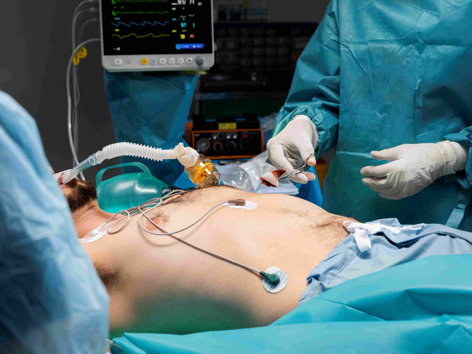
How Is Performed
Preparation: The procedure is done in an emergency setting or OR; the patient is heavily sedated and given blood thinners (Heparin) to prevent clots in the machine.
Percutaneous Access: Using the "Seldinger Technique" where needles and wires guide the cannulas through the skin into the femoral (groin) or jugular (neck) vessels.
Surgical Cut-down: If vessels are too small or damaged, a surgeon makes an incision to directly see and enter the artery or vein.
Imaging Guidance: Real-time Ultrasound and Transesophageal Echo (TEE) are used to ensure the cannula tips are perfectly positioned near the heart.
Connection: Once the tubes are secured, they are connected to the "primed" ECMO circuit, and the machine gradually takes over organ function.
Pre-Procedure Preparation
Emergency Nature: As an emergency life-support measure, formal preparation time is often zero; the medical team acts immediately once the decision is made.
Hemodynamic Stabilization: Medications (vasopressors) are used to keep blood pressure high enough to allow for safe cannula insertion.
Rapid Blood Cross-matching: The procedure involves moving large volumes of blood outside the body, so blood products must be ready.
Anticoagulation Baseline: Checking the patient's clotting status to calibrate the blood-thinning medication required for the ECMO circuit.
Consent: If the patient is unconscious, emergency consent is obtained from the next of kin.
Tests Before ECMO Cannulation
Point-of-Care Ultrasound (POCUS): To check the size and health of the femoral and jugular vessels for the largest possible cannula fit.
Arterial Blood Gas (ABG): To confirm that oxygen levels are critically low despite maximum ventilator support.
Echocardiogram: To evaluate right and left heart function, which determines whether VV or VA ECMO is needed.
Chest X-ray: To assess the severity of lung "white-out" or damage before the procedure begins.
Coagulation Profile: Testing PT/INR and platelet counts to assess the risk of bleeding during the invasive insertion.
Life After ECMO Recovery
ICU Monitoring: Patients are usually kept in a medically induced coma initially, though modern protocols emphasize "Awake ECMO" where possible to keep muscles strong.
Decannulation: Once the heart or lungs show signs of healing (verified by "trialing off" the machine), the cannulas are surgically removed.
Physical Rehabilitation: Because patients are bedbound for days or weeks, intensive physical therapy is required to regain the ability to walk.
Long-term Follow-up: Survivors may experience "Post-ICU Syndrome," requiring respiratory therapy and psychological support.
Organ Monitoring: Regular checks on kidney and liver function are necessary, as these organs can be stressed during the period of support.
Benefits of ECMO Cannulation
The "Ultimate" Life Support: Provides a critical window of time—days to weeks—for the heart and lungs to heal from otherwise fatal injuries.
Restores Oxygen Levels: Immediately corrects life-threatening hypoxia that would otherwise lead to brain death.
Reduces Ventilator Injury: Allows doctors to turn down the pressure on ventilators, preventing further scarring of the lungs (barotrauma).
High Survival Rates: Modern survival rates for neonatal respiratory failure on ECMO are as high as 75%.
Bridge to Permanent Solutions: Acts as a vital safety net for patients waiting for a heart transplant or a long-term LVAD pump.