Medivisor India Treatment Logo
+91 83682 47758
info@medivisorhealth.com
  • Home
    • About Us
    • Our Services
    • Our Team
    • Our Safety Measures
    • Loading treatments...
    • Loading hospitals...
    • Loading doctors...
    • Treatment Cost
    • Treatment Process
    • Visa Process
    • Travel Guide
    • FAQs
    • Why Medivisor
    • Success Stories
    • Patient Activities
    • Social Activities
    • News Coverage
    • Blog
  • Become a Partner

Medivisor India Treatment is a trusted New Delhi–based medical travel company assisting international patients with advanced, affordable, and seamless treatment in India.

  • Medivisor House 359, Sector 1, Vaishali, Ghaziabad, (Delhi/NCR) India
  • +91 8368 247 758
  • info@medivisorhealth.com

About Us

  • About Us
  • Our Services
  • Our Team
  • Our Medical Advisors
  • Our Safety Measures
  • Join Our Network
  • Contact Us

India Treatment

  • Hospitals
  • Treatments
  • Treatment Cost
  • Treatment Process
  • Visa Process
  • Travel Guide
  • FAQs
  • Why Medivisor

Gallery

  • Patient Testimonials
  • Patient Activities
  • News Coverage
  • Blog

Get in Touch

© 2026 Medivisor India Treatment. All rights reserved.

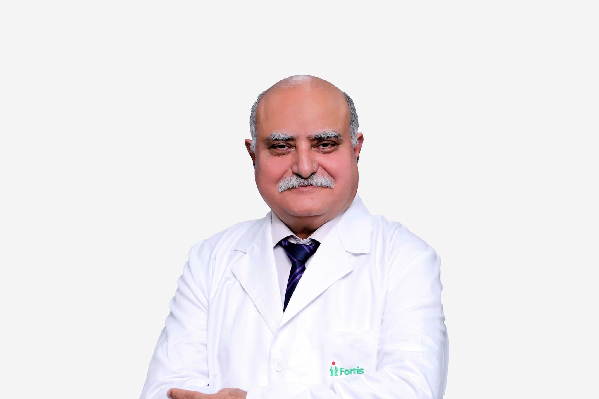
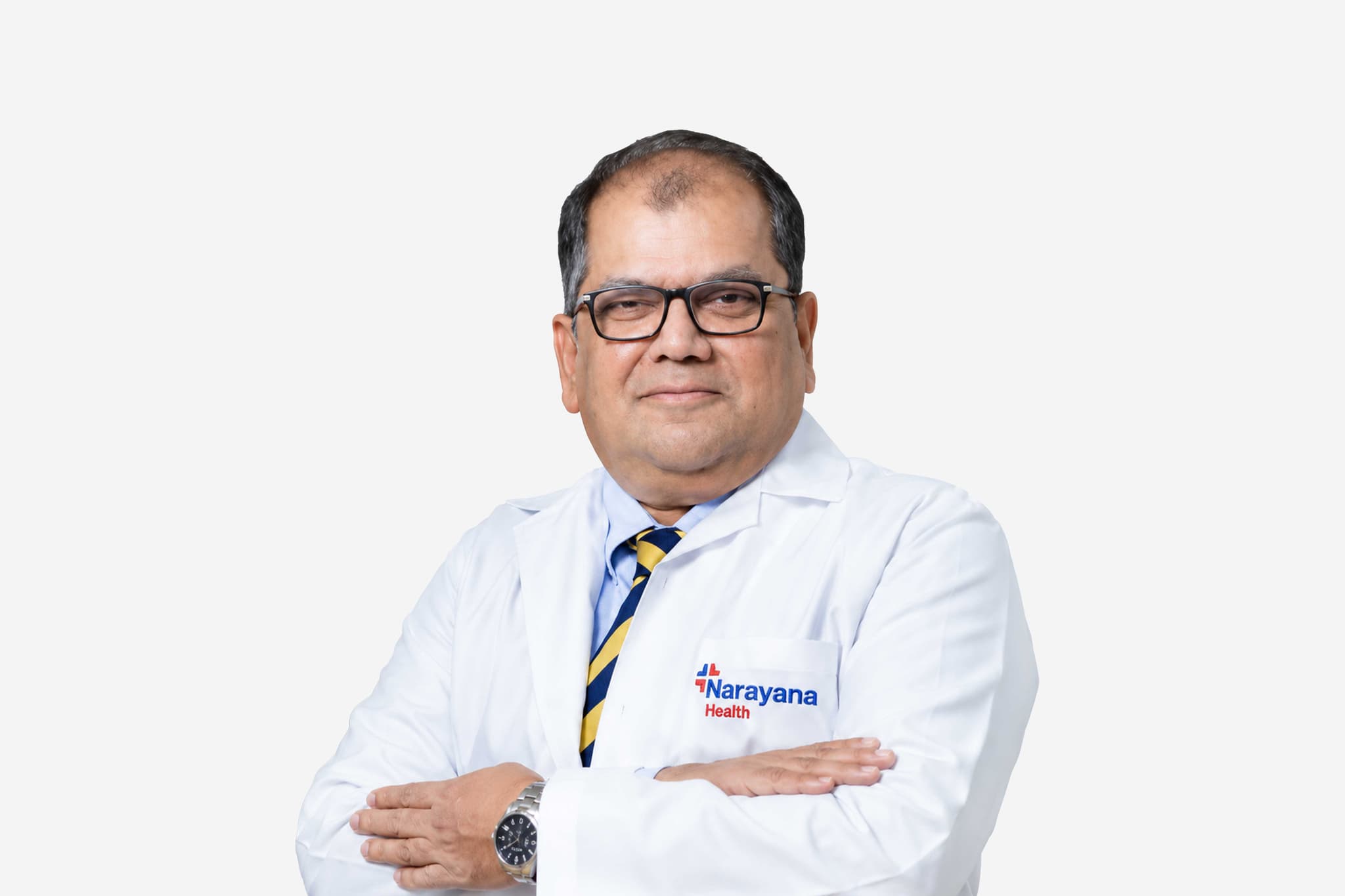
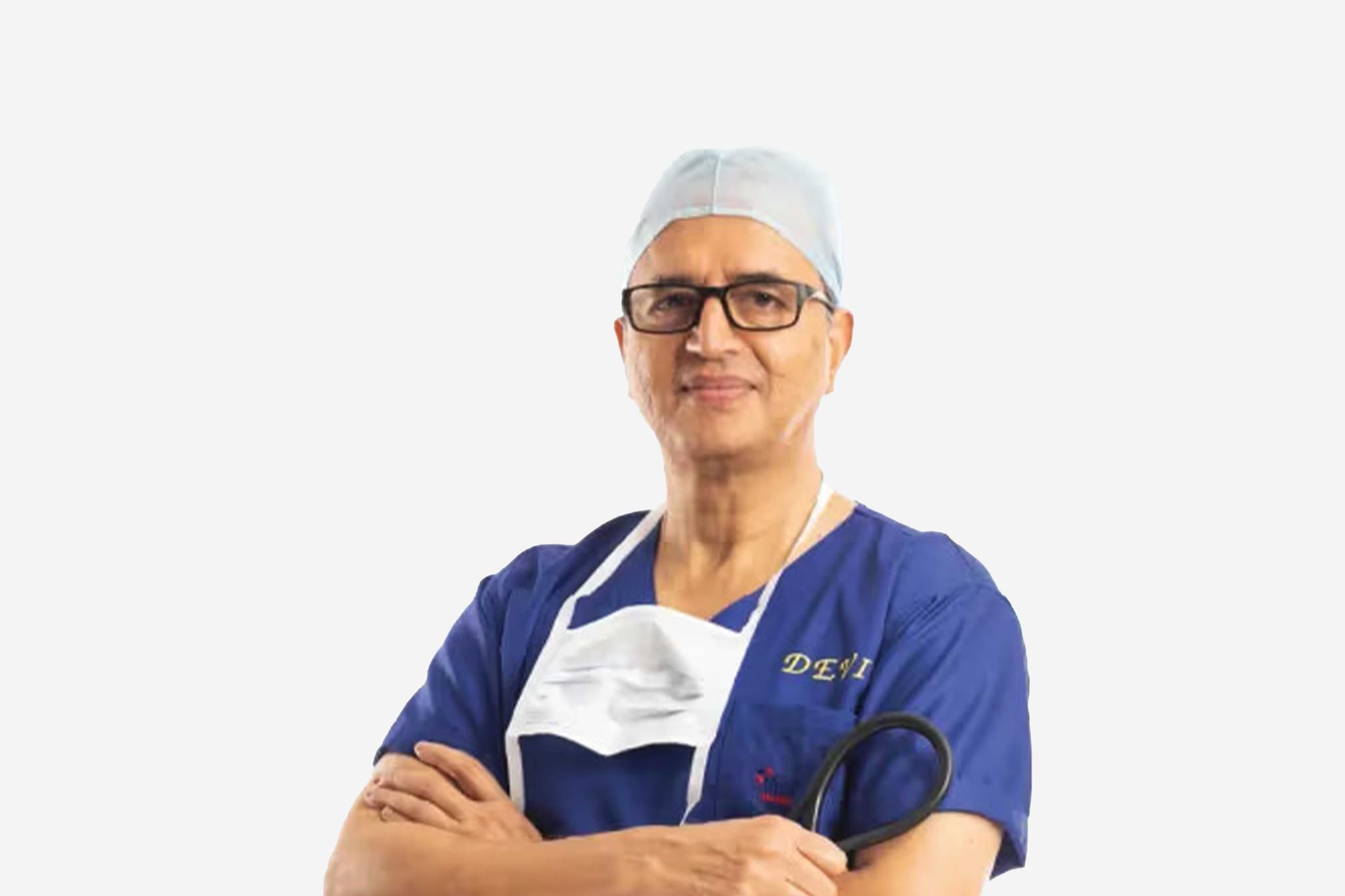
Dr Anil Bhan

Chairman - Cardiac Surgery

Medanta - The Medicity, Gurugram

Cardiac Surgeon

45+ years experience
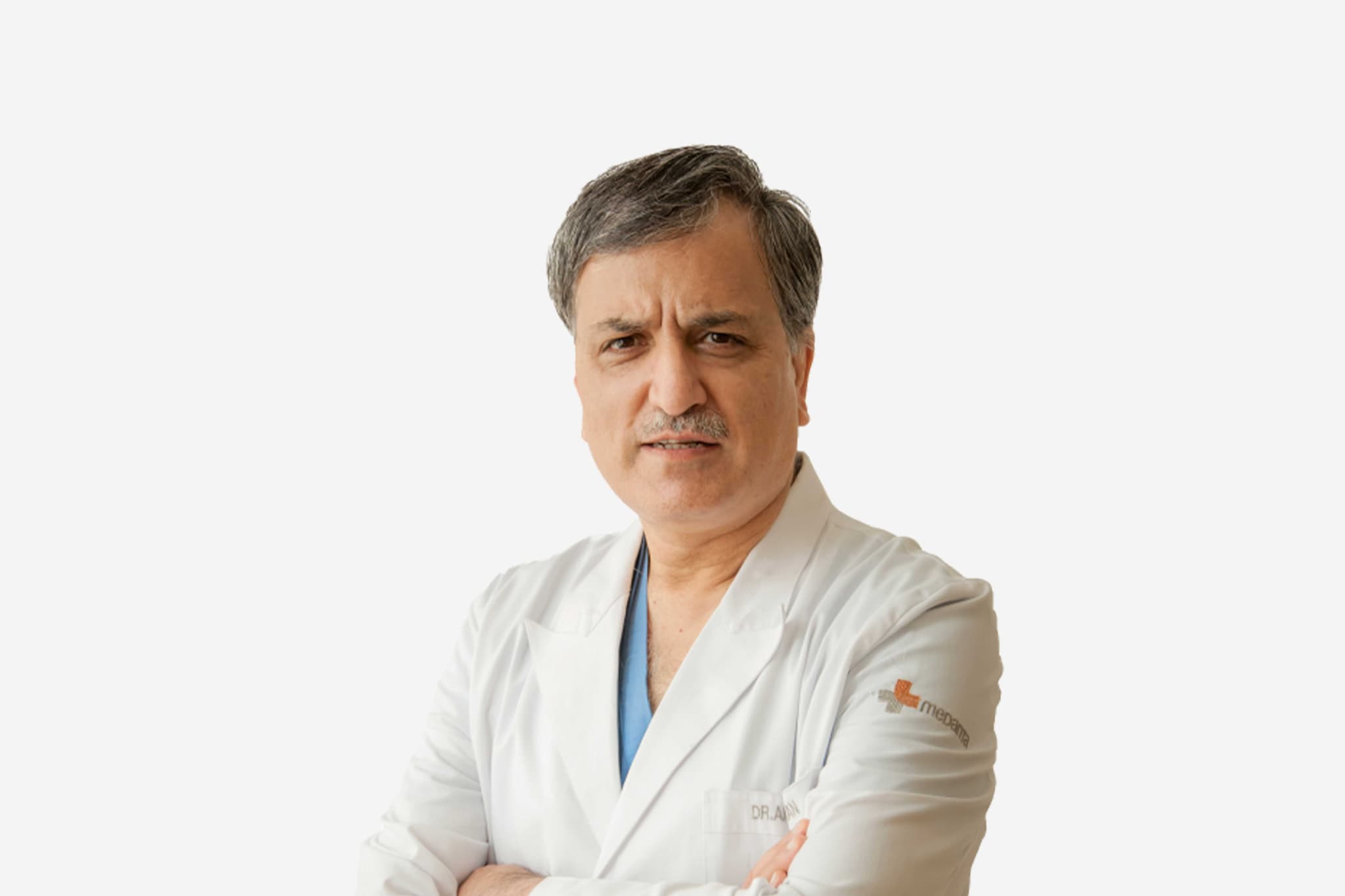
Dr Anil Bhan, Chairman - Cardiac Surgery at Medanta - The Medicity, Gurugram - Medanta Hospitals
  1. Home
  2. Doctors
  3. Dr Anil Bhan

About Dr Anil Bhan

Dr. Anil Bhan is a highly distinguished cardiovascular surgeon and a premier authority in complex heart and vascular procedures. A gold medalist and the top graduate from Medical College Srinagar, he possesses the most extensive experience in aortic surgery in India. Throughout his career, he has successfully performed more than 15,000 cardiac and vascular surgeries, consistently achieving clinical outcomes that rank among the best globally.

Pioneering Breakthroughs in Heart Transplantation and Surgical Infrastructure

Dr. Bhan was an integral member of the surgical team that performed India’s first successful heart transplant in 1994, a landmark achievement in the country's medical history. Beyond his clinical practice, he has been a foundational force in expanding cardiac care infrastructure. He was instrumental in establishing and leading three major cardiac surgical programs: Sri Sathya Sai Institute at Puttaparthi (1992), Whitefield (2001), and the Max Heart and Vascular Institute, Saket (2004).

National Leadership in Medical Innovation

A prolific innovator, Dr. Bhan has designed and developed over 50 specialized surgical instruments used in cardiac surgery today. His contributions have significantly refined surgical techniques and improved precision in the operating room. His leadership extends beyond the hospital, as he continues to shape the standards of cardiovascular care in India through both technological advancement and the scaling of high-quality surgical programs.

Advancing the Future of Aortic and Vascular Care

Dr. Bhan’s clinical focus on aortic surgery has established him as a primary consultant for the most complex vascular pathologies. His deep expertise in this sub-specialty, combined with his role in pioneering transplant medicine, has made him a central figure in the evolution of cardiothoracic surgery in South Asia. He remains dedicated to integrating advanced surgical tools with compassionate patient care to treat end-stage heart and vascular diseases.

Innovation and Clinical Expertise

With a surgical repertoire encompassing over 15,000 procedures, Dr. Bhan specializes in a wide array of adult cardiac surgeries. His expertise includes high-risk aortic repairs, complex vascular reconstructions, and heart failure surgeries. His dual role as a master surgeon and a medical instrument designer allows him to approach cardiac challenges with a unique technical perspective, ensuring the highest standards of safety and surgical efficacy.

Dr. Anil Bhan at a Glance

  • Over 35 years of leadership in Cardiovascular and Thoracic Surgery.

  • Performed more than 15,000 successful cardiac and vascular procedures.

  • Key member of the team that performed India’s first heart transplant (1994).

  • Designed and developed over 50 innovative cardiac surgical instruments.

  • Established major cardiac programs at Puttaparthi, Whitefield, and Max Healthcare.

  • Holds the largest experience in Aortic Surgery in India.

MS - General Surgery | MCh - Cardiothoracic & Vascular Surgery | MBBS
Board Certified in Cardiac Surgeon
2009 Delivered the prestigious P.K. Sen Oration at the CT Conference
2007 Performed the youngest bypass in the world literature with coronary aneurysm repair at the age of 18 months
2000 Did the first extra corporeal membrane oxygenation (ECMO) in India
1995 Used for the first time harmonic scalpel for the harvest of radial/internal mammary artery conduits in India

Affiliated Hospitals

Medanta - The Medicity, Gurugram

Multi-Super Specialty

Gurugram, Delhi NCR

2009

Estd.

1300+

Beds

900+

Doctors

May we help you?

Enter digits only (6-15 digits). We'll add your country code.

Related Treatments

Tetralogy of Fallot Repair

Tetralogy of Fallot (ToF) Repair is a major open-heart surgery performed to correct a combination of four specific heart defects present at birth. The goal of the procedure is to restore normal blood flow to the lungs and ensure that oxygen-rich blood is pumped effectively to the rest of the body. Most infants undergo this definitive correction within their first year of life, typically between 3 to 6 months of age, to prevent long-term damage to the heart muscle and lungs.

When You Should Consider ToF Repair

  • Cyanosis ("Blue Baby" Syndrome): When a newborn has noticeably blue or purple-tinted skin, lips, or nails due to low oxygen levels in the blood.

  • "Tet" Spells: Sudden episodes of profound cyanosis and shortness of breath, often triggered by crying or feeding, which are medical emergencies.

  • Failure to Thrive: When a baby is not gaining weight or growing at a normal rate because the heart is working too hard to circulate oxygen.

  • Heart Murmur: The discovery of a loud, harsh heart murmur during a newborn exam, which often indicates turbulent blood flow through a narrowed pulmonary valve.

  • Low Oxygen Saturation: If pulse oximetry readings consistently show oxygen levels below the normal range, indicating an intracardiac shunt.

Methods Of ToF Repair

  • Complete Intracardiac Repair: The definitive surgical correction involving patching the VSD and widening the pulmonary outflow tract in a single operation.

  • Blalock-Thomas-Taussig (BTT) Shunt: A temporary "palliative" procedure where a small synthetic tube is sewn between a major artery and the pulmonary artery to increase blood flow to the lungs in very small or weak infants.

  • Transannular Patching: A specialized technique used when the pulmonary valve ring is too small, involving a patch that extends across the valve to significantly enlarge the opening.

  • Pulmonary Valve Sparing Repair: A method that focuses on preserving the patient's own pulmonary valve to prevent "leaking" later in life.

  • Monocusp Valve Reconstruction: Using a piece of the patient's own tissue (pericardium) to create a temporary valve leaf to help regulate blood flow immediately after surgery.

How Is Performed

  • Surgical Access: Under general anesthesia, a midline incision is made through the breastbone (median sternotomy) to provide the surgeon with direct access to the heart.

  • Cardiopulmonary Bypass: The child is connected to a heart-lung machine, which takes over the job of circulating and oxygenating the blood so the surgeon can work on a still heart.

  • VSD Patching: The surgeon identifies the large hole between the lower chambers (the Ventricular Septal Defect) and sews a synthetic patch—usually made of Dacron or the patient’s own pericardium—to close it.

  • Relieving Obstruction: Thickened muscle bundles in the right ventricle that block the path to the lungs are carefully cut away.

  • Pulmonary Valve Widening: If the pulmonary valve is narrowed, the surgeon opens it or uses a patch to enlarge the pathway (the pulmonary outflow tract) to ensure easy blood flow to the lungs.

  • Weaning from Bypass: Once the repairs are complete, the heart is restarted, and the heart-lung machine is gradually removed as the heart takes over its new, corrected circulation.

Pre-Procedure Preparation

  • Echocardiogram (Echo): A detailed ultrasound of the heart is mandatory to map the exact size of the VSD and the degree of pulmonary narrowing.

  • Cardiac Catheterization: Occasionally performed to measure the pressures inside the heart chambers and check for any additional abnormal blood vessels.

  • Nutritional Optimization: Many infants are placed on high-calorie formulas or fortified breast milk to ensure they are strong enough for the major surgery.

  • Infection Screening: Ensuring the baby has no signs of a cold, fever, or respiratory infection, which could delay the procedure.

  • Fasting (NPO): Infants must stop feeding several hours before the surgery according to strict hospital guidelines to ensure safety during anesthesia.

Tests Before ToF Repair

  • Chest X-ray: To evaluate the size and shape of the heart (often appearing "boot-shaped" in ToF) and the blood flow patterns in the lungs.

  • Electrocardiogram (EKG): To record the heart's electrical activity and establish a baseline before the VSD patch is placed near the heart’s conduction system.

  • Complete Blood Count (CBC): To check for polycythemia (an abnormally high red blood cell count), which is the body's way of compensating for low oxygen.

  • Cross-match Blood Work: To ensure that appropriately typed blood is available in the operating room for a potential transfusion.

Life After ToF Repair

  • ICU Recovery: Patients usually spend 2 to 4 days in the Pediatric Cardiac ICU for intensive monitoring of heart rhythm, blood pressure, and oxygen levels.

  • Hospital Stay: The typical total stay is 7 to 10 days, depending on how quickly the child transitions back to normal feeding and breathing on their own.

  • Wound Care: The chest incision is closed with dissolvable stitches under the skin; parents are taught how to keep the site clean and dry during the first weeks at home.

  • Activity: Most children recover quickly and are back to their normal baseline activity within a few weeks, though "tummy time" may be restricted to protect the breastbone.

  • Lifelong Follow-up: Regular visits with a Congenital Heart Specialist are mandatory, as some patients may need a pulmonary valve replacement 20–30 years later.

Benefits Of ToF Repair

  • Normal Oxygen Levels: Immediately corrects the "blueness" and allows the child to have normal energy levels and pink skin and lips.

  • Restores Growth: Once the heart is working efficiently, most children experience a "catch-up" period of rapid growth and weight gain.

  • Protects the Heart Muscle: Closing the VSD and relieving the pressure on the right ventricle prevents the heart from becoming dangerously thickened or weak.

  • High Success Rate: With modern surgical techniques, the survival rate for this complex repair is excellent, typically exceeding 95%.

  • Full Active Life: Most children who undergo ToF repair grow up to lead completely normal lives, participating in school, sports, and all regular childhood activities.

PDA Surgical Ligation

Surgical Ligation of a Patent Ductus Arteriosus (PDA) is a definitive procedure to manually close an abnormal, persistent connection between the aorta and the pulmonary artery. While many PDAs are now closed using minimally invasive catheters, surgery remains the primary choice for premature infants, very small babies, or patients with a ductal shape that cannot safely hold a synthetic plug or coil. Closing this "extra" vessel prevents blood from flooding the lungs, which can lead to heart failure and respiratory distress.

When You Should Consider PDA Surgical Ligation

  • Symptomatic Prematurity: For extremely low-birth-weight infants who experience difficulty breathing or feeding and have not responded to medical treatments like Ibuprofen or Indomethacin.

  • Large Ductal Shunt: When the PDA is large enough to cause "volume overload," leading to an enlarged heart and high blood pressure in the lungs (pulmonary hypertension).

  • Anatomical Constraints: If the PDA is too short, wide, or "window-shaped," making it technically difficult or dangerous to place a transcatheter device.

  • Failure of Catheter Closure: When a previous attempt to close the ductus using a catheter-based plug has failed or the device was unable to stay in a stable position.

  • Recurrent Infections: For patients who develop endocarditis (an infection of the heart lining) specifically related to the turbulent blood flow through the PDA.

Methods Of PDA Surgical Ligation

  • Left Posterolateral Thoracotomy: The traditional surgical approach involving a small incision on the left side of the chest, usually between the 4th and 5th ribs.

  • Surgical Clipping: Using a small, permanent titanium clip to pinch the ductus vessel shut, which is often faster and less traumatic than traditional stitching.

  • Suture Ligation: The surgeon uses two thick silk threads to tie the vessel tightly in two places, ensuring no blood can pass through the connection.

  • Ductal Division: A more extensive method where the surgeon ties the vessel in two spots and then cuts the tissue in the middle to ensure it can never reopen.

  • VATS (Video-Assisted) Ligation: A minimally invasive surgical option using a camera and small instruments for older children or larger infants to avoid a full thoracotomy.

How Is Performed

  • Surgical Access: Under general anesthesia, the surgeon makes a small incision on the left side of the chest, reaching the heart from the side rather than through the breastbone.

  • Lung Retraction: The left lung is gently moved aside and protected to provide the surgeon with a clear, direct view of the aorta and the pulmonary artery.

  • Vessel Identification: The surgeon carefully isolates the ductus arteriosus, taking extreme care to identify the nearby nerves that control the voice box and diaphragm.

  • The Closure: Depending on the anatomy, the surgeon either applies a titanium clip or ties two heavy silk sutures around the vessel to "ligate" it.

  • Flow Confirmation: The surgeon confirms that the vessel is completely flattened and that there is no residual "thrill" or vibration, indicating the shunt is closed.

  • Chest Tube Placement: A small drainage tube is often placed in the chest cavity to remove any air or fluid and ensure the left lung re-expands fully after the procedure.

Pre-Procedure Preparation

  • Echocardiogram (Echo): A detailed ultrasound is mandatory to measure the exact diameter of the PDA and assess how much blood is shunting into the lungs.

  • Respiratory Support Optimization: For premature infants in the NICU, ventilator settings are adjusted to ensure the baby is stable enough for the move to the operating room.

  • Infection Screening: Ensuring the patient is free from active pneumonia or other infections that could complicate the surgical recovery.

  • Blood Cross-match: Ensuring that appropriately typed blood is available, as the ductal tissue in premature babies can be extremely fragile and prone to bleeding.

  • Fasting (NPO): Infants must follow strict fasting guidelines before surgery to ensure safety under general anesthesia.

Tests Before PDA Surgical Ligation

  • Chest X-ray: To evaluate the degree of heart enlargement and see how much fluid or "congestion" is present in the lung fields.

  • Electrocardiogram (EKG): To check the heart’s electrical rhythm and look for signs of strain on the left side of the heart caused by the extra blood flow.

  • Complete Blood Count (CBC): To check for adequate hemoglobin and ensures there is no underlying infection before the sterile procedure.

  • Coagulation Profile: To confirm the blood's ability to clot normally, which is vital when working on major blood vessels like the aorta.

Life After PDA Surgical Ligation

  • Chest Tube Removal: The drainage tube is typically removed within 24 to 48 hours once the surgeon confirms the lung is fully expanded and there is no fluid buildup.

  • NICU/Hospital Monitoring: Full-term babies typically stay 2 to 4 days, while premature infants return to the NICU until they reach their original growth and respiratory goals.

  • Pain Management: Discomfort at the rib incision is managed with local nerve blocks and IV medications, transitioning to oral pain relief as the baby begins feeding.

  • Vocal Assessment: Doctors and nurses monitor the baby's cry or voice, as the nerve controlling the left vocal cord is located very close to the ligation site.

  • Activity: Most older children return to normal play and activity within 1 to 2 weeks, with the heart usually returning to its normal size shortly after.

Benefits Of PDA Surgical Ligation

  • Permanent Cure: Surgical ligation has a success rate of nearly 100%; once the vessel is tied or clipped, it is considered permanently closed.

  • Immediate Respiratory Relief: Removing the "flood" of blood to the lungs often allows premature babies to be weaned off ventilators much faster.

  • Protects the Heart: By stopping the volume overload, the surgery prevents the left side of the heart from becoming stretched or weakened.

  • Prevents Lung Damage: Closing the PDA early prevents permanent damage to the small blood vessels in the lungs (pulmonary hypertension).

  • Enables Growth: Many infants experience a rapid improvement in their ability to feed and gain weight once the heart and lungs are no longer struggling.

Complex Congenital Heart Surgery

Complex Congenital Heart Surgery refers to a group of highly specialized operations performed to treat severe, often life-threatening structural heart defects present from birth. Unlike "simple" repairs, such as closing a small hole, complex surgeries often involve rearranging the entire circulatory system. These procedures are frequently performed in multiple stages over several years to allow the heart and lungs to adapt to new blood flow patterns.

When You Should Consider Complex Heart Surgery

  • Hypoplastic Left Heart Syndrome (HLHS): When the left side of the heart is severely underdeveloped and cannot pump enough blood to the body.

  • Transposition of the Great Arteries (TGA): A critical condition where the two main arteries leaving the heart are "switched," sending oxygen-poor blood to the body.

  • Tricuspid Atresia: When a missing heart valve prevents blood from flowing from the right atrium to the right ventricle, resulting in a "single ventricle" circulation.

  • Total Anomalous Pulmonary Venous Return (TAPVR): A defect where the veins bringing blood from the lungs attach to the wrong place in the heart.

  • Truncus Arteriosus: When a single large blood vessel stems from the heart instead of the separate aorta and pulmonary artery.

Common Complex Procedures

  • The Norwood Procedure (Stage 1 of 3): The first step in treating HLHS; the right ventricle is converted into the main pumping chamber, and the aorta is reconstructed to ensure the body receives blood.

  • Arterial Switch Operation (ASO): Performed for TGA; the aorta and pulmonary artery are disconnected and reattached to the correct ventricles, including the delicate transfer of coronary arteries.

  • The Fontan Procedure (Stage 3 of 3): The final stage for single-ventricle defects; oxygen-poor blood from the lower body is connected directly to the pulmonary artery, bypassing the heart.

  • The Glenn Procedure (Stage 2 of 3): Connects the large vein from the upper body (SVC) directly to the pulmonary artery to reduce the workload on a single working ventricle.

  • Ross Procedure: A sophisticated valve replacement where the patient’s own healthy pulmonary valve is moved to the aortic position, allowing it to grow as the child grows.

How Is Performed

  • Median Sternotomy: Under general anesthesia, a midline incision is made through the breastbone to allow the surgical team full access to the heart and great vessels.

  • Advanced Cardiopulmonary Bypass: The patient is connected to a heart-lung machine designed to manage the tiny blood volumes of newborns while maintaining oxygenation to the brain and organs.

  • Deep Hypothermic Circulatory Arrest (DHCA): For the most intricate repairs, the body temperature is lowered to approximately 18°C, and circulation is briefly stopped to provide a still, bloodless field for the surgeon.

  • Anatomical Reconstruction: Using the patient's own tissue or synthetic patches (Dacron/Gore-Tex), the surgeon "re-plumbs" the heart, enlarging vessels and closing internal defects.

  • Coronary Re-implantation: In "switch" procedures, the tiny coronary arteries—often the size of a needle—are meticulously moved to the new aortic root to ensure the heart muscle receives blood.

  • Delayed Chest Closure: In some newborn cases, the chest is left "open" for 2–3 days with a sterile covering to allow the heart to recover from swelling before the final closure.

Pre-Procedure Preparation

  • 3D Anatomical Modeling: Surgeons often use 3D-printed models of the patient's specific heart anatomy to "rehearse" and plan the complex reconstruction before surgery.

  • Prostaglandin Infusion: Many newborns are kept on a continuous IV medication (Alprostadil) to keep the ductus arteriosus open, ensuring survival until surgery can be performed.

  • Nutritional Optimization: Infants may require specialized high-calorie feeding or TPN (IV nutrition) to reach a stable weight and strength for the operation.

  • Cardiac Catheterization: A detailed study to measure internal heart pressures and resistance in the lung vessels, which is critical for planning "staged" procedures.

  • Fasting (NPO): Strict adherence to fasting guidelines is required to ensure safety during the induction of general anesthesia.

Tests Before Complex Heart Surgery

  • Fetal and Neonatal Echocardiogram: The primary diagnostic tool used to visualize the internal structures of the heart and the origin of the great vessels.

  • Cardiac MRI or CT: Provides high-resolution, three-dimensional images of the heart's relationship to the lungs and chest wall.

  • Genetic Screening: To check for associated syndromes (such as DiGeorge Syndrome) that may impact the child's overall surgical risk and recovery.

  • Cross-match Blood Work: Because these surgeries involve significant blood volumes, several units of specifically typed and screened blood are prepared in advance.

Life After Complex Heart Surgery

  • Cardiac ICU (CICU): Patients spend 7 to 21 days in a specialized ICU where heart function, rhythm, and oxygen levels are monitored second-by-second.

  • Inotropic Support: High doses of IV medications are often used for several days to help the "re-plumbed" heart pump effectively as it adapts to the new circulation.

  • Neurological Monitoring: Given the use of bypass and circulatory arrest, the medical team closely monitors for seizures or developmental milestones during recovery.

  • Wound and Bone Healing: For children, the breastbone typically heals within 6 to 8 weeks; parents are taught specific "lifting" techniques to protect the chest.

  • Lifelong CHD Specialist Care: These patients are considered "repaired" rather than "cured" and require lifelong surveillance to monitor for valve issues or rhythm changes.

Benefits Of Complex Heart Surgery

  • Life-Saving Intervention: Provides a definitive chance at survival for infants born with defects that would otherwise be fatal within days or weeks.

  • Improved Oxygenation: Corrects "cyanosis" (blueness), allowing the child’s brain and organs to receive the oxygen necessary for normal development.

  • Restores Physical Potential: Many children grow up to lead active lives, attend school, and participate in sports that would have been impossible without repair.

  • Growth and Development: Relieving the heart's workload allows the body to redirect energy toward physical growth and cognitive milestones.

  • Staged Success: The multi-stage approach (Norwood/Glenn/Fontan) allows the heart to grow and the lungs to mature, leading to better long-term outcomes in single-ventricle patients.

ASD Device Closure
ASD Device Closure

Atrial Septal Defect (ASD) closure is a specialized cardiac procedure performed to repair a hole in the septum, which is the wall separating the heart's upper chambers. This treatment is essential for restoring normal blood flow, preventing the heart from overworking, and reducing the risk of long term complications such as pulmonary hypertension or stroke.

When You Should Consider ASD Closure

  • Persistent shortness of breath, especially during exercise or physical activity.

  • Frequent respiratory infections or lung issues.

  • Chronic fatigue or low energy levels during simple daily tasks.

  • Heart palpitations or the sensation of a skipped heartbeat.

  • Swelling in the legs, feet, or abdomen caused by fluid buildup.

  • Detection of a heart murmur during a routine physical checkup.

Conditions That Require ASD Closure

  • Secundum ASD which is the most common form located in the middle of the atrial wall.

  • Primum ASD which occurs in the lower part of the septum and may affect heart valves.

  • Sinus Venosus ASD located near the entry points of the large veins into the right atrium.

  • Coronary Sinus ASD which involves a defect in the wall between the coronary sinus and the left atrium.

  • Large defects that cause significant blood shunting and heart chamber enlargement.

How ASD Closure Is Performed

  • General anesthesia is administered to ensure the patient is comfortable and pain free.

  • For transcatheter closure, a thin tube is guided through a vein in the groin to the heart.

  • For surgical repair, a chest incision is made to provide direct access to the heart wall.

  • A specialized mesh device or a surgical patch is placed to permanently seal the hole.

  • The heart function is tested using real time imaging to ensure the defect is fully closed.

  • Patients are moved to a specialized recovery unit for continuous monitoring.

Types of ASD Closure

  • Transcatheter Device Closure A minimally invasive method using a catheter to deliver a permanent sealing device to the heart.

  • Open Heart ASD Repair The traditional surgical approach used for very large or complex defects involving a chest incision.

  • Minimally Invasive ASD Surgery Performed through small incisions between the ribs to minimize scarring and speed up healing.

  • Robotic Assisted Repair Uses advanced robotic systems for high precision closure with the smallest possible incisions.

Pre Surgery Preparation

  • Stop smoking at least two to three weeks before the procedure for better lung recovery.

  • Ensure blood pressure and blood sugar levels are well controlled.

  • Follow specific fasting instructions provided by your Medivisor India Treatment coordinator.
  • Adjust or pause blood thinning medications only as advised by your cardiologist.

  • Complete all required cardiac imaging and blood work before the scheduled surgery date.

Pre Surgery Tests

  • ECG to monitor the electrical activity and rhythm of the heart.

  • 2D or 3D Echocardiography to visualize the size and location of the defect.

  • Transesophageal Echo (TEE) for a more detailed view of the heart structures.

  • Chest X ray to evaluate the size of the heart and the condition of the lungs.

  • Routine blood panels including CBC, liver function, and clotting profiles.

Why ASD Closure Is Highly Effective

  • Restores normal blood circulation and prevents oxygen rich blood from mixing with poor blood.

  • Eliminates symptoms like breathlessness and chronic fatigue within weeks.

  • Prevents the right side of the heart from becoming enlarged or failing.

  • Significantly improves daily stamina and long term quality of life.

  • Provides a permanent solution with high success rates in both children and adults.

Recovery After ASD Closure

  • ICU or recovery room stay for one to two days for close observation.

  • Early mobilization and walking are encouraged within twenty four hours.

  • For transcatheter patients, discharge is often possible within forty eight hours.

  • Surgical patients typically require four to seven days of hospital care.

  • Most patients return to school or work within one to four weeks depending on the method.

Life After ASD Closure

  • Exercise tolerance often improves significantly within two to three months of the repair.

  • Follow a heart healthy diet and stay hydrated to support the healing process.

  • Take daily aspirin or blood thinners for six months as prescribed to prevent clots.

  • Use antibiotics before dental procedures for six months to prevent heart infections.

  • Attend regular follow up appointments with a cardiologist to monitor heart health.

Aortic Valve Replacement AVR

Aortic Valve Replacement (AVR) is an advanced cardiac procedure that replaces a damaged, stiff, or leaking aortic valve with a new mechanical or tissue valve. This restores healthy blood flow, improves heart pumping capacity, reduces symptoms, and prevents long-term heart failure or life-threatening complications.

When You Should Consider AVR

  • Severe or persistent shortness of breath that limits walking, climbing stairs, or daily activity.

  • Chest pain, pressure, or heaviness due to the heart struggling to push blood through a narrowed valve.

  • Extreme tiredness or low energy even during simple tasks.

  • Dizziness or fainting episodes, especially during exertion.

  • Irregular heartbeat or noticeable palpitations, indicating the heart is under stress.

  • Swelling in the feet, legs, or ankles, a sign of poor blood circulation or early heart failure.

Conditions That Require AVR

  • Severe Aortic Stenosis – the valve becomes narrowed and heavily calcified, restricting blood flow.

  • Severe Aortic Regurgitation – the valve leaks and allows blood to flow backward into the heart.

  • Congenital valve abnormalities, including bicuspid valves.

  • Infection-related valve damage (endocarditis) that weakens or destroys the valve.

  • Aged, stiff, or heavily calcified aortic valve due to long-term wear and tear.

How Aortic Valve Replacement Is Performed

  • General anesthesia is given to ensure a pain-free and comfortable procedure.
  • The surgeon makes either a full chest incision or a minimally invasive cut depending on your case.

  • The damaged aortic valve is carefully removed.

  • A new mechanical or biological valve is implanted to restore proper blood flow.

  • The heart is restarted, and valve function is tested to ensure smooth operation.

  • You are shifted to the ICU for continuous monitoring and early recovery.

Types of Aortic Valve Replacement

  • Mechanical Valve Replacement
    Long-lasting artificial valve; ideal for younger patients. Requires lifelong blood thinners to prevent clots.

  • Biological (Tissue) Valve Replacement
    Made from natural tissue. Offers natural blood flow and usually requires minimal blood thinner use.

  • Minimally Invasive AVR
    Smaller incisions, less pain, reduced blood loss, and faster healing.

  • Robotic AVR
    Performed using robotic precision tools for high accuracy, minimal scars, and quicker recovery.

  • TAVR (Transcatheter Aortic Valve Replacement)
    A non-surgical, catheter-based procedure performed through the groin. Ideal for elderly or high-risk patients.

Pre-Surgery Preparation

  • Quit smoking at least 2–3 weeks before surgery for better lung function.

  • Keep blood pressure, diabetes, and heart rate well controlled.

  • Follow fasting instructions before the procedure.

  • Stop blood thinners only if your cardiologist advises.

  • Complete all required heart and blood tests before the surgery date.

Pre-Surgery Tests

  • ECG to check heart rhythm.

  • 2D Echocardiography to evaluate valve structure and pumping strength.

  • CT scan or MRI for detailed imaging when needed.

  • Coronary Angiography to detect any artery blockages.

  • Chest X-ray to assess lung health.

  • Routine blood tests including CBC, kidney/liver function, and clotting profile.

Why AVR Is Highly Effective

  • Restores normal forward blood flow from the heart.

  • Reduces breathlessness and chest discomfort.

  • Prevents the heart from becoming enlarged or weak.

  • Improves daily stamina, energy levels, and activity tolerance.

  • Provides long-lasting results with modern valve technology.

Recovery After AVR

  • ICU stay: Usually 1–2 days for close monitoring.

  • Early walking begins within 24 hours.

  • Tubes and drains are removed in 48–72 hours.

  • Home recovery: Typically 4–8 weeks depending on the surgery type.

  • Return to work: Usually within 6–10 weeks.

Life After AVR

  • Avoid smoking permanently to protect the new valve.

  • Follow a heart-healthy, low-salt diet for lifelong cardiac wellness.

  • Exercise daily with light walking, avoid heavy lifting initially.

  • Take medications regularly, especially blood thinners if you have a mechanical valve.

  • Join a cardiac rehabilitation program for guided recovery and long-term heart strength.

Mitral Valve Replacement (MVR)

Mitral Valve Replacement (MVR) is a specialized heart procedure that restores healthy blood flow by replacing a diseased mitral valve with a mechanical or biological valve. This improves heart efficiency, reduces symptoms like breathlessness and fatigue, and prevents long-term complications such as heart failure.

When You Should Consider MVR

  • Shortness of breath during daily activities or while lying down.

  • Chest discomfort or pressure caused by poor blood flow through the heart.

  • Fatigue or low energy during simple tasks.

  • Irregular heartbeat or palpitations due to valve dysfunction.

  • Swelling in feet, legs, or ankles from fluid retention.

  • Fainting or dizziness, especially during physical activity.

Conditions That Require MVR

  • Severe Mitral Stenosis – narrowing of the mitral valve restricting blood flow.

  • Severe Mitral Regurgitation – leaking mitral valve causing backward blood flow.

  • Congenital mitral valve defects present from birth.

  • Valve damage from infection (endocarditis).

  • Calcified or thickened mitral valve leading to poor heart function.

How Mitral Valve Replacement Is Performed

  • General anesthesia is administered for a safe, painless procedure.

  • A chest or minimally invasive incision is made based on patient suitability.

  • The damaged mitral valve is carefully removed.

  • A mechanical or biological replacement valve is implanted.

  • Heart function is tested before closing the incision.

  • Patient is moved to the ICU for monitored recovery.

Types of Mitral Valve Replacement

  • Mechanical Valve Replacement
    Long-lasting artificial valve; requires lifelong blood thinners.

  • Biological (Tissue) Valve Replacement
    Natural tissue valve; usually requires minimal blood thinner use.

  • Minimally Invasive MVR
    Smaller incisions, less pain, quicker healing, and reduced scarring.

  • Robotic MVR
    Uses robotic precision for high accuracy, minimal scarring, and faster recovery.

  • Transcatheter Mitral Valve Replacement (TMVR)
    Non-surgical, catheter-based procedure for high-risk or elderly patients.

Pre-Surgery Preparation

  • Stop smoking 2–3 weeks before surgery.

  • Maintain blood pressure, diabetes, and heart rate within target range.

  • Follow fasting instructions as advised.

  • Pause blood thinners only if instructed by your cardiologist.

  • Complete all cardiac and routine blood tests prior to surgery.

Pre-Surgery Tests

  • ECG to check heart rhythm.

  • Echocardiography (2D/3D) to evaluate mitral valve function.

  • CT or MRI scans for detailed imaging if required.

  • Coronary angiography to detect any blocked arteries.

  • Chest X-ray to assess lung and heart health.

  • Routine blood tests including CBC, kidney/liver function, and clotting profile.

Why MVR Is Highly Effective

  • Restores normal blood flow through the heart.

  • Reduces shortness of breath, fatigue, and chest discomfort.

  • Prevents heart enlargement and failure.

  • Improves daily activity tolerance and quality of life.

  • Provides long-lasting results with modern valve options.

Recovery After MVR

  • ICU stay: 1–2 days for close monitoring.

  • Walking usually begins within 24 hours.

  • Tubes and drains are removed in 48–72 hours.

  • Home recovery: 4–8 weeks depending on the procedure type.

  • Return to work: Typically 6–10 weeks, gradually increasing activity.

Life After MVR

  • Avoid smoking permanently.

  • Follow a heart-healthy, low-salt diet.

  • Engage in daily light exercise; avoid heavy lifting initially.

  • Take prescribed medications regularly, especially blood thinners for mechanical valves.

  • Join a cardiac rehabilitation program for optimal long-term recovery.

Heart Bypass Surgery (CABG)

Coronary Artery Bypass Grafting (CABG), commonly called "heart bypass surgery," is a major surgical procedure used to treat severe coronary artery disease. It creates new pathways for blood to flow to the heart muscle by bypassing clogged or narrowed sections of the coronary arteries. By using healthy blood vessels from elsewhere in the body to "reroute" blood, CABG restores vital oxygen supply to the heart muscle and reduces the risk of a heart attack.

When You Should Consider CABG

  • Left Main Disease: A severe blockage in the main artery supplying the left side of the heart, which is considered high-risk.

  • Triple Vessel Disease: Significant blockages in all three major coronary arteries.

  • Diabetes: Patients with diabetes and multi-vessel disease often have better long-term outcomes with surgery than with stenting.

  • Complex Anatomy: Blockages that are too long, heavily calcified (hardened), or located in areas where a stent cannot be safely placed.

  • Failed Angioplasty: When previous attempts to open arteries with balloons or stents have not been successful or the artery has narrowed again.

Surgical Techniques

  • On-Pump CABG: The traditional method where a heart-lung bypass machine takes over the work of the heart and lungs, allowing the surgeon to operate on a still, non-beating heart.

  • Off-Pump (Beating Heart) CABG: The surgeon uses specialized stabilizers to operate while the heart continues to beat, avoiding the bypass machine. This is often preferred for patients at high risk for stroke.

  • Minimally Invasive (MIDCAB): Small incisions are made between the ribs rather than through the breastbone. This is typically used for bypassing one or two arteries on the front of the heart.

  • Endoscopic Vessel Harvesting (EVH): A 2026 standard where grafts from the leg or arm are removed through tiny incisions using a camera, reducing scarring and pain.

How CABG Is Performed

  • Incision: A midline incision is made, and the breastbone (sternum) is divided to access the heart.

  • Harvesting: Simultaneously, healthy vessels are harvested: the Internal Mammary Artery (chest), Saphenous Vein (leg), or Radial Artery (arm).

  • Bypass: One end of the graft is attached to the aorta (the main artery) and the other end below the blockage, creating a permanent "detour."

  • Restarting: Once the connections are tested for leaks, the heart is restarted (if it was stopped), and the bypass machine is disconnected.

  • Closing: The sternum is secured with permanent stainless steel wires, and the skin is closed with stitches or staples.

Pre-Procedure Preparation

  • Fasting for at least 8–12 hours before surgery, as it is performed under general anesthesia.

  • Extensive blood work, chest X-rays, and an ECG to ensure you are fit for major surgery.

  • Dental clearance is often required to ensure no hidden infections could travel to the heart.

  • Stopping or adjusting certain medications, especially blood thinners like Clopidogrel or Aspirin, as directed.

  • Shaving and surgical scrubbing of the chest, legs, and arms to prevent infection.

Tests Before CABG

  • Coronary Angiogram: The "roadmap" that shows exactly where the blockages are located.

  • Echocardiogram: To assess the heart's pumping strength (Ejection Fraction) and valve function.

  • Carotid Doppler: To check for blockages in the neck arteries that might increase the risk of stroke during surgery.

  • Pulmonary Function Test (PFT): To ensure the lungs are strong enough to handle anesthesia and recovery.

  • Vein Mapping: Ultrasound of the legs or arms to ensure the vessels are healthy enough to be used as grafts.

Life After CABG

  • ICU Stay: Expect to spend the first 24 hours in the Intensive Care Unit for close monitoring of heart rhythm and blood pressure.

  • Hospital Stay: Total recovery in the hospital usually lasts 5 to 7 days.

  • Sternal Precautions: For the first 6 weeks, you must avoid lifting anything heavier than 2–3 kg to allow the breastbone to heal properly.

  • Cardiac Rehabilitation: Starting around week 6, supervised exercise programs are highly recommended to rebuild strength.

  • Long-term Meds: You will likely remain on Aspirin and cholesterol-lowering medications (statins) indefinitely to keep the new grafts clear.

Benefits of CABG

  • Superior Longevity: Provides a long-term solution for complex multi-vessel disease, often outlasting stents.

  • Symptom Relief: Significant reduction or total elimination of chest pain (angina) and shortness of breath.

  • Reduced Heart Attack Risk: By restoring blood flow to large areas of the heart, the risk of a future major cardiac event is lowered.

  • Improved Quality of Life: Most patients return to an active lifestyle and can exercise more effectively than before surgery.

  • 2026 Success Rates: Elective CABG has a high survival rate (approx. 98–99%) due to advanced surgical and anesthesia protocols.

Off-Pump Bypass (Beating Heart Surgery)

Off-Pump Coronary Artery Bypass (OPCAB), also known as "Beating Heart Surgery," is a specialized technique where the surgeon performs the bypass while the heart continues to beat. Unlike traditional CABG, it does not use a heart-lung bypass machine to stop the heart and take over its function. This approach is highly valued for reducing systemic inflammation and protecting vital organs, particularly in high-risk patients.

When You Should Consider OPCAB

  • Elderly Patients (70+ years): Those who may be more vulnerable to the systemic physiological stress of a heart-lung machine.

  • History of Stroke: Patients with a "porcelain" (heavily calcified) aorta where clamping the vessel during traditional surgery increases the risk of a stroke.

  • Chronic Kidney Disease: Maintaining natural blood pressure and pulsatile flow during surgery is generally safer for renal function.

  • Liver Disease or Blood Disorders: Patients who may face higher complications from the intense blood-thinning required for "on-pump" machines.

  • Lung Issues: Those with respiratory compromise who benefit from being taken off a ventilator as quickly as possible following the procedure.

The Core Technology: How It Works

  • Suction Stabilizers: Small, mechanical arms that "grip" a tiny area (1–2 cm) of the heart surface, keeping that specific spot perfectly still while the rest of the heart continues to pump.

  • Intracoronary Shunts: Tiny plastic tubes inserted into the artery during the stitching process to ensure blood continues to flow to the heart muscle while the surgeon sews the graft.

  • Heart Positioners: Suction devices used to gently lift and rotate the beating heart, allowing the surgeon to reach blockages on the side or back walls.

  • Transit Time Flow Measurement (TTFM): A clinical standard used during surgery to verify that blood flow through the new graft is perfect before closing the chest.

  • Deep Pericardial Stay Sutures: Specialized internal stitches that allow the surgeon to maneuver the heart safely into the necessary positions without stopping it.

How Is Performed

  • Surgical Access: Under general anesthesia, a standard midline incision is made through the breastbone (sternotomy) to reach the heart.

  • Graft Harvesting: Healthy vessels are prepared from the chest (internal mammary artery), leg (saphenous vein), or arm (radial artery) to be used as the new bypass routes.

  • Heart Positioning: The surgeon carefully maneuvers the beating heart using positioners to expose the specific blocked coronary arteries.

  • The Bypass: The stabilizer is applied to the target site, and the surgeon meticulously sews the graft onto the artery using ultra-fine sutures.

  • Verification & Closing: After confirming flow with TTFM, the stabilizer is removed, and the breastbone is secured with permanent stainless steel wires.

Pre-Procedure Preparation

  • Fasting (NPO): No food or drink for at least 8–12 hours before surgery to ensure safety during general anesthesia.

  • Baseline Diagnostics: Extensive blood tests, chest X-rays, and an ECG to assess overall surgical readiness and organ function.

  • Dental Clearance: A check to rule out any active oral infections that could travel through the bloodstream and compromise the heart surgery.

  • Medication Adjustment: Reviewing all prescriptions; anti-platelet drugs or blood thinners may need to be paused or adjusted several days prior.

  • Surgical Scrub: Shaving and antiseptic scrubbing of the chest and any potential graft harvest sites on the legs or arms.

Tests Before OPCAB

  • Coronary Angiogram: The essential "roadmap" that identifies the exact location and severity of blockages for the surgical team.

  • Echocardiogram: An ultrasound to evaluate the heart's pumping strength and identify any underlying valve issues.

  • Carotid Ultrasound: To assess stroke risk by checking the health of the arteries supplying blood to the brain.

  • CT Scan of the Aorta: Specifically used to check for heavy calcification (porcelain aorta) that would favor an off-pump approach.

  • Vein/Artery Mapping: Ultrasound imaging to ensure the quality and size of the blood vessels intended for use as bypass grafts.

Life After OPCAB

  • ICU Recovery: Patients typically spend the first 12 to 24 hours in the Intensive Care Unit for close hemodynamic monitoring.

  • Hospital Discharge: The total stay is usually 4 to 5 days, which is often 1–2 days shorter than traditional "on-pump" bypass surgery.

  • Sternal Precautions: To allow the breastbone to heal, patients must avoid lifting anything heavier than 2–3 kg (about 5 lbs) for 6 to 8 weeks.

  • Gradual Recovery: Most patients return to light daily activity quickly but require 2 to 3 months for a full return to strenuous levels.

  • Cardiac Rehab: Participating in a supervised exercise and education program starting around week 6 is vital for long-term cardiovascular health.

Benefits Of OPCAB

  • Reduced Stroke Risk: Avoiding the clamping of a calcified aorta minimizes the chance of dislodging plaque that could travel to the brain.

  • Organ Protection: Shorter ventilator times and more natural, pulsatile blood flow help protect the sensitive kidney and lung systems.

  • Less Bleeding: Beating heart surgery generally requires fewer blood transfusions than procedures involving a bypass machine.

  • Lower Inflammatory Response: Avoiding the heart-lung machine reduces the "whole-body" inflammation often seen after major cardiac surgery.

  • Faster Return to Normalcy: Many patients experience shorter hospital stays and a quicker initial recovery phase compared to traditional methods.

Redo Bypass Surgery
Redo Bypass Surgery

Redo Coronary Artery Bypass Grafting (Redo CABG) is a secondary heart bypass surgery performed on a patient who has already undergone at least one previous bypass. It is technically more demanding than the first surgery because the heart often develops scar tissue (adhesions) that causes it to stick to the underside of the breastbone. In 2026, while complex stenting is often the first choice for failed grafts, Redo CABG remains the definitive solution for patients with extensive new blockages or anatomical challenges that stents cannot fix.

When You Should Consider Redo CABG

  • Graft Attrition: When original vein grafts (typically used 10–15 years ago) have become diseased or completely blocked.

  • Native Disease Progression: New, severe blockages in the heart's original arteries that weren't bypassed during the first surgery.

  • Failed Complex Stenting: When attempts to open old grafts or native arteries with multiple stents have not been successful.

  • Ischemic Heart Failure: When poor blood flow is weakening the heart muscle, and a "complete revascularization" is needed to restore pumping strength.

  • LIMA Failure: In rare cases where the Left Internal Mammary Artery (the "gold standard" graft) has narrowed or failed.

Technical Challenges & Methods

  • The "Re-Entry" Problem: Surgeons use an oscillating saw and extreme caution to open the chest without damaging the heart or old, functioning grafts that may be stuck to the sternum.

  • New Graft Harvesting: Since the best vessels were likely used in the first surgery, surgeons may harvest the Right Internal Mammary Artery, Radial Artery (arm), or additional Leg Veins.

  • Embolic Protection: Old vein grafts are often "crumbly" and can release debris (emboli) if touched; surgeons usually tie these off and replace them to prevent a heart attack during surgery.

  • Retrograde Cooling: A specialized 2026 technique where cooling fluid is pumped backward through the heart's veins (coronary sinus) to protect the muscle while it is stopped.

  • Off-Pump (Beating Heart) Redo: Frequently chosen if the aorta is too calcified to be clamped, reducing the risk of stroke.

How Redo CABG Is Performed

  • Access: The old chest scar is reopened with precision instruments to carefully separate the heart from the surrounding scar tissue.

  • Cannulation: The patient is connected to the heart-lung machine, often through the groin (femoral) vessels for extra safety before the chest is fully opened.

  • Dissection: The surgeon meticulously clears away adhesions to expose the target arteries and the old grafts.

  • Grafting: New bypasses are sewn into place, often using arterial grafts from the arm or chest to ensure better long-term durability.

  • Verification: Transit Time Flow Measurement (TTFM) is used to ensure the new grafts are providing high volumes of blood to the heart muscle.

Pre-Procedure Preparation

  • Multi-Slice CT Scan: A 2026 requirement to map the distance between the heart and the breastbone to plan a safe entry.

  • Fasting: Standard 8–12 hour fast before the surgery, which is always performed under general anesthesia.

  • Blood Cross-Matching: Redo surgeries have a higher chance of needing a blood transfusion, so multiple units of blood are held in reserve.

  • Review of Previous Records: The original "operative note" from the first bypass is essential for the surgeon to know exactly where the old grafts are located.

  • Anticoagulant Adjustment: Blood thinners are carefully managed and often stopped 3–5 days prior to minimize bleeding.

Tests Before Redo CABG

  • Cardiac CT Angiography (CCTA): To visualize the location of old grafts and their proximity to the chest wall.

  • Coronary Angiogram: The essential "roadmap" to identify which old grafts have failed and where new blockages exist.

  • Echocardiogram: To assess current heart function and check for any valve issues that might need fixing at the same time.

  • Carotid Doppler: To ensure there are no blockages in the neck arteries that could increase stroke risk.

  • Viability Study (PET or MRI): To confirm that the heart muscle in the blocked area is still "alive" and will benefit from a new blood supply.

Life After Redo CABG

  • Extended ICU Stay: Patients usually spend 24 to 48 hours in the ICU for closer monitoring of bleeding and heart rhythm.

  • Hospital Stay: Total recovery in the hospital typically lasts 7 to 10 days, slightly longer than the first bypass.

  • Healing Phase: Full recovery can take 8 to 12 weeks. Skin healing may be slower because of the old scar tissue.

  • Cardiac Rehabilitation: Supervised exercise is non-negotiable for redo patients to ensure the new grafts remain open.

  • Aggressive Medical Therapy: High-dose statins and blood thinners are crucial to stop the progression of disease in the new grafts.

Benefits of Redo CABG

  • Complete Revascularization: Unlike stents, which may only fix one spot, a redo bypass can treat all major blockages in one go.

  • Long-Term Durability: Modern arterial grafts used in redos have much higher 10-year success rates than repeat stenting.

  • Symptom Resolution: Provides definitive relief for patients who have "refractory angina" (chest pain that doesn't respond to meds).

  • Improved Life Expectancy: For patients with left main disease or triple vessel disease, surgery offers better survival than medicine alone.

  • 2026 Success Rates: In specialized Indian centers, the success rate for redo CABG now exceeds 93–95% due to better imaging and surgical tech.

Tricuspid Valve Repair

Tricuspid Valve Repair is a surgical or minimally invasive procedure to fix a leaking (regurgitation) or narrowed (stenosis) tricuspid valve, which sits between the right atrium and right ventricle. Repair is increasingly preferred over valve replacement because it preserves the heart's natural anatomy and avoids the need for lifelong, heavy-duty blood thinners. It is a vital intervention for maintaining proper blood flow from the body into the lungs.

When You Should Consider Tricuspid Valve Repair

  • Secondary (Functional) Regurgitation: When the valve leaks because the right side of the heart has stretched (common in patients with left-sided heart disease).

  • Concomitant Repair: When you are already undergoing surgery for a mitral or aortic valve; repairing the tricuspid valve at the same time prevents future heart failure.

  • Severe Right-Sided Symptoms: Such as significant swelling in the legs, abdominal bloating, or unexplained fatigue.

  • Direct Valve Damage: Caused by infection (endocarditis), rheumatic fever, or blunt chest trauma.

  • Pulmonary Hypertension: When high pressure in the lungs forces the tricuspid valve to leak, requiring a surgical "tightening" of the valve base.

Surgical Techniques

  • Annuloplasty (The Ring): The "gold standard" where a cloth-covered medical ring is sewn around the base of the valve to pull the leaflets together for a tight seal.

  • Leaflet Repair: Techniques like "bicuspidization" (tucking a leaflet) or patching holes with a piece of the heart's own sac (pericardium).

  • Neochords: Attaching artificial GORE-TEX strings to support drooping or "flail" leaflets that no longer close properly.

  • Edge-to-Edge Repair (TriClip): A leading-edge, minimally invasive option where a clip is guided through a leg vein to "pin" leaking leaflets together.

  • Minimally Invasive Surgery: Performing the repair through a small incision between the ribs (thoracotomy) rather than opening the breastbone.

How Is Performed

  • Access: Performed via a midline incision (sternotomy) or a minimally invasive side incision.

  • Bypass: The patient is connected to a heart-lung machine, which takes over the work of the heart and lungs during the repair.

  • Inspection: The surgeon opens the right atrium to inspect the valve leaflets and the supporting "annulus" ring.

  • Implantation: The annuloplasty ring or neochords are meticulously sewn into place to restore the valve's shape.

  • Testing: Saline is injected into the ventricle to confirm the valve is leak-proof before the heart is closed and restarted.

Pre-Procedure Preparation

  • Fasting: Required for 8–12 hours before surgery, as it is performed under general anesthesia.

  • Extensive Blood Tests: Including liver and kidney function panels, as these organs are often affected by tricuspid issues.

  • Dental Check-up: To ensure no oral bacteria could cause a post-surgical heart infection.

  • Medication Adjustment: Specifically regarding blood thinners, as directed by your surgical team.

  • Sanitization: Shaving and antiseptic cleaning of the chest and any potential graft sites.

Tests Before Tricuspid Valve Repair

  • Echocardiogram (TTE/TEE): The primary tool used to grade the severity of the leak and measure the size of the heart chambers.

  • Cardiac Catheterization: To check the pressures in the heart and lungs (pulmonary hypertension) and look for coronary artery blockages.

  • Cardiac MRI: To get a high-definition 3D view of the right ventricle's function and volume.

  • Liver Function Tests: To see if the "back-pressure" from the leaky valve has caused liver congestion.

  • Chest X-ray: To evaluate the size of the heart silhouette and the condition of the lungs.

Life After Tricuspid Valve Repair

  • Hospital Stay: Usually lasts 5 to 7 days, with the first 24–48 hours spent in the ICU for close monitoring.

  • Initial Recovery: Most patients are encouraged to sit up and begin walking within 24 hours of surgery.

  • Sternal Precautions: If a sternotomy was performed, no lifting over 3 kg for 6 to 8 weeks to allow the bone to heal.

  • Medication: Most patients take a mild blood thinner (like aspirin) for 3–6 months; lifelong Warfarin is typically not required for a repair.

  • Follow-up: Regular echocardiograms will be scheduled to ensure the repair remains stable and the heart size is shrinking back to normal.

Benefits of Tricuspid Valve Repair

  • High Durability: Over 90% of repairs are successful and significantly reduce leakage for many years.

  • Prevents Heart Failure: Directly reduces the risk of right-sided heart failure and associated liver congestion.

  • Improved Energy: Patients often notice a dramatic reduction in swelling and a significant increase in exercise capacity.

  • Preserves Heart Function: Keeping your natural valve (rather than a replacement) helps the right ventricle maintain its strength.

  • High Success Rates: Elective repairs in specialized centers have low complication rates (1% to 3%) and excellent long-term survival.

Double Valve Replacement (DVR)

Double Valve Replacement (DVR) is a major cardiac surgery where two of the heart's four valves—most commonly the Mitral and Aortic valves—are replaced during a single operation. This is typically required when both valves are severely diseased (stenosed or leaking) and cannot be effectively repaired. DVR remains a life-saving intervention for advanced multi-valve disease, often restoring normal life expectancy for patients with severe heart failure.

When You Should Consider DVR

  • Rheumatic Heart Disease: The leading cause of multi-valve damage, where chronic inflammation scars both the mitral and aortic valves.

  • Calcific Degeneration: Age-related buildup of calcium that stiffens both heart structures simultaneously.

  • Endocarditis: A severe bacterial infection that has spread from one valve to another, causing structural destruction.

  • Left Ventricular Strain: When the failure of one valve causes a "domino effect," putting pressure on the second valve until it also fails.

  • Symptomatic Heart Failure: When symptoms like severe breathlessness, chest pain, and fainting can no longer be managed with medication.

Choosing the Replacement Valves

  • Mechanical Valves: Made of carbon and metal. They are extremely durable and rarely need replacing, but require lifelong blood thinners (Warfarin).

  • Bioprosthetic (Tissue) Valves: Made from pig (porcine) or cow (bovine) tissue. They do not require long-term heavy blood thinners but usually wear out in 10–15 years.

  • On-X Mechanical Valves: A newer generation of mechanical valves that may allow for lower doses of blood thinners.

  • Ross Procedure (Specialized): Using the patient's own pulmonary valve to replace the aortic valve, though less common in a double-replacement scenario.

[Image comparing a mechanical heart valve and a bioprosthetic tissue valve]

How Is Performed

  • Access: A midline incision is made through the breastbone (sternotomy) to provide the surgeon full access to the heart.

  • Cardiopulmonary Bypass: The patient is connected to a heart-lung machine; the heart is temporarily stopped to allow for precise surgery.

  • Valve Removal: The surgeon opens the aorta and the left atrium to meticulously excise the diseased aortic and mitral valves.

  • Implantation: Two new valves are sewn into the heart’s natural rings (annulus) using high-strength sutures.

  • De-airing & Restarting: Air is removed from the heart chambers, blood flow is restored, and the heart is restarted.

Pre-Procedure Preparation

  • Fasting: Required for at least 8–12 hours before surgery, as it is performed under general anesthesia.

  • Blood Prep: Extensive blood work, including cross-matching for several units of blood in case a transfusion is needed.

  • Dental Clearance: Mandatory to ensure no oral bacteria could infect the new prosthetic valves.

  • Medication Adjustment: Adjusting current medications, especially blood thinners and anti-platelet drugs, as directed by the surgeon.

  • System Check: Pulmonary function tests and chest X-rays to ensure the lungs are prepared for recovery.

Tests Before DVR

  • Echocardiogram (TTE/TEE): The primary imaging tool to grade the severity of both valve diseases and measure heart chamber size.

  • Coronary Angiogram: To check for blockages in the heart arteries that might need to be bypassed during the same surgery.

  • Cardiac CT or MRI: To provide 3D anatomical detail of the valves and the surrounding heart structures.

  • Carotid Doppler: To evaluate the risk of stroke by checking the arteries supplying the brain.

  • Organ Function Panels: Including kidney and liver function tests, as these organs are vital for a successful recovery.

Life After DVR

  • ICU Stay: Patients spend 24 to 48 hours in the ICU for intensive monitoring of heart rhythm and blood pressure.

  • Hospital Stay: Total recovery in the hospital usually lasts 7 to 10 days.

  • Anticoagulation: If mechanical valves are used, strict monitoring of blood clotting levels (INR) begins immediately and continues for life.

  • Sternal Precautions: No lifting anything heavier than 3 kg for 8 to 12 weeks to allow the breastbone to heal.

  • Cardiac Rehabilitation: Supervised exercise is critical starting at week 6 to help the heart adjust to the new valves.

Benefits of DVR

  • Corrects Circulation: Immediately corrects the "back-pressure" on the lungs and the rest of the body.

  • Symptom Relief: Drastically reduces shortness of breath, fatigue, and swelling in the legs.

  • Stops Progression: Prevents the progressive stretching and weakening of the heart muscle.

  • Long-Term Durability: Modern prosthetic options allow many patients to experience decades of improved health.

  • Single-Stage Correction: Treating both valves in one surgery avoids the high risk of a "redo" operation later in life.

Cardiac Tumour Removal Surgery

Cardiac Tumour Removal Surgery is a specialized procedure to excise abnormal growths from within or on the heart. While the majority of primary heart tumours (75–80%) are benign (non-cancerous), such as myxomas, they can still be life-threatening. These growths can obstruct blood flow, damage heart valves, or lead to strokes if pieces of the tumour break off and travel to the brain. Advanced imaging and robotic techniques allow for highly precise removal of these rare growths.

When You Should Consider Cardiac Tumour Removal

  • Benign Myxomas: The most common heart tumour, typically found in the left atrium, which requires removal to prevent blood flow obstruction.

  • Papillary Fibroelastomas: Small growths on heart valves that carry a high risk of causing a stroke or heart attack.

  • Symptoms of Obstruction: If a tumour causes dizziness, fainting, or sudden shortness of breath by blocking a heart valve.

  • Embolic Events: If pieces of a suspected tumour have already broken off and caused "mini-strokes" (TIAs) or blood clots in the limbs.

  • Malignant Sarcomas: Rare, aggressive cancers that require surgery to relieve symptoms or as part of a multi-stage treatment plan involving chemotherapy.

Surgical Approaches

  • Open-Heart Surgery (Median Sternotomy): The traditional approach providing the most direct view, necessary for large, complex, or malignant tumours.

  • Minimally Invasive Surgery: Uses small incisions (3–5 cm) between the ribs, often assisted by a 3D endoscope to reduce pain and scarring.

  • Robotically Assisted Surgery: A standard for precision, where surgeons use robotic arms to remove tumours in tight spaces within the heart.

  • Cardiopulmonary Bypass (CPB): Using a heart-lung machine to maintain circulation while the heart is stopped for the safe excision of the growth.

  • Reconstructive Surgery: Using a patch (synthetic or from the patient’s own pericardium) to repair any holes left in the heart wall after the tumour is removed.

How Is Performed

  • Access: The surgeon reaches the heart via a sternotomy or a minimally invasive port-access between the ribs.

  • Bypass: The patient is connected to the heart-lung machine, allowing the surgeon to open the heart chambers in a bloodless environment.

  • Excision: The tumour is meticulously removed, usually along with a small "margin" of healthy tissue to ensure no cells are left behind to regrow.

  • Repair: If the tumour was attached to a valve, the surgeon performs a valve repair or replacement during the same session.

  • Verification: The heart is closed and restarted, and an intraoperative echocardiogram is performed to ensure the tumour is gone and the valves are functioning perfectly.

Pre-Procedure Preparation

  • Fasting: Required for at least 8–12 hours before surgery, as the procedure is performed under general anesthesia.

  • Blood Work: Extensive blood work and cross-matching for blood transfusions, which are common in complex cardiac resections.

  • Dental Clearance: To ensure no bacteria from the mouth could infect the surgical site or any repair patches.

  • Medication Adjustment: Stopping certain medications, particularly blood thinners, several days before the operation.

  • Logistics: Arranging for a hospital stay of roughly one week and a support person for the multi-week recovery at home.

Tests Before Cardiac Tumour Removal

  • Echocardiogram (TTE/TEE): The primary tool used to identify the tumour's size, mobility, and attachment point.

  • Cardiac MRI: Provides high-definition 3D tissue characterization to help distinguish between benign and malignant growths.

  • Cardiac CT Scan: Used to evaluate the tumour’s relationship with the coronary arteries and the chest wall.

  • Coronary Angiogram: Performed in older patients to check for blockages that may need to be bypassed during the same surgery.

  • PET Scan: Occasionally used if a malignant tumour is suspected, to check if the cancer has spread elsewhere in the body.

Life After Cardiac Tumour Removal

  • ICU Stay: Patients spend 1–2 days in the Intensive Care Unit for constant monitoring of heart rhythm and oxygen levels.

  • Hospital Discharge: Most patients go home after 5 to 10 days, depending on whether the approach was open or minimally invasive.

  • Activity Restrictions: No heavy lifting (over 4 kg) for 6 to 12 weeks to allow the breastbone or rib incisions to heal fully.

  • Cardiac Rehabilitation: Supervised exercise is strongly recommended to rebuild physical strength and cardiovascular endurance.

  • Long-term Monitoring: Annual echocardiograms are usually required for several years to ensure the tumour does not recur.

Benefits of Cardiac Tumour Removal

  • Cure for Benign Growths: For tumours like myxomas, surgery is often completely curative with excellent long-term results.

  • Stroke Prevention: Removing highly mobile tumours significantly reduces the risk of life-altering strokes or organ damage.

  • Restores Blood Flow: Eliminates heart failure symptoms caused by tumours obstructing the heart valves.

  • Specialized Outcomes: In-hospital mortality is relatively low (approximately 3%) for such a specialized and complex procedure.

  • Symptom Relief: Most patients experience an immediate improvement in energy levels and a reduction in fainting or palpitations.

Left Ventricular Aneurysm Repair

Left Ventricular (LV) Aneurysm Repair, often called an "Aneurysmectomy" or the "Dor Procedure," is a major surgical operation to correct a "bulge" in the heart's main pumping chamber. This bulge is typically a patch of thin, scarred, non-functioning muscle that forms after a massive heart attack. The focus of this surgery is "Ventricular Restoration"—reshaping the heart from a balloon-like state back into its natural, efficient oval shape to restore pumping power.

When You Should Consider LV Aneurysm Repair

  • Congestive Heart Failure: When the scarred area "balloons" outward, wasting the heart's energy and causing severe breathlessness and fatigue.

  • Recurrent Blood Clots: When blood pools and stagnates inside the bulge, creating clots that carry a high risk of stroke.

  • Refractory Arrhythmias: Life-threatening fast heartbeats (Ventricular Tachycardia) triggered by the border between healthy muscle and scar tissue.

  • Large Aneurysm Size: Even if symptoms are mild, a very large or expanding aneurysm may require repair to prevent progressive heart stretching.

  • Concomitant Surgery: Often performed if you already need a heart bypass (CABG) or mitral valve repair to fully restore heart efficiency.

Surgical Techniques

  • Linear Repair: For smaller aneurysms, the surgeon removes the scarred tissue and sews the healthy muscle edges back together.

  • The Dor Procedure (Endoventricular Circular Patch Plasty): The modern "gold standard" where a synthetic or tissue patch is placed inside the ventricle to rebuild its internal structure.

  • Hybrid LV Restoration: A 2026 approach combining surgical repair with catheter-based techniques for patients who are too high-risk for traditional surgery.

  • Extracellular Matrix (ECM) Patches: A newer option using biological "scaffolding" that may help the heart tissue integrate better than traditional synthetic materials.

  • Ventricular Reconstruction: Using internal sutures to "exclude" the dead tissue from the pumping chamber without actually cutting it out.

[Image showing a synthetic patch being sutured inside the left ventricle during a Dor Procedure]

How LV Aneurysm Repair Is Performed

  • Access: A midline incision is made through the breastbone (sternotomy) to reach the heart.

  • Bypass: The patient is connected to a heart-lung machine; the heart is stopped to allow the surgeon to safely open the ventricle.

  • Clot Removal: Any old blood clots (thrombi) trapped within the aneurysm are carefully removed to prevent future strokes.

  • Reshaping: The surgeon identifies the "border zone" of healthy muscle and secures the patch or sutures to create a new, smaller, and stronger pumping chamber.

  • Verification: An intraoperative ultrasound (TEE) is performed to ensure the heart's "Stroke Volume" (the amount of blood pumped per beat) has significantly improved.

Pre-Procedure Preparation

  • Fasting for at least 8–12 hours before the surgery, which is performed under general anesthesia.

  • Extensive blood work, including kidney function tests and cross-matching for potential blood transfusions.

  • Dental clearance to eliminate any hidden infections that could compromise the surgical site or the patch.

  • Adjusting medications, specifically heart failure drugs like ACE inhibitors and blood thinners, as directed by the surgeon.

  • Review of a "Viability Study" to confirm that the remaining heart muscle is strong enough to support the repair.

Tests Before LV Aneurysm Repair

  • Cardiac MRI: The best tool for mapping the exact size of the aneurysm and distinguishing between scar tissue and healthy muscle.

  • Echocardiogram (TEE): To measure the Ejection Fraction and check if the mitral valve is leaking due to the aneurysm.

  • Coronary Angiogram: To identify blockages in the arteries that will likely be bypassed during the same operation.

  • Cardiac CT Scan: To assess the proximity of the aneurysm to the chest wall, especially important for "redo" surgeries.

  • EP Study (Electrophysiology): Occasionally done if the patient has had life-threatening arrhythmias to locate the "trigger" points.

Life After LV Aneurysm Repair

  • ICU Stay: Usually 2 to 3 days for intensive monitoring of blood pressure, heart rhythm, and fluid levels.

  • Hospital Stay: Total stay typically ranges from 7 to 12 days, depending on the speed of recovery.

  • Mechanical Support: Some patients may briefly require a temporary pump (like an IABP) to help the reshaped heart work in the first 48 hours.

  • Sternal Precautions: No lifting anything heavier than 3 kg for 8 to 12 weeks to ensure the breastbone heals.

  • Long-term Meds: Lifelong use of beta-blockers and blood thinners is often necessary to protect the repair and prevent new clots.

Benefits of LV Aneurysm Repair

  • Improved Pumping Efficiency: Reshaping the heart significantly increases the Ejection Fraction and overall cardiac output.

  • Dramatic Symptom Relief: Most patients report a major decrease in shortness of breath and a return of energy within 4–8 weeks.

  • Reduced Stroke Risk: By removing the "pocket" where blood stagnates, the primary source of heart-related strokes is eliminated.

  • Rhythm Stability: Repairing the "border zone" often resolves or simplifies the management of dangerous heart arrhythmias.

  • 2026 Success Rates: In specialized Indian centers, the success rate for the Dor Procedure is approximately 90–95% for elective cases.

Ventricular Septal Rupture Repair

Ventricular Septal Rupture (VSR) Repair is a high-stakes, emergency surgical procedure to fix a hole in the septum (the wall dividing the left and right ventricles). This rupture is a rare but catastrophic complication of a massive heart attack, occurring when a lack of blood flow causes heart muscle to die and physically tear. Surgical intervention remains the "gold standard," as the condition is almost always fatal without mechanical closure.

When You Should Consider VSR Repair

  • Acute Heart Failure: When the septum tears, oxygen-rich blood surges into the right side of the heart, causing the heart to lose its ability to pump to the rest of the body.

  • Pulmonary Flooding: Sudden, excessive blood flow into the lungs leads to rapid fluid buildup (edema) and severe breathing difficulty.

  • Cardiogenic Shock: If blood pressure drops dangerously low and organs begin to fail due to the massive "shunt" of blood within the heart.

  • Post-Infarction Complication: Typically occurs within the first 24 hours or 3–5 days following a major heart attack.

  • High-Risk Stabilization: If a patient is currently on life support (ECMO) or a balloon pump (IABP) specifically to bridge them to a definitive surgical repair.

Surgical Techniques

  • Infarct Exclusion: The modern standard where a large synthetic patch is "wallpapered" over the hole and anchored to healthy, firm heart muscle away from the fragile tear.

  • Triple Patch Technique: A newer method using three layers of bovine pericardium and surgical glue to ensure a leak-proof seal and minimize the risk of the hole reopening.

  • Extended Sandwich Patch: Using two large Dacron patches to "sandwich" the septum from both the left and right sides, often used for complex or posterior ruptures.

  • Hybrid Repair: A two-stage approach where surgery is followed by a transcatheter "plug" if a small residual leak (shunt) remains after the initial operation.

  • Concomitant CABG: Since a blocked artery caused the rupture, surgeons almost always perform a heart bypass during the same procedure to protect the remaining muscle.

How Is Performed

  • Access: A midline incision is made through the breastbone (sternotomy) for the most direct access to the complex rupture site.

  • Bypass: The patient is connected to a heart-lung machine; the heart is stopped to allow the surgeon to operate on the delicate, damaged tissue.

  • Ventriculotomy: The surgeon opens the scarred area of the left ventricle (the chamber with the highest pressure) to inspect the tear.

  • Debridement: Any "mushy" or dead tissue at the edges of the hole is cleared away to reach firmer muscle that can hold sutures.

  • Patching & Gluing: The synthetic or tissue patch is meticulously secured. Specialized surgical glues are often used to reinforce the suture lines on fragile tissue.

  • Restarting: The heart is carefully restarted, and a transesophageal echo (TEE) is performed immediately to check for any residual leaks.

[Image showing a synthetic patch being sutured over a ventricular septal defect]

Pre-Procedure Preparation

  • Emergency Stabilization: Hemodynamic stabilization is the priority; many patients receive an Intra-aortic Balloon Pump (IABP) to reduce the heart's workload.

  • Fasting: Required, though most patients are already under emergency care and receiving fluids intravenously.

  • Blood Cross-matching: Extensive cross-matching is performed, as these surgeries carry a high risk of bleeding and often require blood transfusions.

  • Tissue Friability Review: Surgeons may delay surgery for 3–7 days if the patient is stable enough to let the heart muscle toughen, which increases suture success.

  • Emergency Consent: Consent is often obtained from family members, as the patient is typically too ill or sedated to provide it themselves.

Tests Before VSR Repair

  • Echocardiogram (TTE/TEE): The essential test to confirm the location and size of the rupture and quantify the "shunt" volume.

  • Coronary Angiogram: Necessary to identify the blocked artery that caused the heart attack and plan the necessary bypass grafts.

  • Cardiac CT Scan: Sometimes used to assess the anatomy of the rupture, especially if it is in a difficult-to-reach posterior location.

  • Swan-Ganz Catheterization: To measure the pressures in the lungs and the degree of oxygen-rich blood mixing in the right side of the heart.

  • Blood Gas Analysis: To monitor how well the lungs are coping with the sudden influx of extra blood.

Life After VSR Repair

  • ICU Stay: Patients typically require 3 to 7 days in the ICU on a ventilator with multiple medications to support blood pressure.

  • Hospital Stay: Total recovery in the hospital usually lasts 2 to 3 weeks due to the severity of the initial heart attack.

  • Long-term Management: Lifelong heart failure medications (such as Beta-blockers and ARNI therapy) are essential to help the heart recover.

  • Residual Shunt Monitoring: 10–20% of cases may have a tiny remaining leak; these are monitored via regular echocardiograms and only repaired if they cause symptoms.

  • Rehabilitation: A slow, medically supervised cardiac rehab program is vital to rebuild strength after such a massive physiological trauma.

Benefits of VSR Repair

  • Life-Saving Intervention: Without surgery, the mortality rate is nearly 90% within weeks; repair offers the only realistic chance for survival.

  • Stops Pulmonary Flooding: Immediately halts the surge of blood into the lungs, allowing for easier breathing and recovery from edema.

  • Restores Systemic Pressure: By closing the hole, the heart can once again send oxygenated blood to the brain, kidneys, and liver.

  • Improved Outcomes: While high-risk, 30-day survival rates in specialized cardiac centers have improved significantly for stable patients.

  • Future Heart Health: For those who survive the initial recovery, long-term heart function can improve significantly with proper care.

LVAD Implantation
LVAD Implantation

Left Ventricular Assist Device (LVAD) Implantation is a major surgical procedure to install a mechanical pump that assists a weakened heart in circulating blood throughout the body. Unlike a total artificial heart, an LVAD works with your existing heart to take over the pumping work of the left ventricle—the heart's main pumping chamber. These devices are increasingly used as "Destination Therapy" for those who may not be eligible for a heart transplant, serving as a long-term life-support system.

When You Should Consider an LVAD

  • End-Stage Heart Failure: When medications and other treatments no longer help and the heart is too weak to support the body's metabolic needs.

  • Bridge to Transplant (BTT): To keep a patient stable and healthy enough to undergo a heart transplant while waiting for a suitable donor organ.

  • Destination Therapy (DT): As a permanent solution to improve quality of life for patients ineligible for a transplant due to age or other medical conditions.

  • Bridge to Recovery (BTR): In cases where heart failure is expected to be temporary (such as viral myocarditis), supporting the heart until it can pump on its own.

  • Severe Symptom Burden: When life is severely limited by extreme fatigue, shortness of breath even at rest, and frequent emergency hospitalizations.

Core Components Of The LVAD System

  • The Internal Pump: Surgically implanted at the apex (tip) of the left ventricle to pull blood out and push it directly into the aorta.

  • The Driveline: A thin, reinforced cable that passes from the internal pump through the skin of the abdomen to connect to the external computer.

  • External Controller: A small computer worn on a belt or harness that monitors the pump's function and provides vital alerts or alarms.

  • Power Source: Uses rechargeable lithium-ion batteries for mobile use or a power cord that plugs into a standard electrical outlet while sleeping.

  • Mobile Monitoring: Modern controllers often sync with smartphone apps to allow caregivers and medical teams to monitor pump flow and battery life remotely.

How Is Performed

  • Surgical Access: The surgeon makes an incision down the center of the chest and separates the breastbone (sternotomy) to reach the heart.

  • Heart-Lung Bypass: A bypass machine takes over heart and lung functions so the surgeon can safely work on a still heart.

  • Implantation: The inflow end of the pump is sewn into the left ventricle, and the outflow graft is meticulously attached to the aorta.

  • Driveline Tunneling: The power cable is carefully tunneled through the abdominal wall to exit the skin at a specific "exit site" on the abdomen.

  • Activation: Once the device is tested and circulating blood, the bypass machine is disconnected and the chest is secured with surgical wires.

Pre-Procedure Preparation

  • Fasting (NPO): No food or drink for 8–12 hours before surgery, as the procedure is performed under general anesthesia.

  • Multidisciplinary Evaluation: Extensive review by a "Heart Failure Team," including cardiologists, surgeons, social workers, and nutritionists.

  • Organ Function Screens: Blood tests to ensure the liver and kidneys are healthy enough to withstand the surgery and the new circulatory demands.

  • Caregiver Training: Both the patient and a designated "caregiver" must learn how to manage the device, change batteries, and handle emergency alarms.

  • Infection Prevention: Dental clearance is required to ensure no oral bacteria could lead to an infection of the mechanical pump components.

Tests Before LVAD Implantation

  • Echocardiogram: To assess the strength of the right ventricle; if the right side of the heart is too weak, a standard LVAD may not be effective.

  • Right Heart Catheterization: To measure the pressures in the heart and lungs to ensure the body can handle the pump's mechanical flow.

  • Cardiac CT Scan: To map the anatomy of the chest and identify the best surgical placement for the pump and the outflow graft.

  • Pulmonary Function Tests: To ensure the lungs are strong enough for the patient to be successfully taken off a ventilator after the procedure.

  • Psychosocial Assessment: To ensure the patient has the necessary support system and cognitive ability to manage the device daily.

Life After LVAD Implantation

  • ICU Recovery: Patients spend the first few days in the Intensive Care Unit for close monitoring of the pump's speeds and blood flow parameters.

  • Hospital Education: Total recovery in the hospital typically lasts 2 to 3 weeks as the patient and family learn to live with the device.

  • Anticoagulation Therapy: Lifelong use of blood thinners (typically Warfarin) is required to prevent blood from clotting inside the mechanical pump.

  • Daily Maintenance: The driveline exit site requires meticulous daily cleaning and sterile dressing changes to prevent serious infections.

  • Activity Restrictions: While most return to an active life, swimming and soaking in baths are prohibited to keep the exit site completely dry.

Benefits Of LVAD Implantation

  • Significant Longevity: One-year survival is approximately 80% to 85%, offering years of life to those with otherwise terminal heart failure.

  • Improved Quality of Life: Most patients see a dramatic reduction in shortness of breath and can return to activities like walking, gardening, and traveling.

  • Organ Protection: By improving systemic blood flow, the LVAD helps protect the kidneys and liver from damage caused by chronic congestion.

  • Advanced Technology: Newer "fully levitated" centrifugal pumps have significantly reduced the risk of stroke and mechanical pump malfunctions.

  • Bridge to Transplant: Successfully keeps patients in peak physical condition so they are ready when a donor heart becomes available.

ECMO Cannulation
ECMO Cannulation

ECMO (Extracorporeal Membrane Oxygenation) Cannulation is a critical surgical or percutaneous procedure where large-bore tubes (cannulas) are inserted into major blood vessels to connect a patient to an ECMO machine. This "heart-lung" bypass technology acts as a temporary life-support system by taking over the work of the heart and/or lungs, allowing these organs to rest and heal. Advances in portable platforms and AI-driven monitoring have expanded the use of this therapy from the ICU to emergency field transport.

[Image comparing VV-ECMO (venous return) and VA-ECMO (arterial return) setups]

When You Should Consider ECMO Support

  • Severe ARDS: When the lungs are so damaged (e.g., from pneumonia) that a ventilator can no longer maintain oxygen levels.

  • Cardiogenic Shock: When the heart is unable to pump enough blood to support the body’s vital organs, often after a massive heart attack.

  • Bridge to Transplant: To keep patients alive and stable while they wait for a donor heart or lung.

  • E-CPR (Extracorporeal CPR): Used during active cardiac arrest in specialized trauma centers to restore circulation when traditional CPR fails.

  • Post-Surgical Recovery: When a patient’s heart or lungs are "stunned" and unable to function independently after complex cardiac surgery.

Major Cannulation Strategies

  • Veno-Venous (VV) ECMO (Lung Support): Blood is drained from a large vein, oxygenated by the machine, and returned to the venous system. It supports the lungs only.

  • Veno-Arterial (VA) ECMO (Heart & Lung Support): Blood is drained from a vein and returned to an artery, bypassing both the heart and lungs to provide full circulatory support.

  • Veno-Arterio-Venous (VAV) ECMO: A hybrid configuration used when a patient needs both the cardiac support of VA and additional oxygenation for the lungs.

  • Dual-Lumen Cannulation: Using a single, specialized tube inserted in the neck that both drains and returns blood, allowing for earlier patient movement.

  • Distal Perfusion Cannula: In leg-based VA ECMO, a smaller third cannula is often added to ensure blood flow reaches the lower leg and prevent limb injury.

How Is Performed

  • Preparation: The procedure is done in an emergency setting or OR; the patient is heavily sedated and given blood thinners (Heparin) to prevent clots in the machine.

  • Percutaneous Access: Using the "Seldinger Technique" where needles and wires guide the cannulas through the skin into the femoral (groin) or jugular (neck) vessels.

  • Surgical Cut-down: If vessels are too small or damaged, a surgeon makes an incision to directly see and enter the artery or vein.

  • Imaging Guidance: Real-time Ultrasound and Transesophageal Echo (TEE) are used to ensure the cannula tips are perfectly positioned near the heart.

  • Connection: Once the tubes are secured, they are connected to the "primed" ECMO circuit, and the machine gradually takes over organ function.

Pre-Procedure Preparation

  • Emergency Nature: As an emergency life-support measure, formal preparation time is often zero; the medical team acts immediately once the decision is made.

  • Hemodynamic Stabilization: Medications (vasopressors) are used to keep blood pressure high enough to allow for safe cannula insertion.

  • Rapid Blood Cross-matching: The procedure involves moving large volumes of blood outside the body, so blood products must be ready.

  • Anticoagulation Baseline: Checking the patient's clotting status to calibrate the blood-thinning medication required for the ECMO circuit.

  • Consent: If the patient is unconscious, emergency consent is obtained from the next of kin.

Tests Before ECMO Cannulation

  • Point-of-Care Ultrasound (POCUS): To check the size and health of the femoral and jugular vessels for the largest possible cannula fit.

  • Arterial Blood Gas (ABG): To confirm that oxygen levels are critically low despite maximum ventilator support.

  • Echocardiogram: To evaluate right and left heart function, which determines whether VV or VA ECMO is needed.

  • Chest X-ray: To assess the severity of lung "white-out" or damage before the procedure begins.

  • Coagulation Profile: Testing PT/INR and platelet counts to assess the risk of bleeding during the invasive insertion.

Life After ECMO Recovery

  • ICU Monitoring: Patients are usually kept in a medically induced coma initially, though modern protocols emphasize "Awake ECMO" where possible to keep muscles strong.

  • Decannulation: Once the heart or lungs show signs of healing (verified by "trialing off" the machine), the cannulas are surgically removed.

  • Physical Rehabilitation: Because patients are bedbound for days or weeks, intensive physical therapy is required to regain the ability to walk.

  • Long-term Follow-up: Survivors may experience "Post-ICU Syndrome," requiring respiratory therapy and psychological support.

  • Organ Monitoring: Regular checks on kidney and liver function are necessary, as these organs can be stressed during the period of support.

Benefits of ECMO Cannulation

  • The "Ultimate" Life Support: Provides a critical window of time—days to weeks—for the heart and lungs to heal from otherwise fatal injuries.

  • Restores Oxygen Levels: Immediately corrects life-threatening hypoxia that would otherwise lead to brain death.

  • Reduces Ventilator Injury: Allows doctors to turn down the pressure on ventilators, preventing further scarring of the lungs (barotrauma).

  • High Survival Rates: Modern survival rates for neonatal respiratory failure on ECMO are as high as 75%.

  • Bridge to Permanent Solutions: Acts as a vital safety net for patients waiting for a heart transplant or a long-term LVAD pump.

Realted Specialist

Dr Naresh Trehan
Dr Naresh Trehan
Cardiac Surgeon
Medanta - The Medicity, Gurugram
57+years experience
View Profile
Dr Ajay Kaul
Dr Ajay Kaul
Cardiothoracic & Vascular Surgeon (CTVS), Heart Transplant Surgeon, Cardiac Surgeon
Fortis Hospital, Noida
38+years experience
View Profile
Dr Anvay Mulay
Dr Anvay Mulay
Cardiothoracic & Vascular Surgeon (CTVS), Lung Transplant Surgeon, Heart Transplant Surgeon, Cardiac Surgeon
Sir H N Reliance Foundation Hospital and Research Centre
30+years experience
View Profile
Dr Arun Garg
Dr Arun Garg
Neurologist
Medanta - The Medicity, Gurugram
41+years experience
View Profile
Dr Arvind Kumar
Dr Arvind Kumar
Lung Transplant Surgeon
Medanta - The Medicity, Gurugram
44+years experience
View Profile
Dr Ashok Kumar Vaid
Dr Ashok Kumar Vaid
Hemato-Oncologist, Medical Oncologist
Medanta - The Medicity, Gurugram
42+years experience
View Profile
Dr Ashok Rajgopal
Dr Ashok Rajgopal
Orthopedic Surgeon, Joint Replacement Surgeon
Medanta - The Medicity, Gurugram
50+years experience
View Profile
Dr Atul Anand Maslekar
Cardiac Surgeon
41+years experience
View Profile
Dr Bhaba Nanda Das
Dr Bhaba Nanda Das
Heart Transplant Surgeon, Cardiac Surgeon
Indraprastha Apollo Hospital, Delhi
37+years experience
View Profile
Dr Deepak Gowda
Dr Deepak Gowda
Cardiothoracic & Vascular Surgeon (CTVS), Cardiac Surgeon
Sakra World Hospital
16+years experience
View Profile
Dr Deepak Sarin
Dr Deepak Sarin
Head & Neck Onco-Surgeon
Medanta - The Medicity, Gurugram
32+years experience
View Profile
Dr Devi Prasad Shetty
Cardiothoracic & Vascular Surgeon (CTVS), Pediatric Cardiac Surgeon, Cardiac Surgeon
Narayana Institute of Cardiac Sciences, Bengaluru
43+years experience
View Profile
Dr Dhiren Shah
Dr Dhiren Shah
Cardiothoracic & Vascular Surgeon (CTVS), Cardiac Surgeon
Marengo CIMS Hospital, Ahmedabad
27+years experience
View Profile
Dr Ganesh Krishnan Iyer
Cardiac Surgeon
Aster CMI Hospital
39+years experience
View Profile
Dr Gaurav Gupta
Dr Gaurav Gupta
Cardiothoracic & Vascular Surgeon (CTVS), Cardiac Surgeon
21+years experience
View Profile
Dr Harsh Vardhan Puri
Lung Transplant Surgeon, Thoracic Surgeon, Thoracic Onco-Surgeon
Medanta - The Medicity, Gurugram
20+years experience
View Profile
Dr Yugal Kishore Mishra
Cardiothoracic & Vascular Surgeon (CTVS), Cardiac Surgeon
Manipal Hospital, Dwarka, Delhi
45+years experience
View Profile
Dr Z S Meharwal
Dr Z S Meharwal
Heart Transplant Surgeon, Cardiac Surgeon
Fortis Escorts Heart Institute, New Delhi
35+years experience
View Profile
Dr Manish Vaish
Dr Manish Vaish
Spine Surgeon, Neurosurgeon
Medanta Super Specialty Hospital, Noida
30+years experience
View Profile
Dr Manoj Kumar Singhal
Nephrologist, Kidney Transplant Surgeon
Medanta Super Specialty Hospital, Noida
39+years experience
View Profile

May we help you?

Enter digits only (6-15 digits). We'll add your country code.