Pituitary Tumor Surgery (Endoscopic)
Endoscopic Pituitary Surgery
Endoscopic Pituitary Surgery, also known as Endoscopic Transsphenoidal Surgery, is a minimally invasive procedure that uses the nostrils as natural pathways to reach and remove tumors from the pituitary gland. Because it avoids large incisions and brain retraction, it typically offers a faster recovery and fewer side effects than traditional open surgery. This approach allows surgeons to access the "master gland" at the base of the brain with extreme precision.
When You Should Consider Endoscopic Pituitary Surgery
Hormone-Secreting Tumors: Such as those causing Cushing’s disease (excess cortisol), acromegaly (excess growth hormone), or prolactinomas.
Non-Functioning Macroadenomas: Large tumors that do not produce hormones but press on the optic nerves, causing vision loss, double vision, or chronic headaches.
Pituitary Apoplexy: An emergency condition where a tumor bleeds or outgrows its blood supply, requiring rapid decompression.
Failed Medical Management: When medications are unable to sufficiently control hormone levels or stop the growth of the tumor.
Rathke’s Cleft Cysts: Benign fluid-filled growths that can interfere with normal gland function or cause pressure symptoms.
How It Is Performed
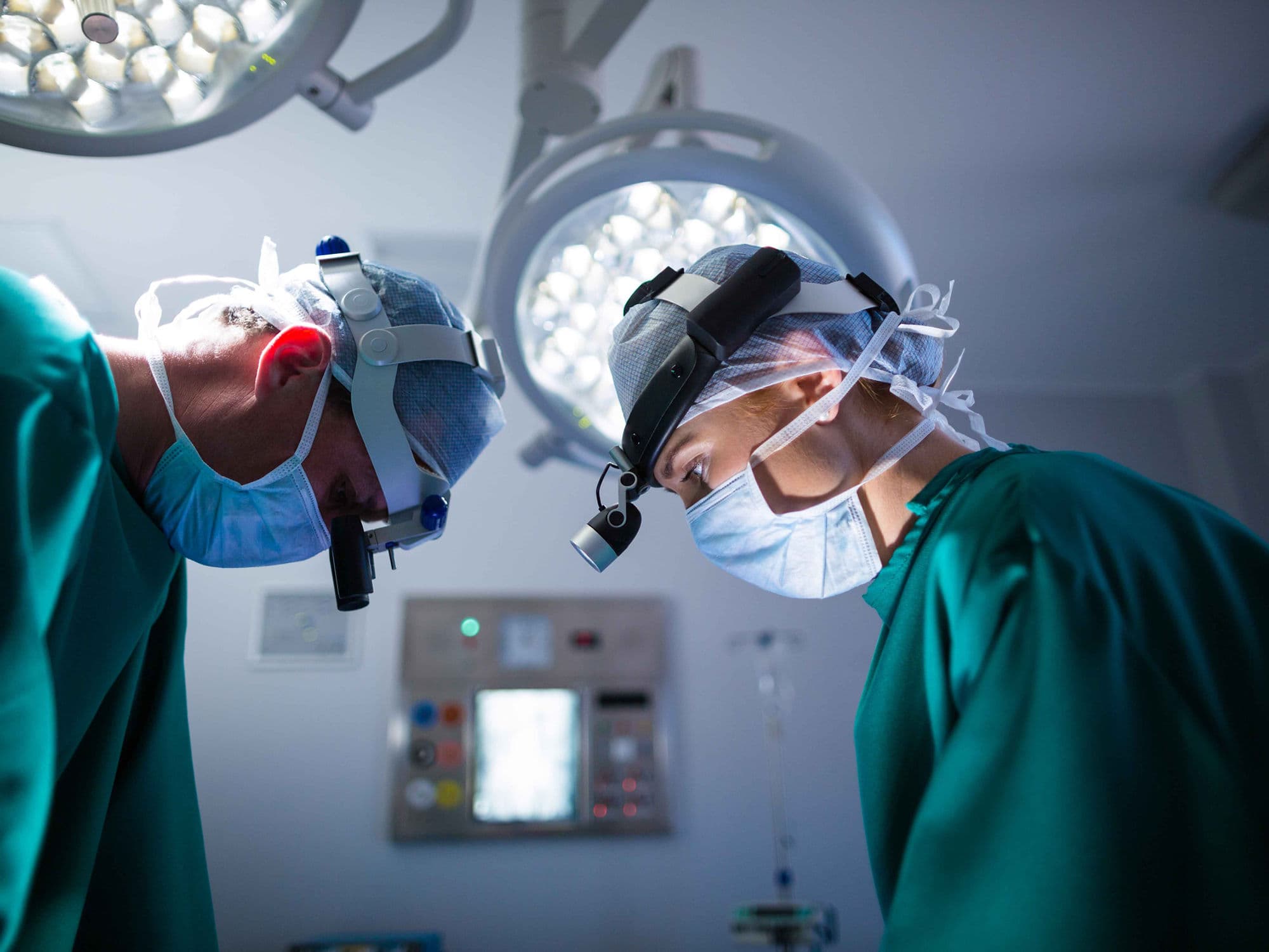
Collaborative Team: The surgery is usually a joint effort between a neurosurgeon and an Ear, Nose, and Throat (ENT) surgeon, taking about 2 to 3 hours under general anesthesia.
Nasal Access: The ENT surgeon inserts a thin, lighted tube with a high-definition camera (endoscope) through one nostril to navigate to the very back of the nasal cavity.
Opening the Sphenoid Sinus: The surgeon opens the sphenoid sinus (an air-filled space behind the nose) to reach the sella turcica, the small bony compartment that houses the pituitary gland.
Tumor Removal: Using specialized long instruments through the other nostril, the neurosurgeon removes the tumor in small pieces. The endoscope provides a panoramic, high-magnification view of the area, including nearby carotid arteries and optic nerves.
Reconstruction: If needed, a small fat graft (often taken from the abdomen) or synthetic material is used to fill the space and seal the area to prevent cerebrospinal fluid (CSF) leaks.
Pre-Procedure Preparation
Endocrine Evaluation: Comprehensive blood and urine tests to establish your baseline hormone levels (growth hormone, ACTH, prolactin, etc.).
High-Resolution MRI: A dedicated "pituitary protocol" scan to map the tumor’s exact size and its relationship to the optic chiasm.
Ophthalmology Exam: A detailed visual field test to document any current vision loss before the surgery.
Nasal Assessment: An ENT evaluation to ensure your nasal passages are clear and suitable for the endoscopic approach.
Fasting: Following "nothing by mouth" instructions for 8 hours prior to your scheduled anesthesia.
Tests Before Endoscopic Pituitary Surgery
Visual Field Testing: To measure peripheral vision, which is often the first thing affected by pituitary tumors.
Dynamic Hormone Testing: Specialized "stimulation" or "suppression" tests to confirm the type of secreting tumor.
Carotid Imaging: Occasionally required if the tumor is very large and wrapping around the main arteries of the brain.
ECG: A standard heart check to confirm cardiovascular stability for the duration of the procedure.
Life After Endoscopic Pituitary Surgery
Hospital Stay: Typically 1 to 3 days, often starting with one night in the Intensive Care Unit (ICU) for close monitoring of your fluid balance and hormone levels.
Immediate Symptoms: It is normal to experience nasal congestion, mild headaches, and "watery" or blood-tinged nasal drainage for 1 to 2 weeks.
The "No" Rules: For 4 to 6 weeks, you must strictly avoid:
Blowing your nose: To prevent pressure buildup that could cause a CSF leak.
Lifting and Straining: No lifting objects over 5 lbs or heavy straining, which increases intracranial pressure.
Drinking through straws: The suction can interfere with the healing of the nasal repairs.Hormone Monitoring: You will work closely with an endocrinologist to check if your gland is producing the correct amount of hormones post-op.
Follow-up MRI: A baseline scan is usually performed 3 months after surgery to ensure the entire tumor was removed.
Why Specialized Treatment Is Highly Effective
No External Scars: By using the natural pathway of the nose, there are no visible incisions on the face or scalp.
Superior Visualization: The endoscope allows surgeons to "see around corners," identifying tumor tissue that might be missed with traditional microscopic surgery.
Rapid Vision Improvement: Decompressing the optic nerves often leads to a quick and significant improvement in peripheral vision and clarity.
Preserves Gland Function: The high-magnification view helps surgeons distinguish between the tumor and the healthy part of the pituitary gland.
Reduced Brain Trauma: Because the brain is not "moved" or retracted to reach the tumor, post-operative headaches and recovery times are greatly reduced.