+91 83682 47758
info@medivisorhealth.com
Medivisor India Treatment Logo
  • Home
    • About Us
    • Our Services
    • Our Team
    • Our Safety Measures
    • Loading treatments...
    • Loading hospitals...
    • Loading doctors...
    • Loading tests & diagnoses...
    • Treatment Cost
    • Treatment Process
    • Visa Process
    • Travel Guide
    • FAQs
    • Why Medivisor
    • Success Stories
    • Patient Activities
    • Social Activities
    • News Coverage
    • Blog
  • Become a Partner

Medivisor India Treatment is a trusted New Delhi-based medical travel company assisting international patients with advanced, affordable, and seamless treatment in India.

  • Medivisor House 359, Sector 1, Vaishali, Ghaziabad, (Delhi/NCR) India
  • +91 8368 247 758
  • info@medivisorhealth.com

About Us

  • About Us
  • Our Services
  • Our Team
  • Our Safety Measures
  • Join Our Network
  • Contact Us

India Treatment

  • Hospitals
  • Treatments
  • Treatment Cost
  • Treatment Process
  • Visa Process
  • Travel Guide
  • FAQs
  • Why Medivisor

Gallery

  • Patient Testimonials
  • Patient Activities
  • News Coverage
  • Blog

Get in Touch

© 2026 Medivisor India Treatment. All rights reserved.

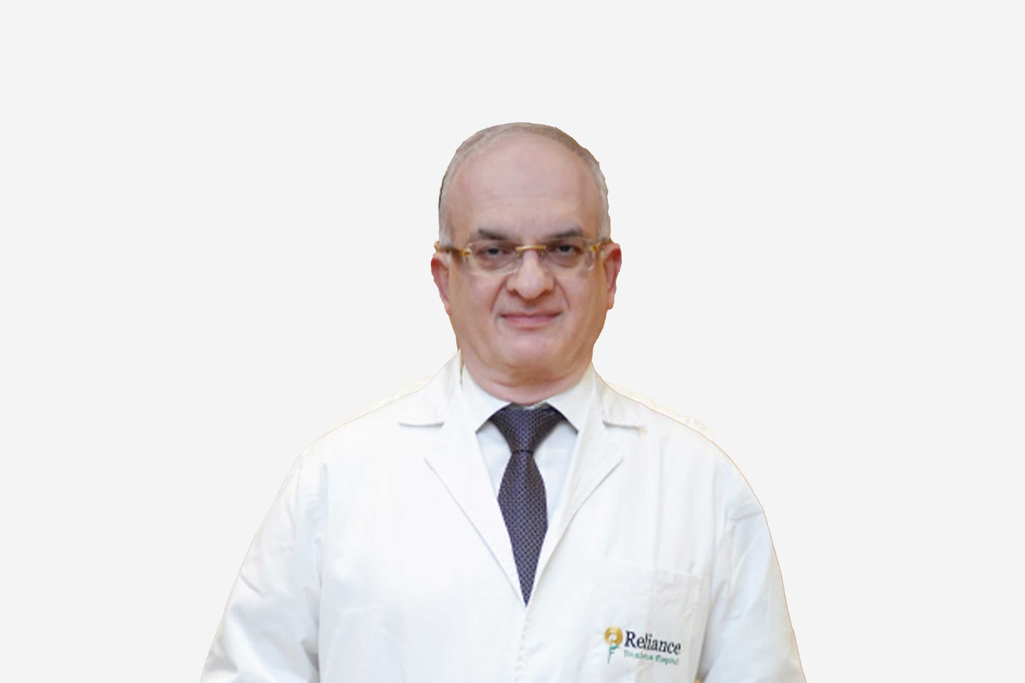
Dr Paresh Doshi

Director, Stereotactic & Functional Neurosurgery

Jaslok Hospital & Research Centre

30+ years experience
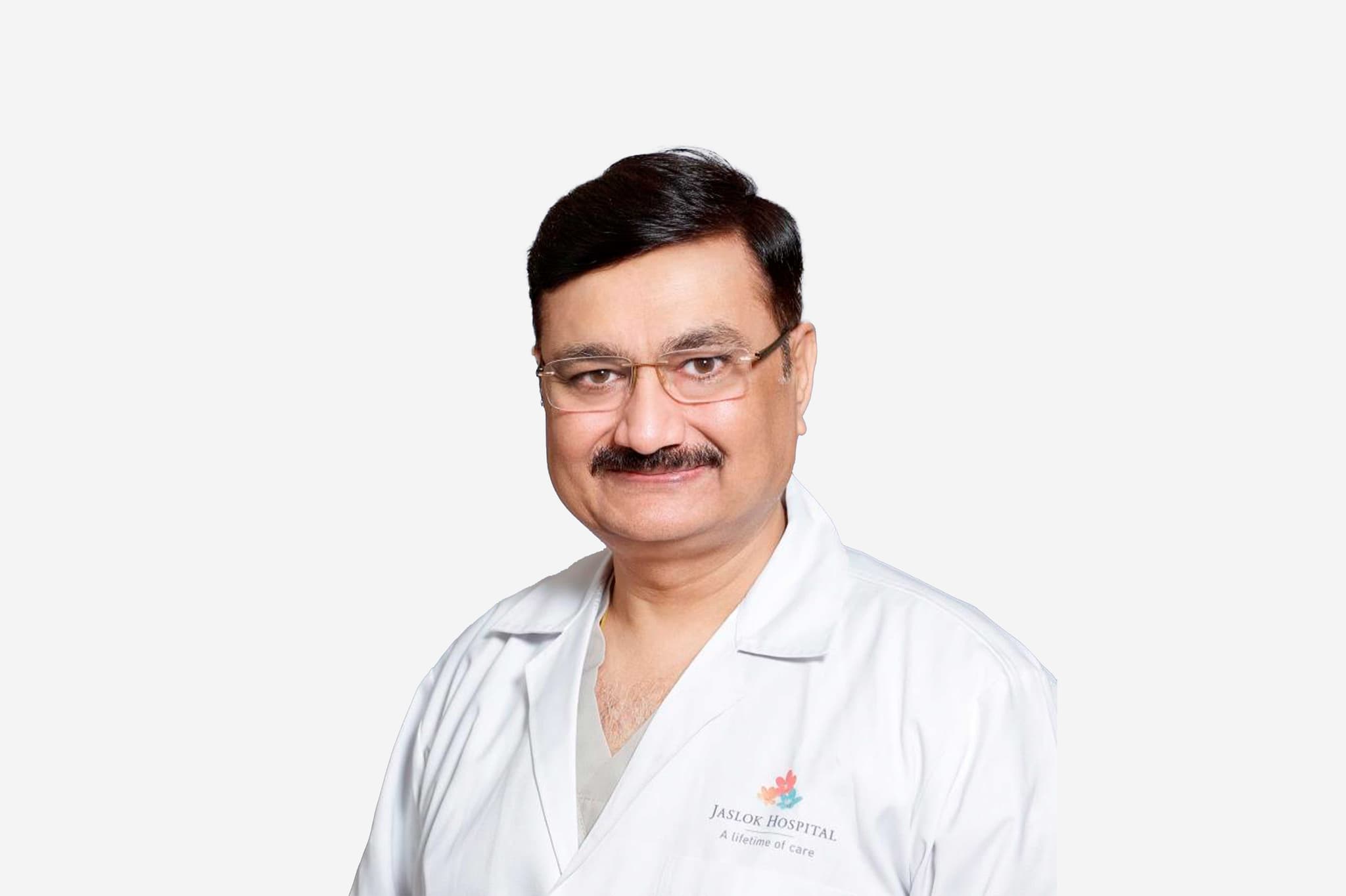
Dr Paresh Doshi, Director, Stereotactic & Functional Neurosurgery at Jaslok Hospital & Research Centre - Jaslok Hospital & Research Centre
  1. Home
  2. Doctors
  3. Dr Paresh Doshi

About Dr Paresh Doshi

Prof. (Dr.) Paresh Doshi is a world-renowned neurosurgeon with over 30 years of experience. He is widely recognized as the pioneer of Deep Brain Stimulation (DBS) in India and a global authority in the field of Functional Neurosurgery. As the Director of the Department of Stereotactic and Functional Neurosurgery at Jaslok Hospital & Research Centre, he has established one of the leading centers in South Asia for the surgical treatment of movement disorders and psychiatric conditions.

Pioneering Milestones and "Firsts"

Dr. Doshi’s career is defined by groundbreaking achievements that have placed India on the global map for advanced neurosurgery:

  • Deep Brain Stimulation (DBS): He performed the first-ever DBS surgery in India. Under his leadership, Jaslok Hospital has completed over 450 DBS procedures, the largest series in South Asia.

  • Global Records: He has operated on the youngest and oldest Parkinson's disease patients in the world, demonstrating the versatility and safety of his surgical techniques.

  • Psychiatric Neurosurgery: He was the first neurosurgeon in the Asian-Australian subcontinent to perform surgery for Depression.

  • Rare Conditions: His department recently published the world’s first series of DBS procedures for patients with Progressive Supranuclear Palsy (PSP).

Global Leadership and Professional Appointments

Dr. Doshi holds several prestigious leadership positions in international and national medical societies:

  • World Society for Stereotactic and Functional Neurosurgery (WSSFN): Director.

  • Asian-Australian Society for Stereotactic and Functional Neurosurgery (AASSFN): Director.

  • The Neuromodulation Society of India: President.

  • Indian Society for Stereotactic and Functional Neurosurgery (ISSFN): Past President.

Academic Contributions and Mentorship

A dedicated academician, Dr. Doshi is committed to the education and training of future neurosurgeons:

  • DNB Neurosurgery Program: He leads the DNB program at Jaslok Hospital, training a specialized neurosurgeon every year.

  • Fellowship Program: He runs an international fellowship program in Functional Neurosurgery, attracting and training specialists from across India and around the world.

  • Research and Publications: His department is actively involved in high-impact clinical research, contributing frequently to top-tier international neurosurgical journals.

Clinical Expertise and Departmental Scope

The Functional Neurosurgery Department under Dr. Doshi offers the largest spectrum of specialized procedures in the region, including:

  • Movement Disorders: Advanced management of Parkinson’s disease, Dystonia, and Essential Tremors.

  • Pain and Epilepsy Surgery: Specialized surgical interventions for chronic pain management and drug-resistant epilepsy.

  • Neuromodulation: Utilizing cutting-edge technology to treat psychiatric conditions and complex neurological deficits.

Prof. (Dr.) Paresh Doshi at a Glance

  • Over 30 years of experience in Neurosurgery.

  • Performed India’s first Deep Brain Stimulation (DBS) surgery.

  • Director of the largest DBS program in South Asia at Jaslok Hospital.

  • First in Asia-Australia to perform surgery for Depression.

  • Global leader holding Directorships in the World and Asian-Australian societies for Stereotactic Neurosurgery.

  • Renowned for operating on the world's youngest and oldest Parkinson’s patients.

  • Leading educator through DNB and international fellowship programs.

MBBS - 1985 (1st Rank), N H L Municipal Medical College, Ahmedabad
MS (General Surgery) - 1989 (Gold Medal), V S Hospital, Ahmedabad
MCh (Neurosurgery) - 1992 (1st Rank), Nair Hospital, Mumbai
Fellowship in Functional Neurosurgery, UK
Young achiever’s Award - Indo-American Society, Mumbai (2002)
Medscape India Award - Excellence in Neurosciences (2012)
Best Neurological Department - Modern Medicare Magazine (2012)
FICCI Award -- Medical Value Travel (Best Case Study). Runners up award for the best case study category for the Autism patient from USA (2019)
Pharma Leaders Brand Award - Indian of the Year in Neurosciences (2019)
Times of India Award - Excellence in Neurosurgery in Mumbai (2021)
May we help you?

Enter digits only (6-15 digits). We'll add your country code.

Related Treatments

Partial Nephrectomy (Kidney-Saving Surgery)

Partial Nephrectomy Partial Nephrectomy, also known as "Kidney-Sparing Surgery," is a complex procedure where only the diseased part of the kidney (usually a tumor) is removed, leaving the healthy, functioning kidney tissue intact. This is the preferred treatment for smaller kidney tumors to preserve as much renal function as possible. When You Should Consider Partial Nephrectomy Small Renal Masses: Typically the first choice for tumors less than 4 cm (Stage T1a) and many tumors up to 7 cm (Stage T1b). Solitary Kidney: When a patient only has one functioning kidney, making it critical to avoid a total removal. Bilateral Kidney Tumors: When tumors are present in both kidneys, requiring a sparing approach on one or both sides. Pre-existing Kidney Disease: For patients with diabetes or hypertension who are at higher risk for future chronic kidney disease. Genetic Predisposition: For patients with conditions like Von Hippel-Lindau (VHL) syndrome who may develop multiple tumors over their lifetime. Methods of Partial Nephrectomy Robotic-Assisted (Da Vinci): The modern standard for precision. It allows the surgeon to perform complex suturing within the tight "ischemia" time window using 3D magnification. Laparoscopic Surgery: A minimally invasive approach using small incisions and specialized instruments to remove the tumor and repair the kidney. Open Surgery: Often reserved for very large or centrally located tumors where the surgeon needs direct physical access to the kidney. Nerve and Vessel Sparing: A technique focusing on identifying the specific arterial branches feeding the tumor to avoid clamping the entire renal blood supply. Renorrhaphy (Repair): The reconstructive phase where the "hole" left in the kidney is stitched closed using specialized sutures and hemostatic agents. How the Procedure Is Performed Anesthesia: Performed under General Anesthesia to ensure total patient comfort and muscle relaxation. The "Clamping" Phase: To prevent heavy bleeding, the surgeon temporarily clamps the Renal Artery. This must usually be completed in under 20–30 minutes to protect kidney health. Tumor Excision: The surgeon cuts out the tumor along with a small "margin" of healthy tissue to ensure no cancer cells remain. Hemostasis: Specialized "bolsters" or glues are often applied to the raw surface of the kidney to stop bleeding instantly. Unclamping: The artery clamp is removed, and the surgeon verifies that the kidney regains its pink color and shows no signs of active bleeding. Drain Placement: A small tube is left in the side for 24–48 hours to monitor for any internal fluid or blood collection. Pre-Procedure Preparation 3D Imaging: A high-resolution CT Scan or MRI with contrast is mandatory to map the "Renal Nephrometry Score" and tumor depth. Kidney Function Test: A DTPA Scan or serum creatinine test is performed to establish a baseline for how well both kidneys are working. Blood Prep: "Cross-matching" for blood units is done in advance due to the highly vascular nature of the kidney. Medication Audit: You must stop all blood thinners (Aspirin, Warfarin, etc.) 7–10 days prior to the operation. Fasting: Maintaining a "nil per oral" status for 8 hours before the procedure for anesthesia safety. Pre-Surgery Tests Complete Blood Count (CBC): To check baseline hemoglobin levels and ensure the body is ready for surgery. Coagulation Profile: Testing PT/INR and Platelet counts to ensure the kidney repair will clot effectively. Chest X-ray and EKG: Standard pre-operative checks to ensure heart and lung fitness for general anesthesia. Urinalysis: To rule out any existing urinary tract infections before the procedure. Electrolyte Panel: Checking sodium, potassium, and calcium levels to ensure the body’s chemistry is balanced. Life After Partial Nephrectomy (Recovery & Risks) Hospital Stay: Usually 2–3 days for Robotic/Laparoscopic surgery and 4–5 days for the Open approach. Catheterization: A Foley catheter is placed in the bladder for 1–2 days to monitor urine output and filtration. Activity Restrictions: No heavy lifting (over 5kg) or strenuous exercise for 6 weeks to prevent the repair from bleeding. Hydration Therapy: Drinking 2–2.5 litres of water daily is recommended to keep the kidneys working at a steady, healthy pace. Urine Leak Risk: If the repair isn't water-tight, a temporary JJ Stent may be placed to allow the kidney to heal. Follow-Up Imaging: A CT scan is typically repeated at 3 or 6 months to monitor the surgical site and ensure no recurrence. Why Specialized Treatment Is Highly Effective Preservation of Function: By saving the healthy part of the kidney, patients have a much lower risk of requiring dialysis in the future. Oncological Equivalent: Modern studies show that for appropriately selected tumors, a partial removal is just as effective as a total removal for cancer control. Robotic Precision: 2026 robotic technology allows for "ultra-selective clamping," where only the tumor's blood supply is stopped, leaving the rest of the kidney "warm." Faster Recovery: Minimally invasive techniques lead to less pain, smaller scars, and a quicker return to normal life compared to traditional surgery. Lower Cardiovascular Risk: Maintaining two functioning kidneys is linked to better long-term heart health and blood pressure control.

Starting Cost: $ 2000

Radical Prostatectomy (Open/Laparoscopic/Robotic)

Radical Prostatectomy (Oncology) Radical Prostatectomy is the surgical removal of the entire prostate gland and surrounding tissues, typically performed to treat Prostate Cancer. Unlike treatments for an enlarged prostate (BPH) which only remove the "core," this treatment removes the entire organ to ensure the cancer is fully eliminated. When You Should Consider Radical Prostatectomy Localized Prostate Cancer: When imaging confirms the cancer is confined within the prostate capsule (Stages T1 or T2). Aggressive Disease: For patients with a higher Gleason Score who require definitive surgical removal rather than "active surveillance." Long Life Expectancy: Typically recommended for patients expected to live 10+ years, where surgical removal offers the best long-term cure rate. Failure of Radiation: As a "salvage" treatment if the cancer returns after previous radiation therapy. Patient Preference: For individuals who prefer the psychological certainty of having the cancerous organ physically removed. Methods of Radical Prostatectomy Robotic-Assisted (Da Vinci): The modern gold standard. The surgeon sits at a console controlling robotic arms with 3D magnification and 360-degree "wristed" instruments for extreme precision. Laparoscopic Surgery: A minimally invasive approach using 5–6 "keyhole" incisions, a camera, and long instruments to operate while viewing a 2D screen. Open Surgery (Retropubic): The traditional method involving a single 4–5 inch incision in the lower abdomen to remove the gland. Nerve-Sparing Technique: A meticulous process where the surgeon peels the delicate "neurovascular bundles" away from the prostate to preserve sexual function. Vesicourethral Anastomosis: The reconstructive step where the bladder is stitched directly back to the urethra to restore the urinary path after the gland is removed. How the Procedure Is Performed Anesthesia: Performed under General Anesthesia to ensure the patient is completely asleep and the abdominal muscles are relaxed. Dissection: The surgeon carefully separates the prostate from the bladder above it and the urethra below it. Lymph Node Removal: Depending on the cancer's aggressiveness, nearby pelvic lymph nodes are often removed to check for microscopic spread. Gland Extraction: The entire prostate and the attached seminal vesicles are removed as a single unit. Catheterization: A Foley catheter is inserted through the penis into the bladder to act as a "splint" while the new connection (anastomosis) heals. Drain Placement: A small suction tube may be left in the abdomen for 24–48 hours to remove excess surgical fluid. Pre-Procedure Preparation Cancer Staging: Includes a Multiparametric MRI (mpMRI) and often a PSMA PET-CT scan to ensure the cancer has not spread. Biopsy Review: The surgical team reviews the Gleason Score and genomic markers to plan the extent of the surgery. Pelvic Floor Training: Patients are taught Kegel exercises weeks before surgery to strengthen the muscles responsible for urinary control. Medication Audit: Patients must stop blood thinners (like Aspirin or Warfarin) 7–10 days prior to the operation. Fasting: Maintaining a "nil per oral" status for 8 hours before the procedure for anesthesia safety. Pre-Surgery Tests PSA Blood Test: To establish the final pre-operative baseline for monitoring future "undetectable" levels. EKG and Chest X-ray: Standard tests to ensure heart and lung fitness for a multi-hour surgical procedure. Basic Metabolic Panel: Checking kidney function (Creatinine) and electrolytes to manage IV fluids during surgery. Coagulation Profile: Testing PT/INR and Platelet counts to ensure safe surgical healing and minimal blood loss. Complete Blood Count (CBC): To check baseline hemoglobin levels in case a blood transfusion is required (rare in robotic cases). Life After Radical Prostatectomy (Recovery & Risks) Hospital Stay: Usually 1–2 days for Robotic/Laparoscopic surgery, or 3–4 days for the Open approach. Catheter Management: The Foley catheter must remain in place for 7 to 14 days to allow the bladder-urethra connection to heal water-tight. Trial of Void: After 1–2 weeks, the catheter is removed in the clinic to ensure the patient can urinate independently. Activity Restrictions: No heavy lifting (over 5kg) or driving for 4 weeks; early walking is encouraged to prevent blood clots. Urinary Incontinence: Most patients experience leaking initially; this typically improves over 3–6 months with consistent pelvic floor exercises. Erectile Dysfunction (ED): It can take 6–18 months for erections to return; doctors often start "Penile Rehabilitation" medication shortly after surgery. Why Specialized Treatment Is Highly Effective Definitive Cure: Offers the highest probability of completely eliminating localized prostate cancer in a single treatment. Pathological Certainty: Removing the gland allows for a total biopsy, giving the most accurate information on the cancer's stage and grade. Robotic Precision: The 3D-high-definition view in 2026 allows surgeons to see nerves and vessels that are nearly invisible to the naked eye. Predictable Monitoring: Post-surgery, the PSA level should drop to "undetectable," making it very easy to monitor for any future recurrence. Limb-Sparing Mindset: Modern techniques focus heavily on "quality of life" preservation, aiming for the "Trifecta" of cancer control, continence, and potency.

Starting Cost: $ 2500

Transurethral Incision of the Prostate (TUIP)

Transurethral Incision of the Prostate (TUIP) Transurethral Incision of the Prostate (TUIP) is a specialized surgical treatment for men with urinary symptoms caused by a moderately enlarged prostate (usually less than 30 grams) or a narrowing of the bladder neck. Unlike TURP or Laser surgery, no tissue is removed; instead, the "tight" area is widened to allow for better flow. When You Should Consider TUIP Small to Moderately Enlarged Prostate: Specifically indicated for prostates under 30 grams where the blockage is at the bladder neck. Bladder Neck Obstruction: When the primary cause of urinary symptoms is a narrowing of the opening between the bladder and the urethra. Preservation of Fertility: For men who wish to minimize the risk of retrograde ejaculation (dry orgasm) often associated with other prostate surgeries. High-Risk Patients: For those who may not tolerate longer surgical procedures, as TUIP is significantly faster than TURP. Failure of Medical Management: When medications like alpha-blockers are no longer providing sufficient relief for urinary flow. Methods of TUIP Cystoscope Access: A thin, lighted scope is inserted through the tip of the penis into the urethra, requiring no external incisions. Electric Knife Incision: The use of a specialized wire or knife to make one or two small, deep cuts (grooves) in the bladder neck. Laser Incision: A modern alternative using a laser fiber to precisely cut the prostate and bladder neck tissue. Channel Widening: Unlike "shaving" tissue, the mechanism relies on making a structural cut that allows the bladder neck to "spring open." Minimal Tissue Trauma: A technique that avoids the removal of prostate mass, leading to significantly less internal wounding. How the Procedure Is Performed Anesthesia: Performed under Spinal Anesthesia (numbing from the waist down) or General Anesthesia for patient comfort. Insertion: The surgeon guides the cystoscope through the urethra until the junction of the bladder and prostate is visualized. Creating Grooves: The surgeon makes precise incisions through the prostate and the muscle of the bladder neck. Widening: By cutting the tight muscle ring, the urethral channel is widened, immediately reducing the resistance to urine flow. Catheterization: A Foley catheter is placed at the end of the 20–30 minute procedure to assist with initial healing and drainage. Pre-Procedure Preparation Prostate Sizing: A Transrectal Ultrasound (TRUS) is essential to confirm the prostate is small enough (under 30g) for this technique. Urodynamic Study: A test to confirm that the blockage is at the bladder neck and not caused by a weak bladder muscle. Medication Review: You must stop blood thinners (like Aspirin or Warfarin) 5–7 days prior to surgery to prevent bleeding. Fasting: Maintaining a "nil per oral" status for 6–8 hours before the procedure for anesthesia safety. Antibiotic Prophylaxis: A dose of antibiotics is given intravenously just before surgery to prevent urinary tract infections. Pre-Surgery Tests Uroflowmetry: To measure the speed and force of the urine stream to establish a baseline for post-operative comparison. Post-Void Residual (PVR): An ultrasound to measure how much urine is left in the bladder after peeing. Urine Culture: To ensure the urinary tract is sterile before the procedure; any infection must be treated first. Basic Metabolic Panel: Checking kidney function (Creatinine) and electrolytes to ensure surgical fitness. Coagulation Profile: Testing the blood's ability to clot (PT/INR) to ensure safe healing of the internal incisions. Life After TUIP (Recovery & Risks) Hospital Stay: Often performed as a Daycare (Same-Day) procedure, or a maximum stay of 24 hours. Catheter Removal: The Foley catheter is typically removed within 24 to 48 hours after the surgery. Hydration Therapy: Drinking 2–3 litres of water daily is essential to flush the urinary tract as the "grooves" heal. Activity Restrictions: Most patients can return to light work within 2–3 days but should avoid heavy lifting for 2 weeks. Urinary Sensations: It is normal to feel a temporary urgency or stinging for the first few days post-surgery. Fertility Preservation: Most men maintain normal ejaculation after TUIP, as the risk of retrograde ejaculation is much lower than with TURP. Why Specialized Treatment Is Highly Effective Rapid Recovery: Due to the minimal tissue trauma, patients experience a much faster return to normal activities than with traditional surgery. High Safety Profile: With very little bleeding and a short operative time, TUIP is an excellent option for patients with minor health concerns. Functional Success: Effectively treats the root cause of bladder neck obstruction, providing a forceful urine stream immediately. No External Scars: The procedure is entirely internal, leaving no visible marks and requiring no wound care on the skin. Preservation of Sexual Health: Offers the best chance of maintaining normal sexual function and fertility compared to other surgical BPH treatments.

Starting Cost: $ 500

Laser Prostate Surgery (HoLEP / ThuLEP)

Laser Prostate Surgery (HoLEP & ThuLEP) Laser prostate surgery, specifically HoLEP (Holmium Laser Enucleation of the Prostate) and ThuLEP (Thulium Laser Enucleation of the Prostate), is a modern, bloodless alternative to traditional TURP. Instead of shaving the prostate in small pieces, the laser "peels" the obstructing tissue away as a whole, making it highly effective for even the largest prostate glands. When You Should Consider Laser Prostate Surgery Very Large Prostate: Especially effective for prostates over 80–100 grams that are too large for standard TURP. High Risk of Bleeding: Ideal for patients who cannot safely stop blood thinners, due to the laser's superior vessel-sealing properties. Severe Urinary Retention: When a patient is completely unable to pass urine and requires a catheter. Recurrent Prostate Issues: For those who have had previous prostate surgery and are experiencing regrowth of obstructing tissue. Desire for Fast Recovery: For patients who want a shorter hospital stay and quicker removal of the urinary catheter. Methods of Laser Prostate Surgery HoLEP (Holmium Laser): Uses a "pulsed" laser beam, which is excellent for "blunt dissection" (peeling) the prostate lobes away from the capsule. ThuLEP (Thulium Laser): Uses a "continuous wave" laser that provides smoother cutting and even better blood vessel sealing (hemostasis). Enucleation Technique: The surgeon uses the laser to peel away the entire obstructing lobe, similar to removing the flesh of an orange from its peel. Morcellation: A specialized "blender-like" device is used to grind the large peeled-off lobes inside the bladder and vacuum them out. Transurethral Access: The entire procedure is performed through the urethra using a laser-integrated scope, requiring no external incisions. How the Procedure Is Performed Anesthesia: Performed under General Anesthesia or Spinal Anesthesia to ensure the patient is completely still and pain-free. Laser Dissection: The laser fiber is used to cut along the "capsule" of the prostate, separating the obstructing tissue from the healthy outer shell. Simultaneous Cauterization: As the laser cuts, it seals every blood vessel it touches, resulting in minimal to no blood loss. Intra-vesical Morcellation: Once the tissue is moved into the bladder, the morcellator sucks up and removes the fragments in a matter of minutes. Final Inspection: The surgeon verifies that the bladder is clear of debris and that there is no active bleeding before finishing. Catheter Placement: A Foley catheter is inserted at the end of the procedure, which is typically removed within 24 hours. Pre-Procedure Preparation Imaging & Size Check: An Ultrasound (TRUS) or MRI is performed to measure the exact prostate volume and map the anatomy. Urine Culture: A sterile urine sample is mandatory; any active infection must be treated with antibiotics before the laser is used. Medication Audit: Patients on Aspirin or Warfarin usually stop these 5–7 days prior, though some laser cases can proceed while on thinners if necessary. Fasting: Maintaining a "nil per oral" (NPO) status for 6–8 hours before the operation for anesthesia safety. Medical Clearance: Ensuring heart and lung fitness for the procedure, especially since it is often performed on older patients. Pre-Surgery Tests Uroflowmetry: To measure the baseline speed and force of the urine stream before surgery. Post-Void Residual (PVR) Volume: An ultrasound test to see how much urine remains in the bladder after peeing. PSA Blood Test: To screen for prostate-specific antigen levels and rule out other underlying conditions. Basic Metabolic Panel: Checking kidney function (Creatinine) and electrolytes to ensure the body can handle the procedure. Coagulation Profile: Testing the blood's ability to clot (PT/INR) to ensure a safe, bloodless surgical outcome. Life After Laser Prostate Surgery (Recovery & Risks) Hospital Stay: Usually a 24-hour stay; many patients are discharged the very next morning after the catheter is removed. Immediate Results: Most patients notice an immediate, forceful urine stream once the initial catheter is taken out. Hydration Therapy: Drinking 2.5–3 litres of water daily for the first week is essential to flush out any minor laser-charred debris. Activity Rules: You can usually return to desk work within 3–5 days, but avoid heavy lifting (over 10 kg) for at least 2 weeks. Retrograde Ejaculation: This is common (90% of cases), where semen travels into the bladder during orgasm; it is harmless but affects fertility. Temporary Urgency: A frequent "urge" to pee or minor leaking may occur for the first few weeks as the internal capsule heals. Why Specialized Treatment Is Highly Effective Superior Hemostasis: The laser's ability to seal vessels as it cuts makes this the safest option for patients with bleeding disorders or heart issues. Complete Tissue Removal: By following the natural "capsule," laser surgery removes more obstructing tissue than TURP, reducing the risk of regrowth. Shorter Catheter Time: Most patients are catheter-free within 24 hours, significantly reducing the risk of catheter-associated infections. Treatment of Any Size: Unlike other minimally invasive methods, HoLEP and ThuLEP can treat prostates of virtually any size (even over 200 grams). Minimal Side Effects: Lower risk of "TURP Syndrome" and other fluid-balance complications due to the use of saline irrigation.

Starting Cost: $ 1000

Transurethral Resection of Prostate (TURP)

Transurethral Resection of the Prostate (TURP) Transurethral Resection of the Prostate (TURP) is a surgical treatment used to treat urinary problems caused by an enlarged prostate (Benign Prostatic Hyperplasia or BPH). It is often called the "Gold Standard" because it is highly effective and requires no external incisions. When You Should Consider TURP Moderate to Severe BPH Symptoms: When urinary frequency, urgency, or a weak stream significantly impact your quality of life. Incomplete Bladder Emptying: If you consistently feel that your bladder is not empty after urinating, which can lead to infections. Recurrent Urinary Tract Infections: When an enlarged prostate causes stagnant urine that leads to frequent UTIs. Bladder Stones or Kidney Damage: When the obstruction is severe enough to cause secondary complications in the urinary system. Failure of Medication: If alpha-blockers or 5-alpha reductase inhibitors are no longer providing sufficient relief. Methods of TURP Monopolar TURP: The traditional method using a wire loop and an electric current with glycine irrigation fluid. Bipolar TURP: A modern advancement using saline irrigation, which significantly reduces the risk of "TURP Syndrome" and allows for safer treatment of larger prostates. Resectoscope Access: A thin, lighted tube is inserted through the tip of the penis into the urethra, requiring no external cuts or stitches. Wire Loop Resection: An electric current is passed through a loop to precisely "shave" away excess tissue blocking the urine flow. Continuous Irrigation: Sterile fluid is pumped through the scope during surgery to wash away blood and tissue fragments. How the Procedure Is Performed Anesthesia: Performed under Spinal Anesthesia (numbing from the waist down) or General Anesthesia to ensure the patient is completely comfortable. Insertion: The surgeon guides the resectoscope through the urethra until it reaches the prostate gland. Tissue Removal: The obstructing lobes of the prostate are shaved into small chips using the electrified wire loop. Fragment Flushing: The shaved tissue pieces are gathered and flushed out of the bladder at the conclusion of the surgery. Catheterization: A three-way Foley catheter is inserted to allow for continuous bladder irrigation immediately following the procedure. Pre-Procedure Preparation Medical Evaluation: Includes a Uroflowmetry test to measure urine speed and an ultrasound to determine the exact prostate size. Medication Adjustment: You must stop blood thinners (like aspirin or warfarin) 5–7 days before surgery to prevent internal bleeding. Fasting: Maintaining a "nil per oral" status for 6–8 hours before the procedure for anesthesia safety. Antibiotic Prophylaxis: A preventative dose of antibiotics is given intravenously just before surgery to minimize infection risks. PSA Testing: A blood test to screen for prostate-specific antigen levels prior to surgical intervention. Pre-Surgery Tests Urine Routine & Culture: To ensure there is no active infection before entering the urinary tract. Prostate Ultrasound (TRUS): To map the dimensions of the prostate and plan the volume of tissue to be resected. Cystoscopy: Occasionally performed beforehand to visualize the narrowness of the urethra and the shape of the prostate. Blood Coagulation Profile: Testing PT/INR and Platelet counts to ensure safe surgical healing. Basic Metabolic Panel: Checking kidney function (Creatinine) and electrolytes, especially important for managing irrigation fluid balance. Life After TURP (Recovery & Risks) Hospital Stay: Most patients stay for 1 to 2 days until the urine is clear and the catheter can be safely removed. Bladder Irrigation: While in the hospital, a sterile fluid "washes" the bladder through the catheter to prevent blood clots. Hydration Therapy: Drinking at least 2–3 litres of water daily at home is essential to flush the healing surgical site. Activity Restrictions: Avoid heavy lifting (over 5 kg) or vigorous exercise for 4–6 weeks to prevent secondary bleeding. Sexual Activity: Avoid sexual intercourse for 4 weeks to allow the internal area to seal completely. Urination Changes: It is normal to feel a temporary burning sensation or urgency for a few weeks as the raw prostatic bed heals. Why Specialized Treatment Is Highly Effective Immediate Symptom Relief: Most patients experience a significantly stronger urine stream and better bladder emptying almost immediately after recovery. No External Scarring: Because the surgery is entirely internal, there are no visible scars or wound care requirements on the skin. Long-Term Durability: TURP results typically last for 15 years or more, often providing a permanent solution for BPH symptoms. Bipolar Safety: Modern Bipolar technology allows surgeons to treat patients with heart conditions or larger prostates more safely than ever before. Diagnostic Benefit: The shaved tissue is always sent for biopsy to confirm the absence of any hidden cancerous cells.

Starting Cost: $ 800

Mini-PCNL / Micro-PCNL

Mini-PCNL and Micro-PCNL (Urology) Mini-PCNL is a "small-access" surgery used for stones between 1 cm and 2.5 cm. Micro-PCNL is an "all-in-one" needle surgery, usually reserved for stones around 1 cm to 1.5 cm. When You Should Consider Mini-PCNL or Micro-PCNL Stone Size: Ideal for stones between 1 cm and 2.5 cm that are too large for ESWL but don't require standard PCNL. Stone Hardness: When stones are too dense to be broken from outside the body by shockwaves. Lower Pole Stones: Particularly effective for stones located in the bottom part of the kidney which are difficult to clear otherwise. Failed Previous Treatments: When non-invasive methods like ESWL have failed to fragment the stone. Patient Preference: For those seeking a higher "stone-free rate" with minimal scarring and a faster return to daily activities. Methods of Mini and Micro-PCNL Mini-PCNL Access: A tiny incision (about 5 mm to 8 mm) is made in the flank area to allow for a thin telescope. Micro-PCNL Access: Uses a specialized 1.5 mm needle—about the thickness of an injection needle—to enter the kidney without a traditional incision. Laser Fragmentation: Utilization of high-power Holmium or Thulium Lasers to break stones into smaller pieces or fine dust. Vortex Suction: A mechanism used in Mini-PCNL to automatically pull fragments out through the sheath using saline irrigation. Dusting Technique: In Micro-PCNL, the laser turns the stone into a fine powder that washes out naturally through urine. How the Procedure Is Performed Anesthesia: Both procedures are performed under General Anesthesia to ensure the patient is completely asleep and still. Imaging Guidance: Surgeons use Live X-ray (Fluoroscopy) and Ultrasound simultaneously to guide the needle or scope to the stone. Nephroscopy: A thin Mini-Nephroscope or a tiny Micro-Lens camera is inserted directly into the kidney to visualize the stone. Fragmentation: The laser fiber is passed through the scope or needle to precisely target and break the stone. Stent Placement: In Mini-PCNL, a small internal JJ stent may be left for 1–2 weeks to ensure proper kidney drainage. Pre-Procedure Preparation Fasting: Maintaining a "nil per oral" status for at least 8 hours before the procedure for anesthesia safety. Medication Review: Suspending blood thinners several days in advance to minimize the risk of bleeding during the kidney puncture. Antibiotic Prophylaxis: Receiving a dose of intravenous antibiotics just before the procedure to prevent urinary tract infections. Hydration: Ensuring adequate fluid intake in the days leading up to the surgery to maintain good kidney function. Clearance: Obtaining medical clearance, especially for patients with pre-existing heart or lung conditions. Pre-Surgery Tests NCCT KUB: A non-contrast CT scan to determine the exact size, location, and hardness (Hounsfield units) of the stone. Urine Culture: To confirm the urine is sterile; surgery is postponed if an active infection is detected. Coagulation Profile: Blood tests (PT/INR) to ensure the blood clots normally before making the kidney access. Renal Function Test: Checking Creatinine and Urea levels to assess how well the kidneys are functioning. ECG and Chest X-ray: Standard pre-anesthetic tests to ensure heart and lung fitness for general anesthesia. Life After Mini and Micro-PCNL Hospital Stay: Typically 24 hours for Mini-PCNL, while Micro-PCNL is often performed as a daycare (same-day) procedure. Hydration Therapy: Drinking 3–4 litres of water daily is essential to flush out laser-dusted particles and fragments. Physical Activity: Most patients return to light desk work within 3–4 days but should avoid heavy lifting for 2 weeks. Urine Appearance: Expect light-pink or blood-tinged urine for 1–2 days as the kidney heals. Stent Removal: If a JJ stent was placed, a minor follow-up procedure is required to remove it after 7–14 days. Benefits of Mini and Micro-PCNL High Stone-Free Rate: Provides a much higher chance of complete stone removal in a single session compared to ESWL. Minimal Pain: Small access points lead to significantly less post-operative pain and a reduced need for painkillers. Cosmetic Results: The incisions are so small (or non-existent in Micro-PCNL) that they often require no stitches and leave no visible scars. Safety: Lower risk of bleeding and kidney damage compared to traditional, large-tract PCNL. Fast Recovery: Allows for a quicker discharge from the hospital and a rapid return to a normal lifestyle.

Starting Cost: $ 1000

Percutaneous Nephrolithotomy (PCNL)

Percutaneous Nephrolithotomy (PCNL) Percutaneous Nephrolithotomy (PCNL) is a minimally invasive "keyhole" surgery specifically used to remove large kidney stones—usually those over 2 cm—that cannot be passed naturally or treated effectively with standard shockwave therapy. This procedure allows surgeons to reach the kidney through a tiny incision in the back, providing a direct route to extract complex or "staghorn" stones that would otherwise require major open surgery. When You Should Consider PCNL Large Kidney Stones: Specifically those larger than 2 cm in diameter which are too substantial for laser ureteroscopy or shockwave lithotripsy. Staghorn Calculi: Complex stones that branch out to fill the various drainage channels (calyces) of the kidney. Failed Previous Treatments: When other non-invasive methods have failed to break or clear the stone. Stones in Diverticula: When a stone is trapped in a small outpouching of the kidney that is difficult to access through the ureter. Cystine or Stones of High Density: Very hard stones that are resistant to being broken by external sound waves. How It Is Performed Anesthesia: The surgery is performed under general anesthesia, ensuring you are completely asleep and comfortable throughout the procedure. Positioning: You are typically placed in a prone (face-down) or supine (face-up) position to allow the surgeon precise access to the kidney through the flank. The Incision: A small, "keyhole" incision (approximately 1 cm) is made in your flank area (the side of your back). Access & Fragmentation: A protective sleeve is inserted through the incision directly into the kidney's collecting system. A nephroscope (a specialized thin camera) is passed through the sleeve to locate the stone under high magnification. If the stone is too large to remove whole, the surgeon uses a lithotripter (a laser, ultrasound, or pneumatic device) to fragment the stone into smaller, manageable pieces. Extraction: The fragments are then physically removed from the kidney using specialized graspers or suction. Drainage: To ensure the kidney heals properly, a nephrostomy tube (draining to an external bag) or an internal ureteral stent is often left in place temporarily. Pre-Procedure Preparation Medical Evaluation: Comprehensive blood and urine tests to rule out active infection and assess kidney function. Advanced Mapping: A high-resolution CT scan or intravenous pyelogram (IVP) to map the exact 3D location of the stones and the surrounding anatomy. Antibiotic Protocol: A preventative dose of antibiotics is administered to minimize the risk of post-surgical infection. Fasting: Adhering to "nothing by mouth" instructions after midnight on the night before your surgery. Medication Audit: You will be asked to stop taking blood thinners (like aspirin, warfarin, or clopidogrel) several days before the procedure to prevent bleeding. Tests Before PCNL Urine Culture: Essential to confirm the urine is sterile before entering the kidney with surgical instruments. CT Stone Protocol: A specialized scan to determine the "Hounsfield units" (density) of the stone, which helps the surgeon choose the right fragmentation tool. Complete Blood Count (CBC): To establish a baseline for your red and white blood cells. ECG: A standard heart check to confirm cardiovascular stability for a procedure that typically lasts 1 to 4 hours. Life After PCNL Hospital Stay: Most patients stay in the hospital for 1 to 2 days for close monitoring of kidney drainage and comfort levels. Tube Management: If a nephrostomy tube was used, it is typically removed before you leave the hospital or at a quick follow-up visit a few days later. Initial Symptoms: It is normal to see small amounts of blood in your urine (hematuria) or feel moderate soreness at the incision site for 1–2 weeks. Activity Restrictions: Avoid strenuous activity, heavy exercise, or lifting anything heavier than 4 kg (approx. 9 lbs) for at least 2 weeks. Hydration: Drinking plenty of fluids is critical to help flush out any remaining tiny microscopic stone fragments. Follow-Up: A check-up is scheduled for 4 to 6 weeks after surgery, often including an X-ray or ultrasound to confirm the kidney is stone-free and functioning correctly. Why Specialized Treatment Is Highly Effective Highest Clearance Rate: PCNL offers the highest "stone-free" rate for large and complex stones compared to any other surgical or non-surgical method. Direct Extraction: Because stones are physically removed rather than just broken, there is a lower risk of fragments getting stuck in the ureter later. Minimally Invasive: Despite treating large stones, the "keyhole" approach means less pain, smaller scars, and a much faster recovery than traditional open kidney surgery. One-Step Solution: Even the largest staghorn stones can often be cleared in a single surgical session. Protects Kidney Health: Rapidly clearing an obstructing stone prevents long-term pressure damage (hydronephrosis) and reduces the risk of recurrent kidney infections.

Starting Cost: $ 800

URS (Ureteroscopy)
URS (Ureteroscopy)

Ureteroscopy (URS) URS (Ureteroscopy) is a minimally invasive surgical procedure used to diagnose and treat issues in the urinary tract, most commonly kidney stones located in the ureter or kidney. Because the procedure utilizes the body's natural openings, it requires no external incisions or surgical cuts. This advanced endoscopic approach allows urologists to reach and treat stones that are too large or too high to pass on their own, often providing immediate relief from renal colic. When You Should Consider Ureteroscopy Obstructing Ureteral Stones: When a stone is stuck in the ureter, causing severe pain, nausea, or potential kidney damage. Large Kidney Stones: For stones located within the kidney that are unlikely to pass naturally or are causing chronic discomfort. Failed Shockwave Therapy: If previous non-invasive treatments (like ESWL) have failed to break the stone into small enough pieces. Diagnostic Necessity: To investigate the cause of blood in the urine (hematuria) or to biopsy suspicious lesions within the ureter or renal pelvis. Bilateral Stones: When stones are present in both ureters, URS can sometimes address both sides in a single surgical session. How It Is Performed Access: A urologist inserts a thin, flexible, or rigid lighted telescope called a ureteroscope through the urethra and bladder, guiding it carefully up into the ureter or kidney. Anesthesia: The surgery is typically performed under general anesthesia and usually takes between 30 minutes to 2 hours, depending on the size and number of stones. Visualization: A sterile saline solution is used to gently expand the bladder and ureter, providing a clear 3D view of the stones and the urinary tract lining. Treatment: Small Stones: These are captured and removed whole using a specialized tiny wire basket device. Larger Stones: These are fragmented into tiny, dust-like pieces using a high-precision Holmium laser beam. Stone Extraction: Once fragmented, the pieces are either extracted with the basket or left to pass naturally and painlessly in the urine. Stent Placement: Frequently, a small temporary tube called a ureteral stent (JJ stent) is placed in the ureter at the end of the procedure to ensure proper urine drainage and to manage internal swelling. Pre-Procedure Preparation Imaging (CT or KUB): High-resolution scans to determine the exact size, density, and location of the stones. Urine Culture: To ensure there is no active urinary tract infection (UTI) before the instruments are introduced. Medication Audit: You may be asked to stop taking blood thinners (like aspirin or warfarin) several days before the procedure to minimize bleeding. Fasting: Following "nothing by mouth" instructions for 8 hours prior to your scheduled anesthesia. Hydration: Your doctor may encourage increased fluid intake in the days leading up to the procedure to help flush the urinary system. Tests Before Ureteroscopy Urinalysis: To check for signs of infection, blood, or crystals in the urine. Blood Panels: A routine check of your blood count, electrolytes, and kidney function (creatinine and BUN levels). ECG: A standard heart check to confirm cardiovascular stability for the duration of the procedure. Coagulation Profile: To ensure your blood clots normally, which is essential for a safe endoscopic procedure. Life After Ureteroscopy Recovery Timeline: URS is often an outpatient procedure, allowing most patients to go home the same day. Most can resume light activities within 2 to 3 days, with a full recovery typically taking one week. Initial Symptoms: For the first 24–48 hours, it is normal to experience mild pelvic pain, pinkish or reddish blood in the urine, and a frequent urge to urinate. Critical Hydration: It is essential to drink 2–3 liters of water daily to flush out any remaining stone fragments and residual blood. Stent Removal: If a stent was placed, it is typically removed in a quick, separate outpatient procedure 4 to 14 days after the surgery. Activity Restrictions: Avoid heavy lifting and strenuous exercise for at least one week to allow the ureter to heal and reduce the risk of bleeding. Why Specialized Treatment Is Highly Effective Immediate Stone Clearance: Unlike medications, URS offers the fastest way to physically remove an obstructing stone and stop the associated pain. No Surgical Scars: By using the body's natural pathways, there is zero impact on your physical appearance and no wound care is required. High Success Rate: Modern laser technology can break even the hardest stones (such as calcium oxalate monohydrate) that other treatments cannot touch. Protects Kidney Function: Rapidly removing an obstruction prevents the backup of urine (hydronephrosis), which can lead to permanent kidney scarring or infection. Precision Mapping: The high-definition cameras on modern ureteroscopes allow for a thorough inspection of the entire upper urinary tract, ensuring no small stones are left behind.

Starting Cost: $ 500

Testicular Cancer Surgery

Testicular Cancer Surgery Testicular cancer surgery is the primary treatment for most stages of the disease. It serves two critical purposes: to physically remove the tumor and to provide a high-quality tissue sample for precise staging and molecular diagnosis. Because testicular cancer is highly treatable, surgery often represents the first and most important step toward a complete cure. When You Should Consider Testicular Cancer Surgery Palpable Mass: The discovery of a painless lump, hardness, or swelling in the scrotum. Elevated Tumor Markers: High levels of Alpha-fetoprotein (AFP), Beta-HCG, or LDH found during blood testing. Suspicious Ultrasound: Imaging that identifies a solid mass within the testicle that is not consistent with a benign cyst. Staging and Diagnosis: When a definitive tissue sample is required to differentiate between seminoma and non-seminoma cell types. Metastatic Spread: If imaging shows the cancer has moved to the retroperitoneal lymph nodes at the back of the abdomen. How It Is Performed Radical Inguinal Orchiectomy: This is almost always the first surgical step. It is an outpatient procedure performed under general or spinal anesthesia and typically takes 30 to 60 minutes. The Incision: Unlike other scrotal surgeries, the incision is made in the groin (inguinal area). This specific approach prevents cancer cells from potentially spreading into the scrotal lymph nodes, protecting the integrity of the lymphatic system. Removal: The entire affected testicle and the spermatic cord—which contains blood vessels and the primary lymph drainage pathway—are removed through this opening. Prosthesis (Optional): If desired, a saline-filled silicone implant can be inserted during the same surgery to maintain a natural aesthetic appearance. Closure: Internal layers are meticulously stitched, and the skin is typically closed with dissolvable sutures or surgical glue for a seamless recovery. Secondary Procedure: RPLND Retroperitoneal Lymph Node Dissection (RPLND): This may be required if imaging shows the cancer has spread to the lymph nodes at the back of the abdomen. Robotic or Open Approach: This can be performed as an open surgery (midline incision) or as a minimally invasive robotic/laparoscopic procedure, depending on the complexity and location of the nodes. Goal: To remove specific lymph nodes to prevent further spread and to determine if follow-up chemotherapy is necessary. Pre-Procedure Preparation Scrotal Ultrasound: To confirm the presence of a solid mass and evaluate the health of the opposite testicle. Sperm Banking: It is highly recommended to bank sperm before surgery or further treatment to ensure future fertility options are preserved. Tumor Marker Blood Tests: Drawing blood for AFP, HCG, and LDH levels to establish a biochemical baseline. Imaging (CT Scan): A scan of the chest, abdomen, and pelvis to determine if the cancer has spread beyond the primary site. Fasting: Adhering to "nothing by mouth" instructions for 8 hours prior to your scheduled anesthesia. Tests Before Testicular Cancer Surgery Comprehensive Metabolic Panel: To check kidney and liver function before the administration of anesthesia. Complete Blood Count (CBC): A routine check of your red and white blood cells and clotting factors. Chest X-ray: To ensure the lungs are clear and the cancer has not affected the thoracic region. ECG: A standard heart check to confirm cardiovascular stability for the procedure. Life After Testicular Cancer Surgery Hospital Stay: Most orchiectomy patients go home the same day. Patients undergoing RPLND typically stay in the hospital for 2 to 4 days for monitoring. Initial Symptoms: Expect bruising, swelling, and mild soreness in the groin and scrotum for 1 to 2 weeks. Supportive underwear or a jockstrap is often recommended for comfort. Activity Restrictions: No heavy lifting (over 5 kg) or strenuous exercise for 2 to 4 weeks. Most men return to light work or school within one week. Hormones and Fertility: Since the remaining testicle usually produces sufficient testosterone and sperm, most men maintain a normal sex drive and fertility. Long-term Surveillance: Regular follow-up appointments, blood tests, and scans are mandatory for several years to ensure continued remission. Why Specialized Treatment Is Highly Effective Curative Potential: Testicular cancer is one of the most curable forms of cancer, with surgery often providing a complete resolution for early-stage cases. Precise Staging: Obtaining the entire tumor allows pathologists to identify the exact cell type, which is vital for tailoring successful follow-up care. Fertility Preservation: Modern surgical techniques and the recommendation of sperm banking ensure that your long-term family planning goals are protected. Minimally Invasive RPLND: Robotic-assisted techniques allow for the removal of lymph nodes with smaller incisions, less pain, and a much faster return to normal activity. Aesthetic Restoration: The availability of high-quality saline implants ensures that the physical changes from surgery do not impact your self-confidence or body image.

Starting Cost: $ 1000

Endoscopic Endonasal Surgery

Endoscopic Endonasal Surgery (EES) Endoscopic Endonasal Surgery (EES) is a minimally invasive surgical technique that uses the nose and sinuses as natural "corridors" to reach and treat conditions at the base of the brain or the top of the spine. Unlike traditional "open" brain surgery, it requires no external incisions on the face or scalp and avoids the need to remove large parts of the skull. This advanced approach allows surgeons to access deep-seated tumors and vascular issues with minimal disruption to healthy brain tissue. When You Should Consider Endoscopic Endonasal Surgery Pituitary Tumors: This is the primary and most common use for EES, particularly for adenomas affecting hormone levels or vision. Skull Base Tumors: Including meningiomas, chordomas, and craniopharyngiomas located at the very bottom of the brain. Cerebrospinal Fluid (CSF) Leaks: To repair physical holes or defects in the skull base where brain fluid is dripping through the nose. Optic Nerve Compression: To decompress nerves that are being pinched by tumors, often leading to rapid vision improvement. Sinus or Nasal Malignancies: For specialized removal of tumors that have invaded the bone separating the nose from the brain. Top-of-Spine Disorders: To treat abnormalities where the skull meets the spinal column without traditional neck surgery. How It Is Performed Collaborative Team: The surgery is typically a joint effort between a neurosurgeon and an ENT (Otolaryngologist), performed under general anesthesia. Access: A high-definition endoscope (a thin tube with a camera) is inserted through the nostrils. This provides a panoramic, high-magnification view of the surgical field. Navigation: Surgeons use an image-guided system (neuronavigation), similar to a GPS for the brain, to map the exact location of the target in real-time using your pre-operative scans. Removal: Specialized long-reach micro-instruments are passed through the nostrils to remove tumors or treat the affected area without any external skin cuts. Reconstruction: To prevent brain fluid leaks, the surgeon may reconstruct the surgical site using a nasoseptal flap (the patient's own nasal tissue with its own blood supply) or specialized synthetic sealants. Closing: Because there are no external incisions, no stitches are needed on the face or scalp; the internal nasal passages are simply cleaned and occasionally padded with temporary sponges. Pre-Procedure Preparation Skull Base MRI and CT: High-resolution scans to provide a 3D "roadmap" of your bone structure and major blood vessels. Endocrine Evaluation: Comprehensive blood tests to check your current pituitary hormone levels. Visual Field Testing: A detailed eye exam to establish a baseline for your vision and peripheral awareness. Nasal Assessment: An ENT check-up to ensure your nasal anatomy (like the septum) is suitable for the endoscopic instruments. Fasting: Following "nothing by mouth" instructions for 8 hours prior to your scheduled anesthesia. Tests Before Endoscopic Endonasal Surgery CT Navigation Scan: A specialized scan performed close to the surgery date to "calibrate" the surgical GPS system. Hormone Panels: Specifically checking thyroid, adrenal, and growth hormones which may be impacted by the surgery. Blood Panels: A routine check of your blood count, electrolytes, and clotting factors. ECG: A standard heart check to confirm cardiovascular stability for the duration of the procedure. Life After Endoscopic Endonasal Surgery Hospital Stay: Patients typically stay in the hospital for 1 to 5 days, often spending the first night in a specialized Neuro-ICU. Immediate Symptoms: It is normal to experience nasal congestion, mild headaches, and blood-tinged nasal drainage for 1 to 2 weeks. Nasal Care: Regular saline nasal rinses are essential to keep the nasal passages clean, moist, and free of crusting. The "No" Rules (4–6 Weeks): To protect the internal seal and prevent a brain fluid leak, you must strictly avoid: Blowing your nose: This can force air into the brain cavity. Straining: Stool softeners are often prescribed to prevent internal pressure. Heavy lifting: Nothing over 5–7 kg (11–15 lbs). Bending over at the waist: Keep your head above your heart level at all times. Follow-up Debridement: You will visit your ENT specialist several times in the first month to have the nasal passages professionally cleaned (debrided). Why Specialized Treatment Is Highly Effective No External Scars: By utilizing natural pathways, there is no impact on your facial appearance or hairline. Enhanced Visualization: The endoscope allows surgeons to see "around corners" and behind critical nerves that would be hidden in traditional surgery. Faster Recovery: Avoiding a craniotomy (opening the skull) significantly reduces post-operative pain and shortens the hospital stay. Direct Access: EES provides the shortest, most direct route to the pituitary gland and skull base, minimizing the "travel distance" through healthy brain tissue. Superior Seal Techniques: The use of vascularized nasal flaps has revolutionized the safety of this procedure, dramatically reducing the risk of post-operative infections and leaks.

Starting Cost: $ 2500

Brainstem Surgery
Brainstem Surgery

Brainstem Surgery Brainstem surgery is one of the most complex and delicate procedures in neurosurgery. The brainstem controls vital life functions—including breathing, heart rate, and consciousness—and acts as the "highway" for all nerve signals traveling between the brain and the body. Because the brainstem is packed with critical nuclei and fiber tracts in a very small space, the surgical goal is usually Maximal Safe Resection or decompression while strictly avoiding these "high-rent" areas. When You Should Consider Brainstem Surgery Focal Brainstem Gliomas: Specific types of tumors that can be safely separated from the surrounding healthy brainstem tissue. Cavernous Malformations (Cavernomas): Small clusters of abnormal blood vessels that have bled or pose a high risk of future hemorrhage within the brainstem. Brainstem Hemangioblastomas: Highly vascular tumors, often associated with Von Hippel-Lindau disease, that cause progressive pressure. Symptomatic Cysts or Abscesses: Collections of fluid or infection that are causing life-threatening pressure on the body's respiratory or cardiac centers. Tectal Plate Tumors: Lesions located in the back of the midbrain that can block the flow of cerebrospinal fluid, causing hydrocephalus. How Brainstem Surgery Is Performed Anesthesia: The surgery is performed under general anesthesia and typically lasts 6 to 10 hours due to the extreme precision required. Surgical Approaches: The entry point is tailored to the exact location of the lesion: Suboccipital/Telovelar: Accessing the back of the brainstem (medulla or pons) through an incision at the very base of the skull. Retrosigmoid: Reaching the side of the brainstem, often used for issues near the cranial nerves. Endoscopic Endonasal: For specific lesions at the very front of the brainstem, surgeons may access the area through the nose. Intraoperative Neuromonitoring (IONM): This is the most critical safety feature. Electrodes continuously monitor motor pathways, sensory pathways, and cranial nerves (eye movement, swallowing, and facial sensation) to warn the surgeon if they are near vital tissue. Microsurgical Dissection: Using a high-powered microscope and specialized lasers or ultrasonic aspirators, the surgeon removes the lesion through "safe entry zones"—specific areas where the density of critical nerve fibers is lowest. Closing: After ensuring all bleeding is controlled, the skull opening is meticulously closed with titanium plates or mesh, and the scalp is stitched. Pre-Procedure Preparation for Brainstem Surgery Advanced Neuroimaging: Utilizing specialized MRI sequences like Tractography (DTI) to map the exact location of the "wiring" inside your brainstem. Steroid Protocol: You will likely be started on high doses of Dexamethasone before surgery to minimize brainstem swelling (edema). Neurological Baseline: A comprehensive assessment of your current motor strength, coordination, and cranial nerve function (swallowing, vision, facial movement). Fasting: Following "nothing by mouth" instructions for 8 hours prior to your scheduled anesthesia. Medication Audit: You must stop all blood thinners and anti-inflammatory medications several days before the procedure to prevent bleeding. Tests Before Brainstem Surgery Brainstem Auditory Evoked Response (BAER): To check the integrity of the hearing pathways through the brainstem. Somatosensory Evoked Potentials (SSEP): To monitor the sensory pathways that travel from your limbs to your brain. Cerebrospinal Fluid (CSF) Study: To rule out active infection or check for specific tumor markers if the diagnosis is unclear. ECG and Chest X-ray: Standard checks to ensure your heart and lungs can handle a long, intensive surgical procedure. Life After Brainstem Surgery Hospital Stay: Typically 7 to 14 days. Most patients spend the first several days in a specialized Neuro-ICU for intensive monitoring of breathing and heart rate. Initial Symptoms: Temporary "cranial nerve palsies" (double vision, facial numbness, or swallowing difficulty) are common as the brainstem heals from surgical manipulation. Post-Op Steroids: Continued use of Dexamethasone is essential to manage swelling within the tight confines of the skull base. Inpatient Rehabilitation: Most patients transition to specialized physical, occupational, and speech therapy for several weeks to regain coordination and strength. Long-term Monitoring: Regular MRI scans and neurological check-ups are mandatory to monitor healing and ensure no recurrence of the lesion. Why Brainstem Surgery Is Highly Effective Protects Vital Functions: The use of real-time IONM monitoring provides an unparalleled safety net, allowing surgeons to stop immediately if vital pathways are at risk. Targeted "Safe Zones": Advanced anatomical knowledge allows surgeons to enter the brainstem through areas that do not contain critical nerve centers, preserving your quality of life. Immediate Decompression: For lesions causing pressure, surgery provides the fastest way to relieve strain on the centers that control breathing and heart rate. Precision Technology: Ultrasonic aspirators allow for the gentle removal of tumor tissue without the "pulling" or "tugging" that can damage delicate brainstem fibers. Multidisciplinary ICU Care: Recovery is managed by a team of neuro-intensivists and specialized nurses trained specifically to handle the unique needs of brainstem surgery patients.

Starting Cost: $ 4000

Acoustic Neuroma Surgery

Acoustic Neuroma Surgery Acoustic neuroma surgery (vestibular schwannoma resection) is a specialized craniotomy performed to remove a benign tumor growing on the hearing and balance nerves. Because these tumors are located in the cerebellopontine angle—a crowded space near the brainstem—the primary goal is to remove the tumor while preserving the facial nerve and, if possible, any remaining hearing. This surgery requires extreme precision to protect the delicate structures that control facial movement and balance. When You Should Consider Acoustic Neuroma Surgery Documented Growth: If follow-up MRI scans show the tumor is enlarging, increasing the risk of future nerve damage. Brainstem Compression: Large tumors that press against the brainstem can become life-threatening and require surgical decompression. Progressive Hearing Loss: When a tumor is still small enough that a hearing-preservation surgical approach is a viable option. Balance Instability: Persistent vertigo or dizziness caused by the tumor’s pressure on the vestibular (balance) nerve. Facial Twitching or Numbness: Signs that the tumor is beginning to impact the adjacent facial or trigeminal nerves. How Acoustic Neuroma Surgery Is Performed Anesthesia: The surgery is performed under general anesthesia and typically lasts 4 to 10 hours, depending on the tumor's size and the chosen surgical approach. Intraoperative Monitoring: Small electrodes are placed on the face and near the ear to provide real-time nerve monitoring. This provides the surgeon with immediate feedback if the facial or hearing nerves are being stressed. Surgical Approaches: Translabyrinthine: The surgeon reaches the tumor through the inner ear bone (mastoid). This provides the best view of the facial nerve but results in permanent, total hearing loss in that ear. Retrosigmoid (Sub-occipital): An incision is made behind the ear to reach the tumor from behind. This offers a chance to preserve existing hearing. Middle Fossa: An opening is made above the ear, used primarily for very small tumors when the primary goal is to save hearing. Tumor Removal: Using a high-powered microscope and an ultrasonic aspirator, the surgeon meticulously peels the tumor away from the brainstem and cranial nerves. Fat Graft: If the inner ear was opened, a small piece of fat (usually taken from the abdomen) is used to seal the area and prevent cerebrospinal fluid (CSF) leaks. Closing: The bone flap is replaced or the opening is covered with a titanium mesh or plate, and the scalp is closed with stitches. Pre-Procedure Preparation of Acoustic Neuroma Surgery High-Resolution MRI: A dedicated "internal auditory canal" protocol scan to map the tumor’s exact relationship to the nerves. Audiogram and ABR: Detailed hearing tests to establish your baseline hearing level and the health of the auditory nerve. Vestibular Testing: To evaluate how much your balance system has already been affected by the tumor. Fasting: Following "nothing by mouth" instructions for 8 hours prior to your scheduled anesthesia. Medication Audit: You may be asked to stop taking blood thinners or anti-inflammatory medications several days before the procedure. Tests Before Acoustic Neuroma Surgery Contrast-Enhanced CT Scan: To provide a detailed map of the bone structures surrounding the inner ear and skull base. Videonystagmography (VNG): A specialized test to record eye movements and determine the extent of balance nerve involvement. Blood Panels: A routine check of your blood count, electrolytes, and clotting factors. ECG: A standard heart check to confirm cardiovascular stability for a multi-hour neurosurgical procedure. Life After Acoustic Neuroma Surgery Hospital Stay: Typically 3 to 5 days, usually including at least one night in the Neuro-ICU for intensive monitoring. Balance and Vertigo: Since the balance nerve is often removed with the tumor, significant dizziness and vertigo are common for the first few days. The brain usually adapts within a few weeks through "vestibular compensation." Activity Restrictions: No heavy lifting or straining for 6 weeks. Most patients return to professional work within 6–12 weeks. Vestibular Rehabilitation: Specialized balance therapy is often started shortly after surgery to speed up the brain's ability to compensate for the lost balance nerve. Facial Nerve Recovery: If the facial nerve was stretched during surgery, temporary facial drooping may occur; specialized facial exercises and eye care (drops/gels) are necessary during the recovery phase. Why Specialized Treatment Is Highly Effective Advanced Nerve Monitoring: The use of real-time electrical feedback significantly increases the chances of preserving the facial nerve and maintaining your natural appearance. Tailored Surgical Approaches: Surgeons can choose the specific "pathway" to the tumor that best balances tumor removal with the goal of hearing preservation. Microsurgical Precision: Utilizing high-definition operating microscopes allows for the safe separation of the tumor from the delicate brainstem surface. Multidisciplinary Expertise: Care is coordinated between neurosurgeons and neuro-otologists (ear specialists) to manage both the neurological and hearing aspects of the condition. Long-Term Tumor Control: For most benign vestibular schwannomas, a complete surgical resection provides a permanent cure with a very low risk of recurrence.

Starting Cost: $ 4000

Endoscopic Skull Base Surgery

Endoscopic Skull Base Surgery Endoscopic Skull Base Surgery is a minimally invasive technique used to reach tumors and abnormalities at the very bottom of the brain and the top of the spine. Instead of utilizing large incisions or traditional "open" craniotomies, surgeons use the nose and sinuses as natural corridors to reach the target area. This advanced approach allows for the treatment of complex conditions located deep within the head without the need to retract or move the brain. When You Should Consider Endoscopic Skull Base Surgery Pituitary Adenomas: The most common use for this approach, particularly for tumors affecting hormone levels or vision. Meningiomas & Chordomas: Tumors located at the base of the skull that would otherwise require highly invasive open surgery. Craniopharyngiomas: Complex tumors located near the pituitary gland and optic nerves. CSF Leaks: Repairing physical holes in the skull base where brain fluid is dripping from the nose. Acoustic Neuromas: Certain tumors affecting the nerves responsible for balance and hearing. Esthesioneuroblastoma: Rare tumors arising from the olfactory (smell) nerves at the roof of the nose. How It Is Performed Team Effort: The surgery is typically a collaborative effort between a Neurosurgeon and an ENT (Otolaryngologist), performed under general anesthesia over 3 to 7 hours. Nasal Access: The ENT surgeon inserts a high-definition endoscope (a thin tube with a camera) through the nostrils. No external skin incisions are made on the face or scalp. Navigation: Using a 3D "GPS" system mapped from your pre-operative MRI, the surgeons navigate through the sphenoid or ethmoid sinuses to reach the skull base bone. Bone Opening: A small, precise window is created in the thin bone at the base of the skull to expose the protective lining of the brain (dura) or the tumor itself. Tumor Removal: Using long, specialized micro-instruments through the other nostril, the tumor is removed in sections. The endoscope provides a close-up, panoramic view of critical structures like the optic nerves and carotid arteries. Reconstruction: To prevent brain fluid leaks, the surgical opening is sealed using a nasoseptal flap (a flap of your own nasal tissue with its own blood supply), fat grafts, or specialized synthetic glues. Pre-Procedure Preparation High-Resolution Imaging: Dedicated skull base MRI and CT scans to map the bone anatomy and vascular structures. Endocrine Testing: Comprehensive blood panels to check pituitary hormone function before the gland is approached. Ophthalmology Review: Detailed visual field and acuity testing if the tumor is near the optic nerves. Nasal Endoscopy: A quick office-based look at your nasal passages to ensure there are no obstructions like polyps or a severely deviated septum. Fasting: Adhering to "nothing by mouth" instructions for 8 hours prior to your scheduled anesthesia. Tests Before Endoscopic Skull Base Surgery CT Navigation Scan: A specialized scan used to "sync" your anatomy with the surgical GPS system in the operating room. Visual Field Test: To establish a baseline for your peripheral vision, which is often improved by the surgery. Blood Panels: A routine check of your blood count, electrolytes, and clotting factors. ECG: A standard heart check to confirm cardiovascular stability for a multi-hour procedure. Life After Endoscopic Skull Base Surgery Hospital Stay: Typically 2 to 4 days, with the first night often spent in the Neuro-ICU for intensive monitoring. Immediate Symptoms: Nasal congestion, "crusting" inside the nose, and mild headaches are normal for 2 to 3 weeks as the sinuses heal. Nasal Care: You will need to perform frequent saline nasal rinses to keep the surgical area clean and moist. The "No" Rules (4–6 Weeks): To protect the internal seal and prevent a CSF leak, you must strictly avoid: Blowing your nose: This can force air into the brain cavity. Straining: Stool softeners are usually prescribed to prevent internal pressure. Heavy lifting: Nothing over 5 kg (11 lbs). Drinking through a straw: The suction pressure can disrupt the nasal flap. Long-term Follow-up: Regular nasal debridement (cleaning) by your ENT and follow-up MRIs are required to monitor healing and ensure no tumor recurrence. Why Specialized Treatment Is Highly Effective No Visible Scars: By using the nose as a natural corridor, there are no incisions on the face, preserving your natural appearance. Faster Recovery: Avoiding a traditional craniotomy means significantly less post-operative pain and a quicker return to daily activities. Panoramic Visualization: The endoscope allows surgeons to see "around the corner" of critical nerves and arteries with better clarity than a traditional microscope. Brain-Sparing Technique: Because the approach is from underneath the brain, there is no need for brain retraction, reducing the risk of post-operative swelling or cognitive changes. Highly Precise Reconstruction: The use of vascularized nasoseptal flaps has revolutionized the success rate of sealing the skull base, making the procedure safer than ever before.

Starting Cost: $ 3000

VSD Device Closure
VSD Device Closure

VSD Device Closure VSD (Ventricular Septal Defect) device closure is a minimally invasive, non-surgical procedure used to seal a "hole in the heart" between the two lower chambers (ventricles). Unlike traditional open-heart surgery, this procedure is performed entirely through a catheter, resulting in no chest scars and a significantly faster recovery. This advanced technique allows for the permanent repair of the heart's internal wall without the need for a heart-lung bypass machine. When You Should Consider VSD Device Closure Muscular VSDs: This is the primary treatment for holes located in the muscular portion of the ventricular septum. Symptom Management: For children or adults experiencing poor weight gain, frequent lung infections, or persistent shortness of breath. Heart Protection: To prevent the left side of the heart from overworking, which can lead to an enlarged heart (cardiomegaly). Pulmonary Hypertension Prevention: To reduce the risk of developing dangerously high blood pressure in the lung arteries. Heart Failure Prevention: Correcting the defect before it leads to more serious long-term cardiac complications. How It Is Performed Access: A small incision is made in the groin to access the femoral vein or artery. No large incisions are made on the chest. Anesthesia: The procedure is performed in a specialized Cardiac Catheterization Lab (Cath Lab) under general anesthesia or heavy sedation, typically taking 1 to 2 hours. Guidance: A thin, flexible tube (catheter) is threaded through the blood vessels into the heart, guided by real-time X-ray (Fluoroscopy) and detailed ultrasound (Transesophageal Echo). Measurement: The specialist measures the exact size and location of the hole to select a custom-sized Nitinol mesh device. Deployment: A folded, umbrella-like device is pushed through the catheter. Once it reaches the hole, it is carefully unfolded to "sandwich" the defect from both sides. Verification: Once the device is securely in place and the hole is confirmed to be sealed, the catheter is removed and the small puncture in the groin is closed. Pre-Procedure Preparation Echocardiogram: A detailed ultrasound of the heart to map the VSD's size and its proximity to the heart's valves. Transesophageal Echo (TEE): A specialized ultrasound performed through the esophagus for high-resolution images of the defect. Dental Clearance: Ensuring there are no active dental infections, which could increase the risk of heart infection (endocarditis) after the device is placed. Fasting: Following "nothing by mouth" instructions for 8 hours prior to the procedure. Medication Audit: You may be asked to adjust or stop certain medications, particularly blood thinners, a few days before the procedure. Tests Before VSD Device Closure Chest X-ray: To evaluate the current size of the heart and check for any fluid in the lungs. Electrocardiogram (ECG): A baseline check of the heart's electrical system to identify any pre-existing arrhythmias. Blood Panels: A routine check of your blood count, electrolytes, and kidney function. Cardiac MRI or CT: Occasionally used to provide a 3D model of the heart for complex or multiple VSDs. Life After VSD Device Closure Hospital Stay: Most patients stay for one night for observation and are discharged the next day. Medication: You will typically take blood-thinning medication (usually Aspirin) for 6 months to prevent clots from forming on the device while the heart lining grows over it. Activity Restrictions: Most patients can return to school or light work within 3 to 5 days. You should avoid strenuous exercise and heavy lifting for at least 2 weeks. Dental Care Precautions: For the first 6 months post-procedure, you must take preventive antibiotics before any dental work to prevent heart infections. Long-term Integration: Over 3–6 months, the heart's natural lining (endocardium) grows completely over the device, making it a permanent and seamless part of your heart. Why Specialized Treatment Is Highly Effective Scar-Free Recovery: By avoiding a sternotomy (opening the chest), patients experience much less pain and have no permanent surgical scars. Rapid Return to Normalcy: Recovery is measured in days rather than the months required for open-heart surgery. High Success Rates: Device closure is a highly reliable method for sealing muscular VSDs with a very low risk of the hole reopening. Protects Electrical System: Advanced imaging ensures the device is positioned to minimize pressure on the heart's natural "wiring." Permanent Solution: The Nitinol mesh is designed to last a lifetime, providing a durable repair that grows with the patient.

Starting Cost: $ 2000

AVM Brain Surgery
AVM Brain Surgery

AVM Surgery (Surgical Resection) AVM (Arteriovenous Malformation) surgery, clinically known as surgical resection, is a major neurosurgical procedure to physically remove an abnormal tangle of blood vessels from the brain or spinal cord. The primary goal is to eliminate the risk of a life-threatening brain hemorrhage. Unlike other treatments that may take years to work, surgical resection provides an immediate and definitive "cure" by removing the malformation entirely in a single session. When You Should Consider AVM Surgery Preventing Hemorrhage: AVMs carry a 2–4% annual risk of bursting; surgery is often the most definitive way to permanently eliminate this risk. Ruptured AVM: Performed as an emergency to remove the malformation and any resulting blood clot (hematoma) to relieve life-threatening pressure on the brain. Seizure Control: If the AVM is irritating the brain's surface and causing frequent seizures that are difficult to manage with medication. Accessible Location: Surgery is highly effective for AVMs located on or near the surface of the brain where they can be reached without disturbing deep, critical structures. Vascular Steal Syndrome: When the AVM "steals" blood from healthy surrounding brain tissue, leading to progressive neurological weakness or cognitive decline. How AVM Surgery Is Performed Craniotomy: The surgeon makes a precise incision in the scalp, usually behind the hairline, and temporarily removes a small section of the skull (bone flap) to access the brain. Anesthesia: The surgery is performed under general anesthesia and typically lasts 4 to 8 hours depending on the size and complexity of the AVM. Microdissection: Using a high-powered operating microscope, the neurosurgeon carefully separates the AVM from the surrounding healthy brain tissue with sub-millimeter precision. Vessel Ligation: The "feeding" arteries that bring high-pressure blood into the AVM are identified and closed with tiny permanent clips or cautery. The "draining" veins are left for last to ensure blood does not back up and cause a rupture during the procedure. Resection: Once the blood supply is completely cut off, the entire malformed tangle is lifted out of the brain cavity. Closing: After confirming there is no remaining bleeding, the bone flap is replaced and secured with small titanium plates, and the scalp is closed with stitches or surgical staples. Pre-Procedure Preparation of AVM Surgery Digital Subtraction Angiography (DSA): A detailed "road map" of the brain's blood vessels to identify every feeding artery and draining vein. Functional MRI (fMRI): To map critical areas of the brain near the AVM responsible for speech, movement, or vision. Pre-Surgical Embolization: In some cases, a catheter procedure is done a day or two before surgery to "glue" some of the vessels, making the final resection safer and reducing blood loss. Fasting: Following "nothing by mouth" instructions for 8 hours prior to your scheduled anesthesia. Anti-Seizure Medication: Often started before the procedure to stabilize the brain's electrical activity. Tests Before AVM Surgery CT and MRI Scans: To provide a 3D view of the AVM's volume and its exact relationship to the surrounding healthy brain tissue. Diffusion Tensor Imaging (DTI): A specialized MRI that shows the white matter "wiring" near the AVM to help the surgeon avoid important pathways. Blood Panels: A routine check of your blood count, electrolytes, and clotting factors to ensure a safe surgical experience. ECG: A standard heart check to confirm cardiovascular stability for a multi-hour neurosurgical procedure. Life After AVM Surgery Hospital Stay: Typically 3 to 7 days. For a ruptured AVM, the stay may extend to 2 or 3 weeks in a specialized Neuro-ICU for intensive recovery. Initial Symptoms: Headaches, fatigue, and "brain fog" are common for the first few weeks. Some patients may also experience temporary scalp numbness or "clicking" sensations as the bone flap heals. Activity Restrictions: No heavy lifting, straining, or vigorous exercise for 6 to 8 weeks. Most patients can return to light work or school within 1–2 months. Final Confirmation: A follow-up angiogram is usually performed before discharge or a few months later to prove the AVM is 100% gone. Rehabilitation: If the AVM was in a functional area, physical or occupational therapy may be recommended to help regain strength or coordination. Why AVM Surgery Is Highly Effective Immediate Risk Elimination: Once the AVM is removed, the risk of a future brain hemorrhage is effectively reduced to zero. Definitive Cure: Unlike radiation therapy, which can take 2–3 years to close an AVM, surgical resection provides an instant resolution. Advanced Micro-Neurosurgery: The use of high-definition microscopes and neuronavigation allows surgeons to navigate the brain's natural folds with minimal impact on healthy tissue. Reduces Brain Irritation: Removing the physical tangle of vessels often leads to a significant reduction in chronic headaches and seizure activity. Integrated Care Teams: Patients benefit from a multidisciplinary team including neurosurgeons, interventional neuroradiologists, and specialized nurses to manage every stage of the journey.

Starting Cost: $ 3000

Aneurysm Clipping
Aneurysm Clipping

Aneurysm Clipping Aneurysm clipping is a specialized neurosurgical procedure used to treat a brain aneurysm by placing a small metal clip across its "neck." This prevents blood from entering the weakened, bulging area of the artery, effectively eliminating the risk of a life-threatening rupture or re-bleeding. Unlike endovascular coiling, which treats the aneurysm from the inside, clipping is an open surgical approach that provides a definitive, mechanical seal. When You Should Consider Aneurysm Clipping Ruptured Aneurysm: Performed as an emergency procedure to stop a subarachnoid hemorrhage (brain bleed) and prevent a second, often fatal, rupture. Large Unruptured Aneurysms: A preventative measure for aneurysms that are growing or have reached a size where the risk of bursting outweighs the risk of surgery. Complex Aneurysm Shape: For wide-necked or irregular aneurysms that may not be suitable for endovascular coiling. Younger Patients: Clipping is often favored for younger patients as it typically offers a highly durable, lifelong solution with a very low rate of recurrence. Mass Effect: When a large aneurysm is pressing on nearby cranial nerves, causing symptoms like double vision or facial pain. How It Is Performed Aneurysm Clipping Craniotomy: A precise incision is made in the scalp, usually behind the hairline. A small section of the skull (bone flap) is temporarily removed to provide the surgeon access to the brain's protective layers. Anesthesia: The surgery is performed under general anesthesia and typically lasts 3 to 5 hours, depending on the aneurysm's location. Microdissection: Using a high-powered operating microscope, the neurosurgeon carefully navigates the natural folds and fluid-filled spaces of the brain to locate the aneurysm without disturbing healthy tissue. Clip Application: A tiny, permanent titanium clip is placed precisely across the neck of the aneurysm. This seals the bulge while allowing blood to flow normally through the main (parent) artery. Flow Verification: Surgeons often use intraoperative fluorescence (ICG dye) or micro-Doppler ultrasound to confirm the aneurysm is completely closed and that all surrounding vessels remain open and healthy. Closure: The bone flap is secured back in place with small titanium plates and screws, and the scalp is closed with stitches or surgical staples. Pre-Procedure Preparation for Aneurysm Clipping Cerebral Angiography: The "gold standard" diagnostic test to map the exact size, shape, and orientation of the aneurysm relative to other blood vessels. Neurological Assessment: A detailed baseline exam of your motor skills, speech, and vision. Steroid/Anti-Seizure Protocol: In some cases, medications are started before surgery to reduce brain irritation or the risk of a seizure. Fasting: Following "nothing by mouth" instructions for 8 hours prior to your scheduled anesthesia. Medication Audit: You will be asked to stop taking blood thinners or anti-inflammatory medications (like aspirin or ibuprofen) several days before the procedure. Tests Before Aneurysm Clipping CTA or MRA Scan: High-resolution 3D imaging used to plan the surgical trajectory and identify the best "angle" for clip placement. Blood Panels: A routine check of your blood count, electrolytes, and clotting factors to ensure a safe surgical experience. ECG and Chest X-ray: Standard checks to confirm your heart and lungs are healthy enough for a multi-hour neurosurgical procedure. Cerebrospinal Fluid (CSF) Analysis: For ruptured cases, this helps determine the extent of the initial bleed. Life After Aneurysm Clipping Hospital Stay: Patients with unruptured aneurysms typically stay 2 to 5 days. For ruptured cases, the stay often extends to 2 to 3 weeks in a specialized Neuro-ICU for intensive monitoring. Initial Symptoms: Headaches, fatigue, and "clicking" or "popping" sensations in the scalp are common as the bone flap heals. Activity Restrictions: No heavy lifting, straining, or vigorous exercise for 6 to 8 weeks. Most patients can return to driving and light desk work within one month. Follow-up Imaging: While the clip is a permanent solution, periodic imaging (MRA or CTA) is performed to monitor the surgical site and ensure no new aneurysms develop. Vasospasm Monitoring: For those who experienced a rupture, the team will monitor closely for "vasospasm" (narrowing of brain vessels) for up to 14 days following the initial bleed. Why Specialized Treatment Is Highly Effective Definitive Mechanical Seal: Once clipped, the risk of the aneurysm ever bleeding again is extremely low, providing long-term peace of mind. Preserves Parent Artery: Advanced micro-surgical techniques ensure that the "hammock" of the clip seals the bulge while maintaining 100% of the normal blood flow to the brain. Real-Time Verification: Intraoperative dye (ICG) allows the surgeon to see blood flow through the vessels in real-time, ensuring the clip is perfectly positioned before the surgery ends. Durability: Titanium clips are MRI-safe and designed to stay in place for a lifetime without needing adjustment or replacement. Addresses Complex Cases: Surgery remains the gold standard for aneurysms that are technically difficult to treat via the "inside" catheter-based methods.

Starting Cost: $ 4000

Transcatheter Mitral Valve Replacement (TMVR/TMVI)

TMVI/TMVR (Transcatheter Mitral Valve Replacement) TMVI (Transcatheter Mitral Valve Implantation) and TMVR (Transcatheter Mitral Valve Replacement) are minimally invasive procedures used to replace a diseased mitral valve without the need for traditional open-heart surgery. These procedures are typically reserved for high-risk patients with severe Mitral Regurgitation (a leaking valve) or Mitral Stenosis (a narrowed valve) who may not tolerate a standard sternotomy. When You Should Consider TMVI / TMVR Severe Mitral Regurgitation: When the mitral valve does not close tightly, causing blood to flow backward into the lungs. Mitral Stenosis: When the valve leaflets become thick or stiff, restricting blood flow from the left atrium to the left ventricle. High Surgical Risk: For patients whose age or underlying health conditions (like lung or kidney disease) make traditional surgery too dangerous. Failed Previous Valve: A "Valve-in-Valve" procedure for patients whose previously implanted surgical biological valve has begun to wear out. Functional Mitral Disease: When heart failure has caused the heart to enlarge, pulling the mitral valve leaflets apart and causing a massive leak. How TMVI / TMVR Is Performed 3D Guidance: The surgical team uses a combination of real-time X-ray (fluoroscopy) and Transesophageal Echocardiography (TEE) to see the heart in three dimensions. Access Routes: * Transseptal: The most common approach; a catheter is guided from the groin vein, through the wall of the heart (septum), and into the mitral position. Transapical: A small incision is made between the ribs to access the valve directly through the tip (apex) of the heart. Valve Positioning: A collapsed artificial valve—constructed from biological tissue on a metal frame—is steered precisely into the center of the diseased native valve. Deployment: The new valve is expanded, either by a balloon or a self-expanding mechanism. This pushes the old valve leaflets aside and anchors the new valve firmly in place. Leak Check: Before finalizing the placement, the team checks for "paravalvular leaks" to ensure blood cannot escape around the edges of the new device. Pre-Procedure Preparation Cardiac CT Scan: A specialized high-resolution scan is mandatory to measure the "neo-LVOT"—ensuring the new valve frame won't block the heart's main exit path. Transesophageal Echocardiogram (TEE): An ultrasound probe passed down the esophagus to provide the clearest possible images of the valve structure. Heart Team Evaluation: A collaborative review by interventional cardiologists and cardiac surgeons to confirm this is the safest treatment path. Dental Clearance: To minimize the risk of bacteria entering the bloodstream and infecting the new heart valve (endocarditis). Fasting (NPO): No food or drink for at least 8 hours prior to the procedure, as it is performed under general anesthesia. Tests Before TMVI / TMVR 3D Cardiac CT: Essential for sizing the valve and mapping the internal dimensions of the left ventricle. Diagnostic Catheterization: To check for blockages in the coronary arteries that might need treatment at the same time. Blood Panels: To assess kidney function and ensure the blood's clotting ability is within a safe range for the procedure. Lung Function Tests: To evaluate the patient's overall respiratory health for anesthesia planning. Life After TMVI / TMVR Hospital Stay: Usually 2 to 5 days, which is significantly shorter than the recovery for open-heart surgery. Medication Adherence: Patients must take anticoagulants (blood thinners) for at least 3 to 6 months—and often indefinitely—to prevent clots from forming on the metal frame. Immediate Improvement: Most patients notice a dramatic reduction in shortness of breath and fatigue almost immediately after the procedure. Activity Restrictions: Heavy lifting and strenuous exercise are restricted for 2 to 4 weeks while the access site in the groin or chest heals. Long-Term Follow-up: Regular echocardiograms are required (at 30 days, 6 months, and annually) to ensure the valve remains functional and secure. Benefits of TMVI / TMVR No Sternotomy: Avoids the need to open the chest bone, resulting in significantly less pain and a lower risk of wound infection. Faster Mobilization: Patients are usually up and walking within a day of the procedure. Effective Symptom Relief: Successfully stops the "back-pressure" on the lungs, allowing for better breathing and increased energy levels. High Success Rate: Modern devices are highly effective at reducing or eliminating mitral leaks, even in the most complex heart geometries.

Starting Cost: $ 10000

Transcatheter Aortic Valve Replacement (TAVR/TAVI)

TAVI/TAVR (Transcatheter Aortic Valve Replacement) (Cardiology) Transcatheter Aortic Valve Implantation (TAVI), also known as TAVR, is a minimally invasive procedure used to treat severe aortic stenosis. As of 2026, it has become a standard of care for patients across all surgical risk categories—from high-risk to low-risk—offering an alternative to traditional open-heart surgery. When You Should Consider TAVI/TAVR Diagnosis of severe aortic stenosis causing restricted blood flow Chest pain (angina) or tightness during physical activity Frequent shortness of breath or feeling easily winded Dizziness, lightheadedness, or fainting spells Symptoms of heart failure, such as swelling in the ankles or feet Key Benefits of TAVI/TAVR Minimally invasive approach with no need for a large chest incision Avoids the use of a heart-lung bypass machine in most cases Significantly shorter recovery time compared to open-heart surgery Faster improvement in breathing and energy levels Lower risk of certain complications like major bleeding or infection How the Procedure Is Performed Access: Usually performed through a tiny incision in the groin (transfemoral approach). Catheterization: A thin tube carries the collapsed replacement valve to the heart. Deployment: The new valve is expanded, pushing the old valve leaflets aside. Immediate Function: The new valve starts working instantly to restore blood flow. Anesthesia: Most procedures use conscious sedation rather than general anesthesia. 2026 Innovations in TAVI Care Universal Risk Application: Now available for low-risk patients as well as high-risk. Advanced Valve Materials: 2026 bioprosthetic valves are designed for greater durability. Conscious Sedation: Improved protocols allow for faster wake-up and recovery times. Cerebral Protection: Specialized filters are used during deployment to reduce stroke risk. Precision Imaging: 3D mapping ensures perfect valve placement and fit. Recovery and Expectations Hospital Stay: Most patients are ready to go home within 1 to 2 days. Post-Op Activity: Walking is encouraged almost immediately after the procedure. Incision Care: The small groin incision heals quickly with minimal scarring. Follow-up: Regular check-ups include an echocardiogram to monitor valve function. Return to Life: Most patients return to normal daily activities within a week. Living with Your New Valve Heart-Healthy Lifestyle: Balanced diet and light exercise support long-term success. Medication Management: Patients typically take blood-thinning medications for a short period. Infection Prevention: Always inform dentists and doctors about your valve before procedures. Regular Monitoring: Periodic imaging ensures the valve remains seated and functional. Immediate Relief: Most patients report a dramatic reduction in symptoms right away.

Starting Cost: $ 15000

Aortic Valve Replacement (AVR)

Aortic Valve Replacement (AVR) Aortic Valve Replacement (AVR) is an advanced cardiac procedure that replaces a damaged, stiff, or leaking aortic valve with a new mechanical or tissue valve. This restores healthy blood flow, improves heart pumping capacity, reduces symptoms, and prevents long-term heart failure or life-threatening complications. When You Should Consider Aortic Valve Replacement Severe or persistent shortness of breath that limits walking, climbing stairs, or daily activity. Chest pain, pressure, or heaviness due to the heart struggling to push blood through a narrowed valve. Extreme tiredness or low energy even during simple tasks. Dizziness or fainting episodes, especially during exertion. Irregular heartbeat or noticeable palpitations, indicating the heart is under stress. Swelling in the feet, legs, or ankles, a sign of poor blood circulation or early heart failure. Conditions That Require Aortic Valve Replacement Severe Aortic Stenosis – the valve becomes narrowed and heavily calcified, restricting blood flow. Severe Aortic Regurgitation – the valve leaks and allows blood to flow backward into the heart. Congenital valve abnormalities, including bicuspid valves. Infection-related valve damage (endocarditis) that weakens or destroys the valve. Aged, stiff, or heavily calcified aortic valve due to long-term wear and tear. How Aortic Valve Replacement Is Performed General anesthesia is given to ensure a pain-free and comfortable procedure. The surgeon makes either a full chest incision or a minimally invasive cut depending on your case. The damaged aortic valve is carefully removed. A new mechanical or biological valve is implanted to restore proper blood flow. The heart is restarted, and valve function is tested to ensure smooth operation. You are shifted to the ICU for continuous monitoring and early recovery. Types of Aortic Valve Replacement Mechanical Valve Replacement Long-lasting artificial valve; ideal for younger patients. Requires lifelong blood thinners to prevent clots. Biological (Tissue) Valve Replacement Made from natural tissue. Offers natural blood flow and usually requires minimal blood thinner use. Minimally Invasive AVR Smaller incisions, less pain, reduced blood loss, and faster healing. Robotic AVR Performed using robotic precision tools for high accuracy, minimal scars, and quicker recovery. TAVR (Transcatheter Aortic Valve Replacement) A non-surgical, catheter-based procedure performed through the groin. Ideal for elderly or high-risk patients. Pre-Surgery Preparation for Aortic Valve Replacement Quit smoking at least 2–3 weeks before surgery for better lung function. Keep blood pressure, diabetes, and heart rate well controlled. Follow fasting instructions before the procedure. Stop blood thinners only if your cardiologist advises. Complete all required heart and blood tests before the surgery date. Pre-Surgery Tests for Aortic Valve Replacement ECG to check heart rhythm. 2D Echocardiography to evaluate valve structure and pumping strength. CT scan or MRI for detailed imaging when needed. Coronary Angiography to detect any artery blockages. Chest X-ray to assess lung health. Routine blood tests including CBC, kidney/liver function, and clotting profile. Why Aortic Valve Replacement Is Highly Effective Restores normal forward blood flow from the heart. Reduces breathlessness and chest discomfort. Prevents the heart from becoming enlarged or weak. Improves daily stamina, energy levels, and activity tolerance. Provides long-lasting results with modern valve technology. Recovery After Aortic Valve Replacement ICU stay: Usually 1–2 days for close monitoring. Early walking begins within 24 hours. Tubes and drains are removed in 48–72 hours. Home recovery: Typically 4–8 weeks depending on the surgery type. Return to work: Usually within 6–10 weeks. Life After Aortic Valve Replacement Avoid smoking permanently to protect the new valve. Follow a heart-healthy, low-salt diet for lifelong cardiac wellness. Exercise daily with light walking, avoid heavy lifting initially. Take medications regularly, especially blood thinners if you have a mechanical valve. Join a cardiac rehabilitation program for guided recovery and long-term heart strength.

Starting Cost: $ 4000

Heart Bypass Surgery (CABG)

Coronary Artery Bypass Grafting (CABG) Coronary Artery Bypass Grafting (CABG), commonly called "heart bypass surgery," is a major surgical procedure used to treat severe coronary artery disease. It creates new pathways for blood to flow to the heart muscle by bypassing clogged or narrowed sections of the coronary arteries. By using healthy blood vessels from elsewhere in the body to "reroute" blood, CABG restores vital oxygen supply to the heart muscle and reduces the risk of a heart attack. When You Should Consider CABG Left Main Disease: A severe blockage in the main artery supplying the left side of the heart, which is considered high-risk. Triple Vessel Disease: Significant blockages in all three major coronary arteries. Diabetes: Patients with diabetes and multi-vessel disease often have better long-term outcomes with surgery than with stenting. Complex Anatomy: Blockages that are too long, heavily calcified (hardened), or located in areas where a stent cannot be safely placed. Failed Angioplasty: When previous attempts to open arteries with balloons or stents have not been successful or the artery has narrowed again. Surgical Techniques On-Pump CABG: The traditional method where a heart-lung bypass machine takes over the work of the heart and lungs, allowing the surgeon to operate on a still, non-beating heart. Off-Pump (Beating Heart) CABG: The surgeon uses specialized stabilizers to operate while the heart continues to beat, avoiding the bypass machine. This is often preferred for patients at high risk for stroke. Minimally Invasive (MIDCAB): Small incisions are made between the ribs rather than through the breastbone. This is typically used for bypassing one or two arteries on the front of the heart. Endoscopic Vessel Harvesting (EVH): A 2026 standard where grafts from the leg or arm are removed through tiny incisions using a camera, reducing scarring and pain. How CABG Is Performed Incision: A midline incision is made, and the breastbone (sternum) is divided to access the heart. Harvesting: Simultaneously, healthy vessels are harvested: the Internal Mammary Artery (chest), Saphenous Vein (leg), or Radial Artery (arm). Bypass: One end of the graft is attached to the aorta (the main artery) and the other end below the blockage, creating a permanent "detour." Restarting: Once the connections are tested for leaks, the heart is restarted (if it was stopped), and the bypass machine is disconnected. Closing: The sternum is secured with permanent stainless steel wires, and the skin is closed with stitches or staples. Pre-Procedure Preparation Fasting for at least 8–12 hours before surgery, as it is performed under general anesthesia. Extensive blood work, chest X-rays, and an ECG to ensure you are fit for major surgery. Dental clearance is often required to ensure no hidden infections could travel to the heart. Stopping or adjusting certain medications, especially blood thinners like Clopidogrel or Aspirin, as directed. Shaving and surgical scrubbing of the chest, legs, and arms to prevent infection. Tests Before CABG Coronary Angiogram: The "roadmap" that shows exactly where the blockages are located. Echocardiogram: To assess the heart's pumping strength (Ejection Fraction) and valve function. Carotid Doppler: To check for blockages in the neck arteries that might increase the risk of stroke during surgery. Pulmonary Function Test (PFT): To ensure the lungs are strong enough to handle anesthesia and recovery. Vein Mapping: Ultrasound of the legs or arms to ensure the vessels are healthy enough to be used as grafts. Life After CABG ICU Stay: Expect to spend the first 24 hours in the Intensive Care Unit for close monitoring of heart rhythm and blood pressure. Hospital Stay: Total recovery in the hospital usually lasts 5 to 7 days. Sternal Precautions: For the first 6 weeks, you must avoid lifting anything heavier than 2–3 kg to allow the breastbone to heal properly. Cardiac Rehabilitation: Starting around week 6, supervised exercise programs are highly recommended to rebuild strength. Long-term Meds: You will likely remain on Aspirin and cholesterol-lowering medications (statins) indefinitely to keep the new grafts clear. Benefits of CABG Superior Longevity: Provides a long-term solution for complex multi-vessel disease, often outlasting stents. Symptom Relief: Significant reduction or total elimination of chest pain (angina) and shortness of breath. Reduced Heart Attack Risk: By restoring blood flow to large areas of the heart, the risk of a future major cardiac event is lowered. Improved Quality of Life: Most patients return to an active lifestyle and can exercise more effectively than before surgery. 2026 Success Rates: Elective CABG has a high survival rate (approx. 98–99%) due to advanced surgical and anesthesia protocols.

Starting Cost: $ 3000

Related Specialists

Dr Manoj R Mashru
Dr Manoj R Mashru
M.B.B.S M.D - Medicine D.M - Cardiology
Saifee Hospital, Mumbai
38+years experience
View Profile
Dr Gautam Zaveri
Dr Gautam Zaveri
Director - Spine Surgery
Sir H N Reliance Foundation Hospital and Research Centre
30+years experience
View Profile
Dr Percy J Chibber
Dr Percy J Chibber
Mentor - Urology
Sir H N Reliance Foundation Hospital and Research Centre
40+years experience
View Profile

May we help you?

Enter digits only (6-15 digits). We'll add your country code.