+91 83682 47758
info@medivisorhealth.com
Medivisor India Treatment Logo
  • Home
    • About Us
    • Our Services
    • Our Team
    • Our Safety Measures
    • Loading treatments...
    • Loading hospitals...
    • Loading doctors...
    • Treatment Cost
    • Treatment Process
    • Visa Process
    • Travel Guide
    • FAQs
    • Why Medivisor
    • Success Stories
    • Patient Activities
    • Social Activities
    • News Coverage
    • Blog
  • Become a Partner

Medivisor India Treatment is a trusted New Delhi-based medical travel company assisting international patients with advanced, affordable, and seamless treatment in India.

  • Medivisor House 359, Sector 1, Vaishali, Ghaziabad, (Delhi/NCR) India
  • +91 8368 247 758
  • info@medivisorhealth.com

About Us

  • About Us
  • Our Services
  • Our Team
  • Our Medical Advisors
  • Our Safety Measures
  • Join Our Network
  • Contact Us

India Treatment

  • Hospitals
  • Treatments
  • Treatment Cost
  • Treatment Process
  • Visa Process
  • Travel Guide
  • FAQs
  • Why Medivisor

Gallery

  • Patient Testimonials
  • Patient Activities
  • News Coverage
  • Blog

Get in Touch

© 2026 Medivisor India Treatment. All rights reserved.

Gastroenterology & Hepatology

Gastroenterology & Hepatology

  1. Home
  2. Departments
  3. Gastroenterology & Hepatology

Department of Gastroenterology & Hepatology

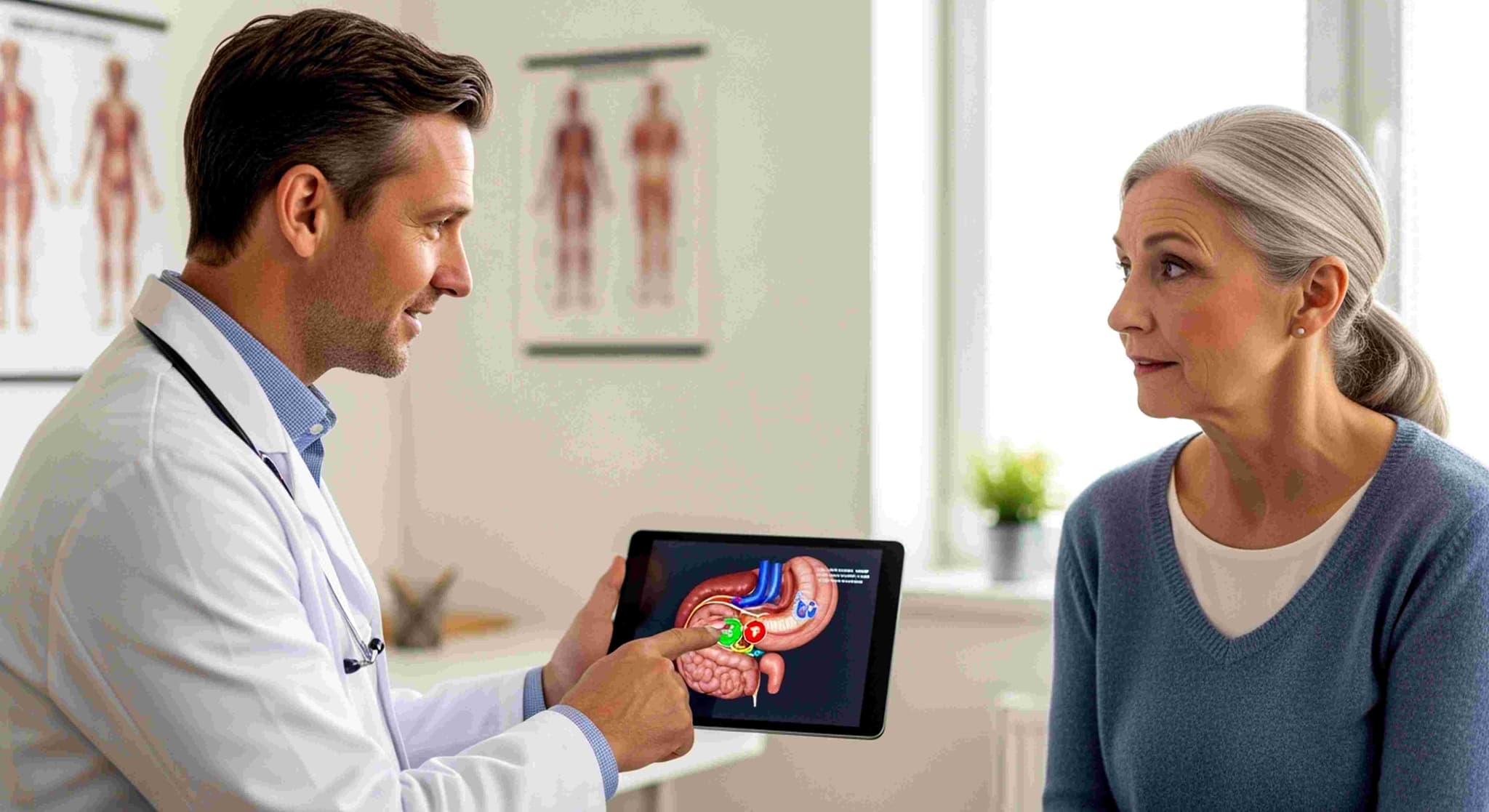
The Department of Gastroenterology & Hepatology—frequently known as the Center for Digestive & Liver Health—is a specialized wing dedicated to the comprehensive care of the entire digestive tract, liver, pancreas, and biliary system. This department seamlessly balances non-invasive medical management with high-tech endoscopic procedures for complex conditions like inflammatory bowel disease, liver cirrhosis, and digestive tract cancers. For international and local patients alike, this wing offers an integrated care model, combining advanced diagnostic precision with elite "incisionless" therapeutic expertise.
A Multidisciplinary Circle of Digestive & Liver Experts

Our department is staffed by a specialized team of clinicians who manage digestive and metabolic health across all stages of life:

  • Medical Gastroenterologists: Primary experts focused on diagnosing and managing disorders of the esophagus, stomach, intestines, and colon, such as IBS, IBD, and severe acid reflux.

  • Hepatologists: Specialized physicians dedicated exclusively to the study and treatment of liver diseases, including hepatitis, fatty liver disease, and liver cirrhosis.

  • Advanced Therapeutic Endoscopists: Highly skilled proceduralists who perform complex, minimally invasive interventions inside the GI tract without external incisions.

  • Gastrointestinal Surgeons & Transplant Specialists: Expert surgeons who perform surgical resections and work alongside hepatologists for liver transplantation.

  • Pancreatobiliary Specialists: Clinicians dedicated to treating complex disorders of the pancreas, gallbladder, and bile ducts, including pancreatitis and gallstones.

  • GI Clinical Nutritionists: Specialized dietitians who create targeted medical nutrition plans for gut rehabilitation, metabolic liver care, and post-procedural recovery.

Advanced Technology and "Incisionless" Care

The department utilizes specialized high-definition imaging and miniaturized endoscopic instrumentation designed for the delicate GI tract:

  • High-Definition Chromoendoscopy: Advanced video endoscopy that uses specialized light filters and dyes to detect early precancerous mucosal lesions in real time.

  • Endoscopic Ultrasound (EUS): Miniaturized ultrasound probes attached to endoscopes to capture detailed images and perform fine-needle biopsies of deep pelvic and abdominal organs.

  • Capsule Endoscopy: A vitamin-pill-sized wireless camera that patients swallow to capture thousands of images of the deep small intestine that traditional scopes cannot reach.

  • Transient Elastography (FibroScan): Non-invasive ultrasound-based technology that measures liver stiffness and fat accumulation without requiring a physical needle biopsy.

  • ERCP (Endoscopic Retrograde Cholangiopancreatography): Specialized fluoroscopic endoscopic technology used to remove bile duct stones, place stents, and clear blockages without open surgery.

Specialized Functional and Diagnostic Areas

To support the journey from initial symptom evaluation to long-term disease management, the department integrates specialized recovery and diagnostic zones:

  • Advanced Endoscopy & Suite Lounge: State-of-the-art procedure rooms equipped with carbon dioxide insufflation and real-time imaging for safe, comfortable endoscopic procedures.

  • The Liver Care & Transplant Clinic: A dedicated outpatient hub for long-term monitoring of chronic liver disease, pre-transplant evaluations, and post-transplant follow-up care.

  • GI Motility & Physiology Lab: A specialized diagnostic zone for high-resolution esophageal and anorectal manometry to evaluate swallowing and digestive movement disorders.

  • Day-Care Infusion Center: A comfortable environment designed for patients receiving biological therapies and infusions for Inflammatory Bowel Disease (Crohn's and Ulcerative Colitis).

  • International Digestive Health Suite: A private sanctuary for global patients to coordinate travel logistics, multi-specialty consultations, and personalized care pathways.

A Focus on Gut Health and Patient-Centered Care

The modern gastroenterology experience prioritizes early detection, organ preservation, and patient comfort:

  • Painless Endoscopy Protocols: 24/7 access to specialized monitored anesthesia care ensures smooth, comfortable, and memory-free endoscopic procedures.

  • Third-Space Endoscopy: Cutting-edge procedures (such as POEM and ESD) that allow for the removal of deep tumors or treatment of swallowing disorders entirely through internal mucosal tunnels.

  • Comprehensive Cancer Screening: Combining routine colonoscopies with genetic risk assessments to catch colorectal cancers in their earliest, most treatable stages.

  • Holistic Gut-Brain Care Pathways: Integrated care plans that combine medical treatment, dietary therapy, and lifestyle modification to manage functional bowel disorders effectively.

May we help you?

Enter digits only (6-15 digits). We'll add your country code.

Gastroenterology & Hepatology Treatments in India

Colectomy (Colon Cancer)

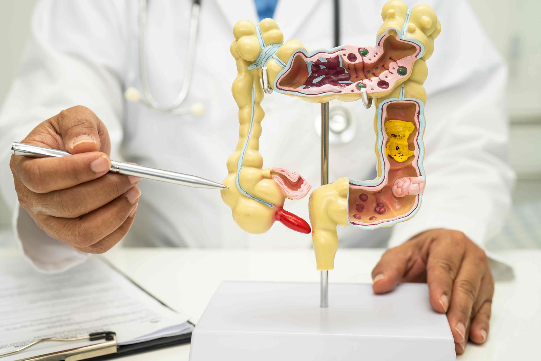
Colon Cancer Treatment involves a combination of specialized surgical, medical, and radiological interventions to remove malignant tumors from the large intestine. As of 2026, clinical standards emphasize a precision-based approach, utilizing robotic surgery for localized tumors and advanced immunotherapy for cases with specific genetic biomarkers (MSI-H/dMMR), aiming to achieve long-term remission while preserving bowel function.

  • Persistent changes in bowel habits, such as diarrhea, constipation, or narrowing of the stool.

  • Presence of bright red blood or dark, tarry stools during bowel movements.

  • Ongoing abdominal discomfort, including persistent cramps, gas, or localized pain.

  • A frequent feeling that the bowel does not empty completely after a movement.

  • Unexplained weight loss accompanied by chronic fatigue or weakness.

  • Signs of anemia, such as paleness and lack of energy, due to hidden internal bleeding.

  • Adenomatous Polyps that show signs of high-grade dysplasia or early transformation.

  • Localized Colon Adenocarcinoma confined to the inner layers of the intestinal wall.

  • Hereditary Syndromes including Lynch Syndrome or Familial Adenomatous Polyposis (FAP).

  • Inflammatory Bowel Disease (IBD) such as long-term Ulcerative Colitis or Crohn’s disease.

  • Advanced or metastatic colon cancer that has spread to the liver, lungs, or peritoneum.

  • Diagnostic colonoscopy is utilized to visualize the colon and remove precancerous polyps.

  • High-resolution CT, MRI, or PET scans are performed to map the tumor’s size and spread.

  • A partial or total colectomy is surgically performed to remove the malignant section of the colon.

  • Adjuvant chemotherapy is administered after surgery to eliminate any remaining microscopic cells.

  • Systematic immunotherapy is introduced for tumors with specific genetic MSI-H or dMMR markers.

  • Targeted drug therapy is used to block the proteins that allow cancer cells to grow and spread.

  • Laparoscopic or Robotic Colectomy A minimally invasive surgery to remove the cancerous part of the colon through small abdominal incisions.

  • Polypectomy The removal of small, early-stage cancerous growths directly through a colonoscope without external incisions.

  • Immunotherapy (Pembrolizumab/Nivolumab) Advanced biological treatments that help the immune system identify and attack cancer cells.

  • Targeted Therapy (Bevacizumab/Cetuximab) Precision medications that target specific growth factors or blood vessel formation in the tumor.

  • Cytoreductive Surgery with HIPEC A specialized procedure where the abdominal cavity is bathed in heated chemotherapy after tumor removal.

  • Colostomy or Ileostomy A surgical procedure to create a stoma (opening) in the abdomen to allow waste to exit the body temporarily or permanently.

  • Complete a "bowel prep" to thoroughly clear the colon of stool before the procedure.

  • Consult with a dietitian to establish a low-residue diet in the days leading up to surgery.

  • Adjust or stop blood-thinning medications as instructed by the surgical oncology team.

  • Undergo a preoperative physical to ensure readiness for general anesthesia and recovery.

  • Meet with an ostomy nurse if there is a possibility of needing a temporary or permanent stoma.

  • Colonoscopy with biopsy to confirm the specific pathology and grade of the cancer.

  • CT Scan of the chest, abdomen, and pelvis to determine the clinical stage of the disease.

  • CEA (Carcinoembryonic Antigen) Blood Test to establish a baseline for post-treatment monitoring.

  • MSI/dMMR Biomarker Testing to identify if the tumor will respond to modern immunotherapies.

  • Complete Blood Count (CBC) to check for anemia or other blood-related issues before surgery.

  • Features a 91% five-year survival rate for localized cases through early detection and surgery.

  • Utilizes 2026 precision medicine to match treatments to the tumor's unique genetic profile.

  • Offers minimally invasive robotic options that significantly reduce recovery time and pain.

  • Employs neoadjuvant therapies to shrink large tumors, making them easier to remove safely.

  • Integrates multidisciplinary care to manage complex cases that have spread to other organs.

  • In-hospital monitoring for 3–5 days to ensure the return of normal bowel function.

  • Personalized pain management and early mobilization to prevent blood clots and lung issues.

  • Gradual transition from a liquid diet to soft foods as the digestive tract heals.

  • Specialized wound care and training for patients who require a temporary or permanent stoma.

  • Follow-up visits with the surgical and oncology teams to monitor healing and plan next steps.

  • Regular surveillance through colonoscopies and blood tests every 6–12 months for the first few years.

  • Adoption of a high-fiber diet rich in fruits, vegetables, and whole grains to support colon health.

  • Commitment to regular physical activity and maintaining a healthy weight to reduce recurrence risk.

  • Avoidance of tobacco products and limiting alcohol consumption as part of a cancer-prevention lifestyle.

  • Participation in survivorship programs to manage the emotional and physical transition after treatment.

Endoscopic Retrograde Cholangiopancreatography (ERCP)

Endoscopic Retrograde Cholangiopancreatography (ERCP) is a highly specialized procedure used primarily to treat disorders of the bile ducts and pancreatic ducts. It uniquely combines advanced endoscopy with real-time X-ray imaging (fluoroscopy). By 2026, ERCP has evolved from a diagnostic tool into a powerful therapeutic intervention, allowing physicians to clear obstructions, place stents, and repair leaks within the delicate ductal system of the liver and pancreas without the need for invasive abdominal surgery.

  • Common Bile Duct Stones: Removing gallstones that have migrated out of the gallbladder and are blocking the main drainage duct.

  • Malignant Obstructions: Relieving blockages caused by tumors in the pancreas, bile ducts, or liver.

  • Biliary Strictures: Opening narrowed areas of the ducts caused by scar tissue or chronic inflammation.

  • Post-Surgical Bile Leaks: Sealing leaks that can occasionally occur following gallbladder or liver surgery.

  • Chronic Pancreatitis: Treating complications such as pancreatic stones or narrowings that cause severe, recurring pain.

  • Sphincter of Oddi Dysfunction: Managing issues with the muscular valve that controls the flow of digestive juices.

  • Massive Stone Burden: Cases with very large or numerous stones that may require advanced "fragmentation" techniques.

  • Anatomical Variations: Patients who have had previous gastric bypass or stomach surgery, requiring a modified "enteroscopy" approach.

  • Primary Sclerosing Cholangitis (PSC): A chronic condition requiring careful, repeated dilations of the bile ducts.

  • High-Risk Pancreatitis Patients: Individuals with a history of post-procedure inflammation who require preventative "stenting" of the pancreatic duct.

  • Pediatric ERCP: Rare cases in children involving congenital ductal abnormalities that require specialized pediatric endoscopes.

[Image showing the endoscope positioned in the duodenum with the catheter entering the bile duct]

  • Deep Sedation or Anesthesia: To ensure absolute stillness and comfort, the procedure is performed under deep sedation or general anesthesia.

  • Side-Viewing Endoscopy: A specialized duodenoscope is passed through the mouth and stomach to reach the "papilla"—the small opening where the ducts meet the intestine.

  • Cannulation: A tiny catheter is threaded into the papilla to gain access to the biliary or pancreatic tree.

  • Retrograde Contrast: A radio-opaque dye is injected "backward" into the ducts, highlighting the internal anatomy on a live X-ray monitor.

  • Sphincterotomy: Using a small electrified wire, the surgeon may make a tiny cut in the muscle valve (sphincter) to widen the opening for stone removal.

  • Mechanical Intervention: Tools such as baskets, balloons, or stents are passed through the scope to clear blockages or provide structural support.

  • Disposable DuodenoscopesThe widespread adoption of single-use scopes to eliminate the risk of bacterial transmission and ensure the highest mechanical reliability for every patient.

  • SpyGlass™ CholangioscopyA "micro-scope" that is passed through the main endoscope, allowing the doctor to see directly inside the bile duct in high-definition to biopsy tumors or break up stones with lasers.

  • AI-Assisted FluoroscopyArtificial intelligence that enhances X-ray images in real-time, helping the doctor identify "shadows" or subtle strictures with greater clarity.

  • Biodegradable Biliary StentsNext-generation stents that provide support to a narrowed duct for several months and then dissolve, avoiding the need for a second procedure to remove them.

  • Laser Lithotripsy (EHL)Using targeted laser energy to "shatter" stubborn gallstones into tiny fragments that can be easily swept out of the duct.

  • Automated Risk-Prediction ModelsSoftware that analyzes a patient's health data before the procedure to suggest the exact dose of preventative medication needed to stop post-op inflammation.

  • Fasting (NPO): A strictly empty stomach is required, usually meaning no food or drink for 8 to 12 hours prior to the procedure.

  • Antibiotic Prophylaxis: Certain patients may be given a dose of antibiotics before the start of the procedure to prevent ductal infection.

  • Blood Work: Checking coagulation (clotting) factors to ensure it is safe to perform a sphincterotomy or biopsy.

  • Medication Coordination: Careful management of blood thinners and diabetic medications is essential for a safe recovery.

  • Post-Op Plan: Because of the intensive nature of ERCP, patients must have a designated driver and a quiet place to recover for the first 24 hours.

  • Pancreatitis Prevention: Use of rectal medications or temporary "protective" pancreatic stents to lower the risk of post-ERCP inflammation.

  • Amylase/Lipase Checks: Blood tests performed if a patient experiences unusual pain after the procedure to check for pancreatic irritation.

  • Continuous Vital Monitoring: Specialized anesthesia teams monitor heart rhythm and oxygen levels throughout the entire intervention.

  • Radiation Safety: Using "low-dose" pulsed fluoroscopy to minimize X-ray exposure for both the patient and the medical team.

  • Avoids Major Surgery: ERCP provides a "keyhole" alternative to traditional open-belly surgery for clearing bile duct obstructions.

  • Immediate Obstruction Relief: Patients with jaundice (yellowing of the skin) often see a rapid improvement in symptoms once the duct is cleared or stented.

  • High Precision: The combination of live video and live X-ray ensures that the doctor knows exactly where the instruments are at all times.

  • Life-Saving for Sepsis: In cases of "cholangitis" (infected bile), ERCP provides urgent drainage that is critical for survival.

  • Dual Functionality: It allows for the immediate transition from identifying a problem to fixing it within the same 30-to-60-minute session.

  • Immediate Observation: Most patients stay in a recovery unit for 1 to 2 hours to ensure the sedation has worn off and no immediate pain is present.

  • Sore Throat and Bloating: These are the most common minor side effects and typically resolve within 24 to 48 hours.

  • Dietary Progression: Patients usually start with clear liquids and move to a low-fat diet as tolerated by their digestive system.

  • Activity Limits: No driving or making major decisions for 24 hours. Most patients return to full activity within 2 to 3 days.

  • Follow-up Imaging: A simple ultrasound or blood test is often scheduled a few weeks later to confirm the ducts remain clear.

  • Rapid resolution of painful symptoms like biliary colic (gallstone pain) or chronic abdominal pressure.

  • Successful management of complex conditions like cancer-related blockages, allowing for a better quality of life.

  • Permanent removal of dangerous "trapped" stones that could otherwise lead to severe liver damage.

  • A return to a normal, healthy diet once the flow of digestive enzymes and bile has been restored.

  • Empowerment through the successful treatment of a condition that previously would have required a long, difficult surgical recovery.

Laparoscopic Polypectomy

Laparoscopic Polypectomy generally refers to the minimally invasive removal of polyps from the gallbladder or, in specific gynecological cases, subserosal uterine polyps. While most uterine polyps are removed via hysteroscopy (through the cervix), the laparoscopic approach is reserved for polyps or small growths located on the outer surface of an organ or within the abdominal cavity.

  • Symptomatic Polyps: Growths causing localized pain or persistent digestive issues.

  • Suspect Features: Polyps showing irregular shapes or rapid growth during an ultrasound.

  • Size-Dependent Risks: Gallbladder polyps exceeding 1 cm in size, which is a standard surgical indication in 2026 guidelines.

  • Outer Organ Growth: When a polypoid growth is located on the exterior of the uterus or attached to pelvic ligaments.

  • Cancer Mitigation: To prevent the risk of malignancy in polyps that show signs of growth.

  • Laparoscopic Cholecystectomy: Removal of the entire gallbladder for polyps larger than 10mm, as they cannot be safely scraped off the wall.

  • Laparoscopic Excision: The use of precision shears or lasers to excise a growth at its base from the exterior of an organ.

  • High-Definition Visualization: Utilizing zoomed-in views to identify a polyp’s blood supply and seal vessels before cutting.

  • Oncological Safety Protocols: The use of specimen bags during removal to ensure tissue does not contact the abdominal wall.

  • Hysteroscopy (Alternative): The standard approach for internal uterine polyps, distinct from the laparoscopic method used for external growths.

  • Anesthesia: The surgery is performed under general anesthesia to ensure the patient is comfortable and still.

  • Access Incisions: The surgeon makes 1 to 3 tiny incisions, ranging from 5–10 mm, in the abdominal wall.

  • Instrument Insertion: A laparoscope and specialized micro-instruments are inserted through the small incisions.

  • Vessel Sealing: Surgeons use high-definition cameras to seal blood vessels before removal, resulting in near-zero blood loss.

  • Abdominal Inflation: Carbon dioxide gas is used to create space, though this can lead to temporary post-operative bloating.

  • Fasting Requirements: Patients must fast for 8-12 hours before the surgery or catheterization.

  • Medical Assessments: Blood tests, ECG, and chest X-rays are conducted to assess overall health.

  • Medication Adjustments: Current medications must be adjusted as directed by the medical team.

  • Allergy Discussion: Patients must discuss any allergies, particularly to materials used in surgical devices or dyes.

  • Post-Op Planning: Arranging for post-operative care and a support person for the recovery period.

  • Ultrasound Imaging: Used to monitor the size and shape of the polyp and detect rapid growth.

  • Blood Tests: To assess overall health and check for markers of inflammation or infection.

  • ECG: To monitor the heart's electrical rhythm and check for conduction issues.

  • Pulse Oximetry: Used to evaluate oxygen saturation levels in the blood before anesthesia.

  • CT Scan: For detailed mapping of the abdominal cavity and complex growths.

  • Hospital Stay: Usually a same-day discharge or a short 23-hour observation stay.

  • Immediate Recovery: Walking is encouraged immediately to aid digestion and prevent blood clots.

  • Pain Management: Discomfort is typically managed with over-the-counter anti-inflammatories.

  • Return to Work: Patients can generally return to work within 5 to 7 days post-procedure.

  • Activity Restrictions: Heavy lifting must be avoided for approximately 4 weeks to ensure proper healing.

  • Minimal Tissue Disruption: Rapid recovery due to the use of tiny incisions rather than large open cuts.

  • Precise Visualization: High-definition cameras allow for the zoom-in on blood supplies for safer removal.

  • Near-Zero Blood Loss: Advanced sealing techniques minimize bleeding during the excision.

  • Strict Safety: The use of specialized bags prevents tissue fragments from remaining in the pelvic cavity.

  • Long-Term Cure: Provides a definitive treatment with high technical success rates for symptomatic growths.

Liver Biopsy
Liver Biopsy

A liver biopsy is a medical procedure used to remove a small piece of liver tissue for laboratory examination. It is considered the definitive "gold standard" for identifying the precise cause of liver disease and determining the extent of organ damage. By 2026, advances in real-time imaging and needle technology have made the procedure faster and safer, allowing clinicians to distinguish between various types of inflammation, scarring, and tumors that blood tests or standard scans might miss.

  • Unexplained Liver Enzymes: When blood tests show persistent elevations in ALT or AST that cannot be explained by standard diagnostic sets.

  • Staging Fibrosis: To determine the exact degree of scarring (cirrhosis) in patients with chronic conditions like Fatty Liver Disease (NASH/MASH).

  • Hepatitis Monitoring: Evaluating the severity of chronic Hepatitis B or C to decide on the most effective antiviral therapy.

  • Autoimmune Disorders: Diagnosing conditions where the body's immune system attacks the liver, such as Primary Biliary Cholangitis (PBC).

  • Transplant Monitoring: Checking a transplanted liver for early signs of organ rejection or medication toxicity.

  • Lumps or Masses: Investigating a suspicious nodule found on an ultrasound or MRI to rule out malignancy.

  • Percutaneous Biopsy: The standard approach. Under the guidance of ultrasound or CT, a specialized needle is inserted through the skin between the ribs directly into the liver to collect a tissue core.

  • Transjugular Biopsy: A specialized technique for patients with blood clotting issues or excess abdominal fluid (ascites). A catheter is threaded through the jugular vein in the neck and guided down to the liver's internal veins.

  • Laparoscopic Biopsy: Tissue is collected during a minimally invasive surgical procedure, allowing the doctor to visually inspect the liver surface and sample specific, hard-to-reach areas.

  • Fine-Needle Aspiration (FNA): Often used for specific liver tumors to collect cells for cytology, though it provides less structural information than a core biopsy.

  • Positioning: The patient typically lies on their back with the right arm raised above the head to widen the space between the ribs.

  • Imaging Guidance: A physician uses an ultrasound probe to identify the safest "window" for the needle, avoiding the gallbladder and major blood vessels.

  • Local Anesthesia: The skin and the lining of the chest wall are numbed; most patients feel a sensation of pressure rather than sharp pain.

  • The "Click": Many modern biopsy needles use a spring-loaded mechanism that captures the tissue sample in a fraction of a second.

  • Breath-Hold: The patient is asked to hold their breath for a few seconds during the needle insertion to keep the liver perfectly still.

  • Sample Verification: The tissue core is immediately inspected to ensure it is of sufficient length (usually 1.5 to 2 cm) for an accurate diagnosis.

  • AI-Enhanced HistopathologyArtificial Intelligence algorithms that analyze biopsy slides to provide a more objective "score" for inflammation and fat content, reducing human variability.

  • Molecular ProfilingUsing biopsy tissue for genetic sequencing to identify the exact subtype of liver cancer, allowing for highly targeted chemotherapy.

  • Digital "Tension" MeasurementNeedles equipped with sensors that can measure liver stiffness (elasticity) at the exact moment the biopsy is taken.

  • Multiparametric MRI-Guided BiopsyA high-tech approach that overlays MRI data onto ultrasound monitors, helping surgeons target "hot spots" of metabolic activity.

  • Micro-RNA AnalysisAnalyzing the sample for specific RNA markers that can predict how fast a patient's liver disease might progress over the next decade.

  • Transjugular Pressure MonitoringMeasuring the pressure within the liver veins (HVPG) during a transjugular biopsy to provide critical data on portal hypertension.

  • Coagulation Testing: Essential blood tests (INR and Platelets) are performed to ensure the blood clots normally before the procedure.

  • Medication Adjustment: Patients must stop taking aspirin, ibuprofen, and other blood thinners for 5–7 days prior to the biopsy.

  • NPO Guidelines: Standard fasting (no food or drink) for 6–8 hours before the appointment is typically required.

  • Observation Planning: Arranging for someone to drive you home, as you will need to remain under observation for several hours post-procedure.

  • Ultrasound Mapping: A pre-procedure scan to confirm the liver size and the absence of fluid that might interfere with the needle path.

  • Post-Biopsy Recovery Position: Patients must lie on their right side for 2–4 hours immediately after the procedure. This uses the body's own weight to apply pressure to the biopsy site, preventing bleeding.

  • Vital Sign Checks: Nurses monitor blood pressure and heart rate every 15 minutes for the first hour to detect any signs of internal bleeding.

  • Pain Assessment: Monitoring for "referred pain" in the right shoulder, which is a common but harmless sign of diaphragm irritation.

  • Follow-up Ultrasound: In rare cases of suspected bleeding, a quick bedside scan can confirm the stability of the liver.

  • Definitive Accuracy: While scans show the "shape" of the liver, a biopsy shows the "activity" of the cells, providing an answer that imaging cannot.

  • Personalized Care: Knowing the exact stage of disease allows doctors to prescribe the correct dosage of medication and avoid unnecessary treatments.

  • Cures Through Discovery: Often identifies rare, treatable conditions like Wilson’s disease or Hemochromatosis that might otherwise go undetected.

  • Proven Safety Record: With modern imaging guidance, the risk of serious complications has dropped to less than 1% in most clinical centers.

  • Minimal Recovery: Most patients return to their daily routine within 48 hours, making it a low-impact procedure with high-impact results.

  • Activity Limits: Avoid heavy lifting, intense exercise, or "straining" the abdominal muscles for at least one week following the procedure.

  • Wound Care: The small puncture site usually only requires a simple bandage; it should be kept dry for 24 hours.

  • Pain Management: Over-the-counter acetaminophen is usually sufficient for any lingering site soreness; aspirin and NSAIDs should still be avoided for a few days.

  • Signs to Watch For: Patients are instructed to contact their doctor immediately if they experience severe abdominal pain, fainting, or fever.

  • Results Timeline: A preliminary report may be available in 2–3 days, while complex stains and genetic markers typically take 7–10 business days.

  • A clear, evidence-based diagnosis that ends the "diagnostic mystery" of elevated liver enzymes.

  • A personalized roadmap for health, including specific dietary, lifestyle, and medication recommendations.

  • Peace of mind for transplant recipients knowing their new organ is being monitored with the highest level of detail.

  • Improved long-term outcomes through the early detection of fibrosis before it progresses to irreversible cirrhosis.

  • The opportunity to participate in clinical trials for new liver therapies based on the specific tissue characteristics found.

Top Doctors for Gastroenterology & Hepatology in India

Dr Rajnesh Chander Reddy
Gastroenterologist
Apollo Health City, Jubilee Hills, Hyderabad
30+years experience
Dr Kona Lakshmi Kumari
Gastroenterologist
Yashoda Hospital, Somajiguda, Hyderabad
27+years experience
Dr Vishal Soni
Dr Vishal Soni
Gastroenterologist
Apollo Hospital International Ltd, Ahmedabad
15+years experience
Dr Harit Kumar Chaturvedi
Surgical Oncologist
Max Super Specialty Hospital, Vaishali, Ghaziabad
38+years experience
Dr Vedant Kabra
Dr Vedant Kabra
Surgical Oncologist
Fortis Memorial Research Institute, Gurugram
25+years experience
Dr Ramesh Sarin
Dr Ramesh Sarin
Surgical Oncologist
Indraprastha Apollo Hospital, Delhi, New Delhi
40+years experience
Dr Rajeev Agarwal
Dr Rajeev Agarwal
Surgical Oncologist
Medanta - The Medicity, Gurugram
47+years experience
Dr Rajesh Mistry
Dr Rajesh Mistry
Surgical Oncologist
Kokilaben Dhirubhai Ambani Hospital, Mumbai
39+years experience
Dr P Jagannath
Dr P Jagannath
Surgical Oncologist
Fortis S L Raheja Hospital, Mumbai
35+years experience
Dr Vijay V Haribhakti
Surgical Oncologist
Sir H N Reliance Foundation Hospital and Research Centre, Mumbai
40+years experience
Dr M Chandrashekhar
Dr M Chandrashekhar
Surgical Oncologist
Apollo Hospital, Bannerghatta Road, Bengaluru
36+years experience
Dr Sandeep Nayak P
Dr Sandeep Nayak P
Surgical Oncologist
Fortis Hospital, BG Road, Bengaluru
24+years experience
Dr Sharat Damodar
Dr Sharat Damodar
Surgical Oncologist
Manipal Hospital, Millers Road, Bengaluru
30+years experience
Dr Vivek Mangla
Dr Vivek Mangla
Surgical Oncologist
Max Super Specialty Hospital, Vaishali, New Delhi
21+years experience
Dr Sowrabh Kumar Arora
Surgical Oncologist
Max Super Specialty Hospital, Vaishali, New Delhi
20+years experience
Dr Vimalathithan S
Dr Vimalathithan S
Surgical Oncologist
Gleneagles Hospital, Perumbakkam, Chennai
17+years experience
Dr Sujay Susikar
Dr Sujay Susikar
Surgical Oncologist
Kauvery Hospital, Chennai
15+years experience
Dr Chinnababu Sunkavalli
Surgical Oncologist
Yashoda Hospital, Hitec City, Hyderabad
23+years experience
Dr Trilok Pratap Singh Bhandari
Surgical Oncologist
Apollo Health City, Jubilee Hills, Hyderabad
24+years experience
Dr Ch Mohana Vamsy
Dr Ch Mohana Vamsy
Surgical Oncologist
Omega Hospital, Gachibowli, Hyderabad
35+years experience
Dr Joy Ghose
Dr Joy Ghose
Surgical Oncologist
Sahyadri Super Specialty Hospital, Deccan Gymkhana, Pune
15+years experience
Dr Snita Sinukumar
Dr Snita Sinukumar
Surgical Oncologist
Apollo Hospital, Pune
11+years experience
Dr Amit Parasnis
Dr Amit Parasnis
Surgical Oncologist
Manipal Hospital, Baner, Pune
25+years experience
Dr Shaikat Gupta
Dr Shaikat Gupta
Surgical Oncologist
Apollo Multispecialty Hospital, Kolkata
34+years experience
Dr Suvadip Chakrabarti
Surgical Oncologist
Apollo Multispecialty Hospital, Kolkata
16+years experience
Dr Nitin Singhal
Dr Nitin Singhal
Surgical Oncologist
Sterling Hospitals, Ahmedabad
15+years experience
Dr Nikunj Patel
Dr Nikunj Patel
Surgical Oncologist
Marengo CIMS Hospital, Ahmedabad
9+years experience
Dr Pradeep Chowbey
Dr Pradeep Chowbey
General Surgeon
Max Super Specialty Hospital, Saket, New Delhi
50+years experience
Dr B M L Kapoor
Dr B M L Kapoor
General Surgeon
Indraprastha Apollo Hospital, Delhi, New Delhi
50+years experience
Dr Pawanindra Lal
Dr Pawanindra Lal
General Surgeon
Indraprastha Apollo Hospital, Delhi, Kochi
31+years experience
Dr Neeraj Goyal
Dr Neeraj Goyal
General Surgeon
Max Super Specialty Hospital, Patparganj, New Delhi
26+years experience
Dr Vivek Bindal
Dr Vivek Bindal
General Surgeon
Max Super Specialty Hospital, Vaishali, New Delhi
17+years experience
Dr Neel Shah
Dr Neel Shah
General Surgeon
Indraprastha Apollo Hospital, Delhi, New Delhi
30+years experience
Dr Rajan Madan
Dr Rajan Madan
General Surgeon
BLK-Max Super Specialty Hospital, Delhi, New Delhi
42+years experience
Dr Sushil Kumar Jain
General Surgeon
Indraprastha Apollo Hospital, Delhi, New Delhi
39+years experience
Dr Hemanth Kumar
Dr Hemanth Kumar
General Surgeon
Fortis Flt. Lt. Rajan Dhall Hospital, Vasant Kunj, Greater Noida
20+years experience
Dr Muffazal Lakdawala
General Surgeon
Sir H N Reliance Foundation Hospital and Research Centre, Mumbai
19+years experience
Dr Deepraj Bhandarkar
General Surgeon
P D Hinduja Hospital, Mahim, Mumbai
35+years experience
Dr Shirish Bhagvat
Dr Shirish Bhagvat
General Surgeon
Sir H N Reliance Foundation Hospital and Research Centre, Mumbai
20+years experience
Dr Narendra Nikam
Dr Narendra Nikam
General Surgeon
Lilavati Hospital & Research Centre, Mumbai
15+years experience
Dr Sumit Talwar
Dr Sumit Talwar
General Surgeon
Manipal Hospital, Old Airport Road, Bengaluru
30+years experience
Dr Madva Jayashankar
General Surgeon
Manipal Hospital, Old Airport Road, Bengaluru
38+years experience
Dr Deepak Subramanian
General Surgeon
MGM Healthcare, Chennai
15+years experience
Dr Jameel Akhter
Dr Jameel Akhter
General Surgeon
Apollo Hospital, Greams Road, Chennai
21+years experience
Dr Venkatasubramanian Rangarajan
General Surgeon
Apollo Hospital, Greams Road, Chennai
20+years experience
Dr Girish Bapat
Dr Girish Bapat
General Surgeon
Jehangir Hospital, Pune
33+years experience
Dr Ramesh Agarwalla
Dr Ramesh Agarwalla
General Surgeon
Fortis Hospital, Anandpur, Kolkata
36+years experience
Dr Sumantra Ray
Dr Sumantra Ray
General Surgeon
Woodlands Multispecialty Hospital, Kolkata
24+years experience
Dr Subhash Gupta
Dr Subhash Gupta
Gastro Onco-Surgeon
Max Super Specialty Hospital, Vaishali, New Delhi
38+years experience

May we help you?

Enter digits only (6-15 digits). We'll add your country code.