Medivisor India Treatment Logo
  • Home
  • About Us
    • About Us
    • Our Services
    • Our Team
    • Our Medical Advisors
    • Our Safety Measures
  • Treatment
    • Loading treatments...
  • Hospitals
    • Loading hospitals...
  • Patient Guide
    • Treatment Cost
    • Treatment Process
    • Visa Process
    • Travel Guide
    • FAQs
    • Why Medivisor
  • Gallery
    • Success Stories
    • Patient Activities
    • Social Activities
    • News Coverage
    • Blog
  • Become a Partner

+91 83407 80250

Medivisor Logo

Medivisor India Treatment is a trusted New Delhi–based medical travel partner connecting global patients to advanced, affordable, high-quality healthcare in India.

  • Medivisor House 359, Sector 1, Vaishali, Ghaziabad, (Delhi/NCR) India
  • +91 8340 780 250
  • info@medivisorhealth.com

About Us

  • About Us
  • Our Services
  • Our Team
  • Our Medical Advisors
  • Our Safety Measures
  • Join Our Network
  • Contact Us

India Treatment

  • Hospitals
  • Treatments
  • Treatment Cost
  • Treatment Process
  • Visa Process
  • Travel Guide
  • FAQs
  • Why Medivisor

Gallery

  • Patient Testimonials
  • Patient Activities
  • News Coverage
  • Blog

Get in Touch

© 2026 Medivisor India Treatment. All rights reserved.

General Surgery hospital

General Surgery

  1. Home
  2. Departments
  3. General Surgery

General Surgery Department

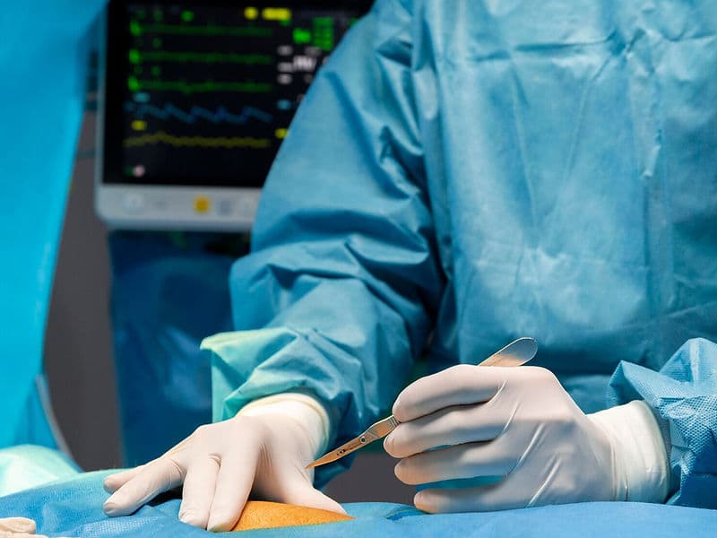
The General Surgery Department serves as the foundational pillar of modern clinical care. Far from being "basic," this department represents a high-level specialty focused on the comprehensive management of the abdominal organs, digestive tract, and endocrine system. For international patients, the department offers a sophisticated blend of traditional surgical wisdom and cutting-edge "keyhole" technology, ensuring that both elective procedures and emergency interventions are handled with elite precision and a focus on rapid recovery.

A Versatile Team of Surgical Experts

Our general surgery wing is staffed by a multidisciplinary team trained in the most advanced surgical modalities:

  • General & Laparoscopic Surgeons: Specialists in "Minimal Access" techniques who perform major abdominal operations through tiny, high-precision incisions.

  • Colorectal Specialists: Experts focused on the lower digestive tract, specializing in the treatment of colorectal cancers, complex fistulas, and inflammatory bowel diseases.

  • Endocrine Surgeons: A focused team dedicated to the surgical management of the thyroid, parathyroid, and adrenal glands.

  • Trauma & Emergency Surgeons: Front-line experts trained to perform life-saving interventions for internal injuries and acute abdominal crises.

  • Wound Care & Clinical Fellows: A dedicated support team that manages preoperative optimization and meticulous postoperative recovery.

Advanced Surgical Technology and Tools

The department utilizes a high-tech "Surgical Tower" environment to minimize downtime and maximize patient safety:

  • High-Definition Laparoscopic Systems: Advanced camera platforms that provide surgeons with a crystal-clear, magnified view of the internal abdominal workspace.

  • Ultrasonic "Harmonic" Scalpels: Cutting-edge tools that use high-frequency vibrations to cut tissue and seal blood vessels simultaneously, ensuring virtually bloodless surgery.

  • Precision Surgical Stapling: Automated devices used to securely reconnect intestinal segments with greater consistency than traditional hand-suturing.

  • Reinforced Surgical Mesh: Medical-grade, bio-compatible nets used to provide a permanent, low-recurrence fix for all types of hernias.

  • Electrocautery Precision: Sophisticated instruments that instantly seal small blood vessels during surgery, maintaining a dry and sterile surgical field.

Specialized Functional and Diagnostic Units

To provide seamless care, the department integrates several specialized zones for diagnosis and recovery:

  • The Endoscopy & Colonoscopy Suite: A diagnostic hub where surgeons use flexible cameras to identify ulcers, polyps, or early-stage cancers.

  • Minor Procedure Rooms: Dedicated suites for localized surgical tasks such as cyst removals or abscess drainage under local anesthesia.

  • Surgical Intensive Care (SICU): A high-intensity recovery environment for patients undergoing complex abdominal reconstructions or trauma care.

  • Advanced Wound Clinic: A specialized facility for managing surgical scars and complex healing requirements, such as diabetic foot care.

  • International Patient Support Office: A central point for global families to coordinate surgical bookings, pathology reports, and travel logistics.

A Focus on Rapid Recovery and Wellness

The modern surgical experience is designed to get patients back to their lives as quickly as possible:

  • "Keyhole" Advantages: Most procedures are performed via minimally invasive routes, resulting in less post-operative pain and minimal scarring.

  • Early Ambulation Protocols: A structured recovery plan that encourages movement shortly after surgery to promote digestion and prevent complications.

  • Integrated Care Pathways: From initial ultrasound and CT diagnostics to follow-up pathology, the entire process is managed under one specialized roof.

  • Global Safety Standards: Every procedure follows international protocols for sterilization and surgical safety, ensuring peace of mind for those traveling for care.

May we help you?

Enter digits only (6-15 digits). We'll add your country code.

General Surgery Treatments

Appendectomy (Open/Laparoscopic)

Appendectomy (Open/Laparoscopic)

An Appendectomy is the surgical removal of the appendix, a small, finger-shaped pouch attached to the large intestine. It is the standard treatment for appendicitis, an inflammation of the organ that can lead to a life-threatening rupture if left untreated.

When You Should Consider Appendectomy

  • Pain that starts near the belly button and moves to the lower right abdomen.

  • "Rebound tenderness" or sharp pain in the lower right abdomen when pressure is released.

  • Fever and chills accompanying abdominal discomfort.

  • Nausea, vomiting, or loss of appetite.

  • High white blood cell counts detected during blood testing.

Methods of Appendectomy

  • Laparoscopic Appendectomy: The modern minimally invasive standard using three small incisions and a camera (laparoscope).

  • Open Appendectomy: A traditional approach using a single 2- to 4-inch incision, typically reserved for ruptured cases.

  • Emergency Appendectomy: Immediate surgical intervention performed to prevent or treat an appendix rupture.

  • Interval Appendectomy: A delayed surgery performed after a severe infection has been initially managed with antibiotics.

How Appendectomy Is Performed

  • Insufflation: In laparoscopic cases, carbon dioxide gas is used to inflate the abdomen for better visibility.

  • Appendix Identification: The surgeon locates the appendix and carefully ties off its base to separate it from the large intestine.

  • Irrigation: If the appendix has ruptured, the abdominal cavity is washed with saline to remove bacteria and pus.

  • Drainage: A small tube may be placed in the incision site for a few days to allow excess fluid to escape in complex cases.

  • Closure: Incisions are secured using absorbable sutures, surgical glue, or sterile strips.

Pre-Procedure Preparation

  • Maintaining NPO (nothing by mouth) status for several hours prior to the operation.

  • Starting Intravenous (IV) fluids to ensure proper hydration.

  • Administration of prophylactic antibiotics to reduce the risk of surgical site infection.

  • Undergoing general anesthesia to remain completely unconscious throughout the procedure.

Tests Before Appendectomy

  • Physical Examination: To check for localized pain and abdominal wall tension.

  • Ultrasound or CT Scan: Imaging used to confirm the inflammation or see if the appendix has burst.

  • Complete Blood Count (CBC): To identify signs of infection through elevated white blood cell levels.

  • Urine Test: Often performed to rule out a urinary tract infection or kidney stones which can mimic appendicitis symptoms.

Life After Appendectomy

  • For uncomplicated cases, hospital discharge usually occurs within 24 hours.

  • Recovery for laparoscopic surgery includes avoiding heavy lifting (over 5kg) for 1–2 weeks.

  • Recovery for open surgery requires avoiding strenuous exercise for 4–6 weeks to prevent hernia.

  • Diet begins with clear liquids and progresses as bowel function returns to normal.

  • Patients should monitor for signs of abscess, such as new fevers or persistent bloating.

Benefits of Appendectomy

  • Provides a permanent cure for appendicitis by removing the source of inflammation.

  • Prevents life-threatening complications associated with an appendix rupture or peritonitis.

  • Minimal scarring and faster recovery times when performed using laparoscopic techniques.

  • Rapidly alleviates severe abdominal pain and restores normal digestive health.

Cholecystectomy (Open/Laparoscopic)

Cholecystectomy (Open/Laparoscopic)

A Cholecystectomy is the surgical removal of the gallbladder, a small organ located under the liver that stores bile. It is primarily performed to treat gallstones (cholelithiasis), gallbladder inflammation (cholecystitis), or biliary colic, which is intense pain caused by stones blocking the bile ducts.

When You Should Consider Cholecystectomy

  • Sudden and rapidly intensifying pain in the upper right portion of the abdomen.

  • Nausea or vomiting following high-fat meals.

  • Yellowing of the skin and the whites of the eyes (jaundice).

  • Fever or chills, which may indicate a gallbladder infection.

  • Confirmation of gallstones or a non-functional gallbladder through diagnostic testing.

Methods of Cholecystectomy

  • Laparoscopic Cholecystectomy: The modern "gold standard" minimally invasive approach used in over 90% of cases, involving four small incisions.

  • Open Cholecystectomy: A traditional surgical method using a larger incision, typically reserved for complex cases or severe scarring.

  • Emergency Cholecystectomy: Immediate removal performed when acute inflammation or infection poses a significant health risk.

  • Single-Incision Laparoscopic Surgery (SILS): A specialized technique where the gallbladder is removed through one small incision in the umbilicus.

How Cholecystectomy Is Performed

  • Pneumoperitoneum: During laparoscopic surgery, the abdomen is inflated with carbon dioxide gas to provide a clear workspace for the surgeon.

  • Duct and Artery Clipping: The surgeon identifies the cystic duct and cystic artery and secures them with permanent clips before separation.

  • Gallbladder Detachment: The organ is carefully separated from the liver bed using specialized surgical instruments.

  • Organ Extraction: The gallbladder is placed in a sterile bag and removed through one of the small abdominal incisions.

  • Drainage: In open or complex cases, a Jackson-Pratt (JP) drain may be placed temporarily to remove excess fluid or bile.

Pre-Procedure Preparation

  • Fasting (NPO) for at least 6 to 8 hours prior to the operation.

  • Discontinuing blood-thinning medications several days before surgery as directed by the surgical team.

  • Receiving general anesthesia and the placement of a breathing tube for the duration of the procedure.

  • Undergoing a pre-operative evaluation to check liver enzymes and white blood cell counts.

Tests Before Cholecystectomy

  • Abdominal Ultrasound: The "gold standard" imaging test used to identify gallstones and gallbladder wall thickening.

  • HIDA Scan: A specialized nuclear medicine test used to evaluate the functional emptying of the gallbladder.

  • Blood Panels: To check for elevated liver enzymes or signs of infection (leukocytosis).

  • MRCP or ERCP: Advanced imaging or endoscopic procedures used to check for stones that may have moved into the common bile duct.

Life After Cholecystectomy

  • Most laparoscopic patients return home the same day or after one overnight stay.

  • Recovery for open surgery involves a hospital stay of 3 to 5 days and restricted activity for 4 to 6 weeks.

  • A low-fat diet is recommended for the first few weeks as the body adjusts to bile flowing directly into the small intestine.

  • Patients may experience temporary shoulder pain or bloating due to the gas used during laparoscopic procedures.

  • Monitoring for Post-Cholecystectomy Syndrome (PCS), which affects 10–15% of patients and involves lingering indigestion or nausea.

Benefits of Cholecystectomy

  • Provides permanent relief from the recurring pain of biliary colic and gallstones.

  • Prevents serious complications such as gallbladder rupture, gangrene, or severe pancreatitis.

  • Minimally invasive techniques result in smaller scars, less post-operative pain, and a faster return to daily activities.

  • Eliminates the risk of future gallbladder infections and inflammation.

Hernia Repair (Inguinal/Umbilical)

Hernia Repair (Inguinal/Umbilical)

Hernia repair is a surgical procedure to correct protrusions of tissue or organs through weakened areas of the abdominal wall. An inguinal hernia occurs in the groin area (the inguinal canal), while an umbilical hernia occurs at or near the navel (belly button). By reinforcing these weak points, surgery prevents the risk of "incarceration" or "strangulation," where the blood supply to the trapped tissue is cut off, making it an essential intervention for both comfort and long-term health.

When You Should Consider Hernia Repair

  • Persistent pain or a "dragging" sensation in the groin or abdomen, especially when lifting or coughing.

  • A visible bulge that may disappear when lying down but protrudes during physical activity.

  • Umbilical protrusions that become larger, more painful, or aesthetically concerning.

  • Inguinal hernias that cause discomfort during daily movements or exercise.

  • As a preventive measure to avoid the emergency complications of a trapped (strangulated) intestine.

  • When a hernia interferes with professional duties that require physical labor or heavy lifting.

Conditions That Require Specialized Care

  • Incarcerated Hernia: Tissue that is trapped in the abdominal wall and cannot be pushed back in.

  • Strangulated Hernia: A medical emergency where the blood supply to the trapped tissue is restricted.

  • Recurrent Hernia: A hernia that has returned after a previous surgical repair.

  • Bilateral Inguinal Hernias: When hernias are present on both the left and right sides of the groin simultaneously.

  • Large Incisional Hernias: Weakness occurring at the site of a previous surgical scar.

How Hernia Repair Is Performed

  • The procedure begins with Reduction, where the protruding fat or intestine is gently pushed back into the abdominal cavity.

  • Open Surgery involves a single incision made directly over the bulge to access the muscle defect.

  • Laparoscopic Surgery utilizes three to four "keyhole" incisions and a camera to repair the wall from the inside.

  • Robotic-Assisted Surgery provides the surgeon with high-definition 3D visualization and robotic precision for complex cases.

  • In most adult cases, a Synthetic Mesh is placed over the weak spot to act as a permanent reinforcement.

  • The muscle layers are sutured, and the skin is closed with dissolvable stitches, surgical tape, or skin glue.

Innovations in Hernia Surgery

  • Bio-Absorbable MeshesAdvanced mesh materials that provide a scaffold for the body’s own tissue to grow into before eventually dissolving, reducing long-term foreign body sensation.

  • Self-Gripping (ProGrip) MeshSpecialized mesh with micro-hooks that eliminate the need for traditional tacks or sutures, significantly reducing post-operative nerve pain.

  • 3D-Mapped Mesh ContouringThe use of meshes designed to anatomically fit the specific curvature of the groin or abdominal wall for a more natural feel.

  • Single-Port Robotic RepairThe ability to perform complex hernia reconstructions through just one tiny incision, further minimizing scarring.

  • Advanced Nerve MappingIntraoperative tools that help surgeons identify and avoid sensory nerves, lowering the risk of chronic post-surgical pain.

  • Glue-Based FixationUsing medical-grade adhesives instead of mechanical tacks to secure the mesh, leading to a smoother recovery.

Pre-Procedure Preparation

  • Physical examination and medical history to determine the most effective surgical approach (Open vs. Laparoscopic).

  • Guidance on smoking cessation, as nicotine can significantly weaken tissue healing and increase recurrence risk.

  • Review of medications, particularly anticoagulants (blood thinners) that may need to be paused.

  • Fasting (NPO) instructions starting the night before surgery to ensure a safe anesthetic experience.

  • Coordination for a "day-case" discharge, as most patients return home within hours of the procedure.

Pre-Procedure Tests

  • Ultrasound or CT Scan: To determine the exact size of the hernia and identify if any vital organs are involved.

  • Routine Blood Work (CBC/Electrolytes): To ensure general health and proper clotting function.

  • Electrocardiogram (ECG): For patients over 40 or those with cardiac history to ensure fitness for anesthesia.

  • Cough Test: A physical assessment to judge the stability of the hernia under abdominal pressure.

  • Urinalysis: To rule out any underlying infections prior to the placement of surgical mesh.

Why This Treatment Is Highly Effective

  • Prevents Emergencies: Repairing a hernia electively removes the life-threatening risk of bowel strangulation.

  • Minimal Recurrence: The use of modern mesh technology has reduced the risk of a hernia returning to less than 5%.

  • Rapid Return to Activity: Laparoscopic and robotic techniques allow most patients to return to desk work within a week.

  • Restored Core Strength: Reinforcing the abdominal wall improves overall stability and comfort during physical movement.

  • Cosmetic Improvement: Corrects the visible bulge, restoring the natural contour of the navel or groin.

Recovery and Monitoring

  • Patients are encouraged to walk within a few hours of surgery to promote blood flow and lung function.

  • Pain management typically transitions from clinical care to over-the-counter medications within 48 hours.

  • Heavy lifting (typically over 10–20 lbs) is restricted for 4 to 6 weeks to allow the mesh to integrate fully.

  • Most patients can resume driving once they can perform an emergency stop without abdominal discomfort.

  • Follow-up visits are scheduled to monitor the incision sites and ensure there is no fluid buildup (seroma).

Life After Hernia Repair

  • Freedom from the chronic aching or "heaviness" associated with the hernia.

  • Ability to return to high-impact sports and strenuous gym activities once full clearance is granted.

  • Permanent structural support of the abdominal wall through the integrated mesh.

  • Significant reduction in the psychological stress associated with a visible or painful bulge.

  • Long-term protection against the risk of intestinal complications.

Incisional Hernia Repair

Incisional Hernia Repair

An Incisional Hernia Repair is a surgical procedure to fix a protrusion of tissue or an organ (usually the intestine) through a previously made surgical scar. This occurs when the abdominal wall muscles fail to heal completely or weaken over time following an earlier surgery.

When You Should Consider Incisional Hernia Repair

  • A visible bulge or lump at the site of a previous surgical scar.

  • A bulge that becomes more prominent or painful when coughing, straining, or lifting.

  • Persistent aching or pressure at the previous incision site.

  • Nausea or vomiting if the protruding tissue becomes trapped (incarcerated).

  • Confirmation of a muscle "defect" via CT scan or physical examination.

Methods of Incisional Hernia Repair

  • Open Repair: A traditional approach involving an incision over the old scar, often used for very large hernias or complex cases.

  • Laparoscopic Repair: A minimally invasive method using small "keyhole" incisions, CO2 gas, and a camera to repair the defect from the inside.

  • Robotic-Assisted Repair: A modern variation of laparoscopic surgery offering enhanced precision for medium to large hernias.

  • Tension-Free Mesh Repair: The use of synthetic or biologic mesh to reinforce the abdominal wall, significantly reducing the risk of recurrence.

How Incisional Hernia Repair Is Performed

  • Sac Reduction: The surgeon identifies the hernia sac and carefully pushes the protruding tissue or intestine back into the abdominal cavity.

  • Adhesiolysis: Freeing the intestine from old scar tissue or adhesions that may have formed since the previous surgery.

  • Mesh Placement: A flexible, permanent screen (mesh) is secured over or under the muscle layer to act as a scaffold for new tissue growth.

  • Fixation: The mesh is secured using sutures, tacks, or specialized surgical staples to ensure it remains in a "tension-free" position.

  • Abdominal Wall Reconstruction: In complex cases, the surgeon may rearrange muscle layers to achieve a more natural and strong closure.

Pre-Procedure Preparation

  • Quitting smoking for several weeks to improve blood flow and tissue healing.

  • Weight management and stabilization of blood sugar levels to minimize the risk of repair failure.

  • Fasting (NPO) as directed before undergoing general anesthesia.

  • Evaluation of the old surgical site to plan the best approach (open vs. minimally invasive).

Tests Before Incisional Hernia Repair

  • Physical Exam: To feel for the bulge and assess the strength of the surrounding abdominal wall.

  • CT Scan: The primary imaging tool used to measure the exact size of the muscle defect and identify the contents of the hernia.

  • Blood Panels: To check for signs of infection or uncontrolled diabetes that could affect recovery.

  • Pre-Surgical Optimization Review: Assessment of lifestyle factors (BMI, smoking status) that impact surgical success.

Life After Incisional Hernia Repair

  • Hospital stays range from same-day discharge for laparoscopic cases to 3–5 days for large open repairs.

  • Use of an abdominal binder (elastic wrap) for 4–6 weeks to support the new repair.

  • Strict activity restrictions, including no heavy lifting (>5 kg) for 6–8 weeks.

  • Immediate encouragement of gentle walking to prevent blood clots during the early recovery phase.

  • Monitoring for seromas (fluid collection) or signs of mesh infection at the incision site.

Benefits of Incisional Hernia Repair

  • Significantly reduces the risk of hernia recurrence from 30–50% (without mesh) to below 10% (with mesh).

  • Restores the structural integrity and strength of the abdominal wall.

  • Eliminates the risk of life-threatening complications such as bowel obstruction or strangulation.

  • Relieves chronic pain and discomfort associated with the protruding tissue.

Laparoscopic Hernia Repair

Laparoscopic Hernia Repair

Laparoscopic Hernia Repair, also known as minimally invasive or keyhole surgery, uses specialized tools and a camera to repair a weakness in the abdominal wall from the inside out. It is most commonly used for inguinal (groin), umbilical (navel), and ventral hernias.

When You Should Consider Laparoscopic Hernia Repair

  • A visible bulge in the groin or abdomen that may become more prominent when standing or coughing.

  • Pain or pressure at the hernia site during physical activity or lifting.

  • Inguinal hernias that occur on both sides (bilateral) of the groin.

  • Recurrent hernias where a previous open surgical repair has failed.

  • Desire for a faster return to work and daily activities with minimal scarring.

Methods of Laparoscopic Hernia Repair

  • TAPP (Transabdominal Preperitoneal): The surgeon enters the peritoneal cavity where the organs are located to reach and repair the hernia.

  • TEP (Totally Extraperitoneal): The surgeon operates in the space between the muscle and the abdominal lining, avoiding the organ cavity entirely.

  • IPOM (Intraperitoneal Onlay Mesh): Primarily used for ventral hernias, where a specialized mesh is placed on the inside of the abdominal wall.

  • Robotic-Assisted Laparoscopy: Using a robotic interface for enhanced precision and 3D visualization during the repair.

How Laparoscopic Hernia Repair Is Performed

  • Insufflation: The abdominal cavity is inflated with Carbon Dioxide (CO2) gas to create a dome-shaped workspace.

  • Port Insertion: Three to four small incisions (0.5–1 cm) are made to allow the insertion of a laparoscope and long, thin instruments.

  • Reduction: Protruding tissue, such as fat or a loop of intestine, is carefully pulled back into the abdominal cavity from the inside.

  • Mesh Reinforcement: A lightweight, flexible synthetic mesh is unfurled over the defect to reinforce the weakened wall.

  • Fixation: The mesh is secured in place using surgical tacks, staples, or specialized surgical glue to prevent shifting.

Pre-Procedure Preparation

  • Fasting (NPO) for 6–8 hours prior to the surgery to ensure safety during anesthesia.

  • Pausing blood-thinning medications several days in advance as coordinated with the surgical team.

  • Medical clearance to ensure the patient can tolerate general anesthesia and abdominal CO2 inflation.

  • Discussing the specific approach (TAPP vs. TEP) based on the hernia's location and surgical history.

Tests Before Laparoscopic Hernia Repair

  • Physical Examination: The primary method to determine if the hernia is "reducible" or "incarcerated."

  • Ultrasound or CT Scan: Imaging used to confirm the diagnosis and map the size of the abdominal wall defect.

  • Electrocardiogram (ECG): Often required for patients over a certain age to ensure heart health for general anesthesia.

  • Blood Panels: To check for infection markers and ensure proper kidney and liver function.

Life After Laparoscopic Hernia Repair

  • Most patients return home the same day as the procedure (day-care surgery).

  • Walking is encouraged immediately following surgery to prevent blood clots.

  • Desk-based work can typically be resumed within 3 to 7 days.

  • Strict lifting restrictions—usually no more than 5–10 kg—must be followed for 4–6 weeks.

  • Monitoring for temporary shoulder pain, which is a common side effect of the CO2 gas used during surgery.

Benefits of Laparoscopic Hernia Repair

  • Results in significantly smaller scars and a lower risk of wound infection compared to open surgery.

  • Offers a faster recovery timeline and a quicker return to professional and athletic activities.

  • Provides a superior view for the surgeon to identify and repair multiple hernia defects through the same incisions.

  • Utilizes a "tension-free" mesh technique that lowers the risk of the hernia returning.

Mesh Hernioplasty

Mesh Hernioplasty

Mesh Hernioplasty is the surgical repair of a hernia using a synthetic or biological flat sheet (mesh) to reinforce a weakened abdominal wall. Unlike a traditional herniorrhaphy, which simply stitches the muscle together, a hernioplasty is a tension-free repair. This approach is considered the global gold standard for significantly reducing the likelihood of a hernia returning.

When You Should Consider Mesh Hernioplasty

  • Presence of a visible bulge in the abdomen or groin that increases with physical strain.

  • Recurrent hernias where previous non-mesh (stitch-only) repairs have failed.

  • Large abdominal wall defects that cannot be closed securely with stitches alone.

  • Pain, pressure, or a pulling sensation at the site of a previous surgical scar or natural weakness.

  • Confirmation of a hernia defect through a physical exam or diagnostic imaging like a CT scan.

Methods of Mesh Hernioplasty

  • Open Hernioplasty: A traditional approach where a single incision is made directly over the hernia to place and secure the mesh.

  • Laparoscopic Hernioplasty: A minimally invasive "keyhole" technique using a camera and small ports to place mesh from the inside.

  • Robotic Hernioplasty: Utilizing robotic assistance for high-precision mesh placement and complex abdominal wall reconstruction.

  • Tension-Free Repair: A specialized technique where the mesh bridges the gap in the muscle without pulling the edges together, minimizing post-op pain.

How Mesh Hernioplasty Is Performed

  • Tissue Reduction: The bulging fat or intestine is gently returned to its proper position inside the abdominal cavity.

  • Mesh Customization: A mesh is selected and often trimmed to overlap the edges of the muscle defect by at least 3–5 cm.

  • Layered Placement: Depending on the technique, mesh is placed on top of the muscle (onlay), between muscle layers (sublay), or under the muscle (underlay).

  • Fixation: The mesh is secured using permanent sutures, specialized tacks, staples, or medical-grade surgical glue.

  • Integration: The surgeon ensures the mesh is flat and secure to allow natural fibrous tissue to grow into the mesh pores over time.

Pre-Procedure Preparation

  • Fasting (NPO) for 6–8 hours prior to the scheduled surgery.

  • Pausing blood-thinning medications to prevent the formation of hematomas around the mesh site.

  • Pre-operative evaluation to determine the most appropriate type of anesthesia (General, Spinal, or Local).

  • Measurement of the hernia defect via ultrasound or CT scan to ensure the correct mesh size is available.

Tests Before Mesh Hernioplasty

  • Physical Evaluation: To check the size of the bulge and determine if the tissue is "incarcerated" or trapped.

  • CT Scan or Ultrasound: Imaging used to measure the exact dimensions of the hole in the abdominal wall.

  • Blood Panels: Routine screens to ensure the patient is fit for the selected type of anesthesia.

  • Electrocardiogram (ECG): Often performed for older patients to assess heart health before the procedure.

Life After Mesh Hernioplasty

  • Most patients return home the same day, as the procedure is typically performed on a day-care basis.

  • Strict activity restrictions are required, including no heavy lifting (>5 kg) for at least 6 weeks.

  • Use of an abdominal binder or compression garment for approximately 4 weeks to support the repair.

  • Patients may feel "twinges" or minor soreness for a few months as the body integrates the mesh.

  • Return to light activities is encouraged early to promote circulation and healing.

Benefits of Mesh Hernioplasty

  • Significantly lowers the recurrence rate from up to 30% (with stitches alone) to less than 5%.

  • Provides a permanent, lifelong "scaffold" that reinforces the weakened abdominal wall.

  • The tension-free nature of the repair results in less post-operative pain compared to traditional methods.

  • High technical success rates and durability, as the mesh is designed to stay in the body indefinitely.

Ventral Hernia Repair

Ventral Hernia Repair

A Ventral Hernia Repair is a surgical procedure to fix a hole or weakness in the abdominal wall muscles through which fat or organs (such as the intestine) bulge. A "ventral" hernia specifically refers to any hernia occurring on the front (anterior) of the abdomen, excluding the groin area.

When You Should Consider Ventral Hernia Repair

  • A visible bulge on the abdomen that may disappear when lying down or appear when straining.

  • Pain or discomfort at the hernia site, especially when lifting or coughing.

  • Nausea or vomiting, which may indicate that the bowel is trapped (incarcerated).

  • A bulge that becomes hard, red, or purple, signifying a potential medical emergency.

  • Significant epigastric or umbilical discomfort that interferes with daily physical activities.

Methods of Ventral Hernia Repair

  • Open Repair: A traditional method where an incision is made directly over the hernia to push the bulge back and close the muscle.

  • Laparoscopic Repair: A minimally invasive approach using "keyhole" incisions and CO2 gas to repair the defect from inside the abdominal cavity.

  • Robotic-Assisted Repair: Utilizing advanced technology to perform complex muscle rearrangements (like TAR) through small incisions.

  • Suture (Primary) Repair: Simply stitching the muscle together, typically reserved only for very small holes (less than 1–2 cm).

  • Mesh Reinforcement: Using a synthetic or biologic screen to provide a "tension-free" bridge across the weakened muscle wall.

How Ventral Hernia Repair Is Performed

  • Abdominal Contents Reduction: The surgeon carefully returns the protruding fat or organs to their proper place inside the abdomen.

  • Pneumoperitoneum: In laparoscopic or robotic cases, the abdomen is inflated with gas to provide a clear view and workspace.

  • Mesh Placement: A permanent synthetic mesh is secured on top of, between, or under the muscle layers to act as a scaffold.

  • TAR (Transversus Abdominis Release): A complex robotic or open technique used to close very large gaps by rearranging the abdominal muscle layers.

  • Closure: The incisions are closed with sutures or surgical glue, and the reinforced wall begins the process of growing into the mesh.

Pre-Procedure Preparation

  • Optimization of health factors, including quitting smoking and stabilizing blood sugar for diabetic patients.

  • Weight management, as a BMI over 35 can significantly increase the risk of the hernia returning.

  • Fasting (NPO) for a specified period before the administration of general anesthesia.

  • Discussion with the surgeon regarding the specific type of ventral hernia (epigastric, umbilical, or incisional).

Tests Before Ventral Hernia Repair

  • Physical Examination: To classify the hernia and check for "reducible" versus "incarcerated" tissue.

  • CT Scan: The primary diagnostic tool used to measure the exact size of the muscle defect and assess the involvement of bowel loops.

  • Blood Panels: To ensure the patient is fit for general anesthesia and to check for signs of inflammation.

  • Medical Clearance: Evaluation of heart and lung health, particularly for older patients or those undergoing complex robotic repairs.

Life After Ventral Hernia Repair

  • Small or simple repairs are often same-day procedures, while complex repairs may require a 3–5 day hospital stay.

  • Patients must wear an abdominal binder (compression belt) for 4–6 weeks to support the repair and reduce swelling.

  • Strict activity restrictions: no heavy lifting (>5 kg) for at least 6–8 weeks.

  • Avoidance of core-heavy exercises like planks or crunches until cleared by the surgical team.

  • Close monitoring for seromas (fluid pockets) or chronic pain at the mesh fixation site.

Benefits of Ventral Hernia Repair

  • Reduces the recurrence rate from approximately 30–40% (with stitches alone) down to 5–10% with mesh.

  • Prevents life-threatening emergencies such as bowel strangulation where blood supply is cut off.

  • Restores the strength and appearance of the abdominal wall.

  • Effectively relieves chronic pain and discomfort associated with the protrusion of abdominal contents.

May we help you?

Enter digits only (6-15 digits). We'll add your country code.

Get Treated

Join thousands of patients who trust us with their health. Get started today and experience world-class medical care at your fingertips.

Trusted by 2,500+ patients worldwide 🌍