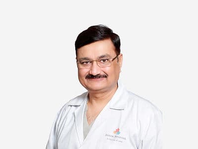
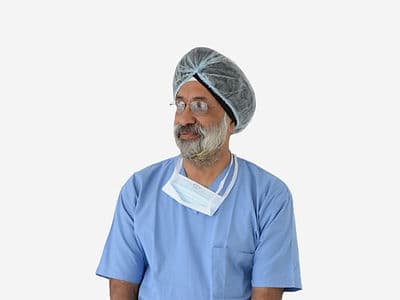
+91 83682 47758
info@medivisorhealth.com
Medivisor India Treatment Logo
  • Home
    • About Us
    • Our Services
    • Our Team
    • Our Safety Measures
    • Loading treatments...
    • Loading hospitals...
    • Loading doctors...
    • Treatment Cost
    • Treatment Process
    • Visa Process
    • Travel Guide
    • FAQs
    • Why Medivisor
    • Success Stories
    • Patient Activities
    • Social Activities
    • News Coverage
    • Blog
  • Become a Partner

Medivisor India Treatment is a trusted New Delhi–based medical travel partner connecting global patients to advanced, affordable, high-quality healthcare in India.

  • Medivisor House 359, Sector 1, Vaishali, Ghaziabad, (Delhi/NCR) India
  • +91 8368 247 758
  • info@medivisorhealth.com

About Us

  • About Us
  • Our Services
  • Our Team
  • Our Medical Advisors
  • Our Safety Measures
  • Join Our Network
  • Contact Us

India Treatment

  • Hospitals
  • Treatments
  • Treatment Cost
  • Treatment Process
  • Visa Process
  • Travel Guide
  • FAQs
  • Why Medivisor

Gallery

  • Patient Testimonials
  • Patient Activities
  • News Coverage
  • Blog

Get in Touch

© 2026 Medivisor India Treatment. All rights reserved.

Dr Amitabha Chanda

Director - Neurosurgery

36+ years experience

 Dr Amitabha Chanda
  1. Home
  2. Doctors
  3. Dr Amitabha Chanda

About Dr Amitabha Chanda

Dr. Amitabha Chanda is a distinguished neurosurgeon with over 36 years of experience in India. He boasts an impressive academic record, having excelled in various medical examinations and earned multiple gold medals during his MBBS at Medical College Kolkata. Dr. Chanda was the top candidate in both his M.S. and MCh programs.

Pioneer in Awake Brain Surgery

A pioneer in "Awake Brain Surgery" for brain tumors, he has successfully performed over 100 such procedures, earning extensive media coverage. His expertise is further enhanced by fellowship training in the U.S., Canada, and Europe. Dr. Chanda has authored numerous articles and contributed to international textbooks. He has received several accolades, including the Bengal Excellence Award in 2024 and the Healthcare Leader Award in 2023. His recent book, "Neurosurgeoner Diary," has become a bestseller, showcasing his unique experiences in neurosurgery.

Dr. Amitabha Chanda at a Glance

  • Distinguished neurosurgeon with over 36 years of extensive clinical experience.

  • Earned multiple gold medals during his MBBS and topped both his M.S. and MCh programs.

  • Pioneer in "Awake Brain Surgery" with over 100 successful procedures performed.

  • Completed advanced neurosurgical fellowship training across the U.S., Canada, and Europe.

  • Published author of numerous medical articles, international textbooks, and the bestseller "Neurosurgeoner Diary."

  • Recipient of the Healthcare Leader Award (2023) and the Bengal Excellence Award (2024).

MBBS at Medical College Kolkata
M.S. (Master of Surgery) exam in University of Calcutta.
MCh. in Neurosurgery at PGIMER, Chandigarh

No awards & achievements available

Affiliated Hospitals

CMRI Hospital | CK Birla Hospitals, Kolkata

Multi-Super Specialty

Kolkata, West Bengal

1969

Estd.

440+

Beds

210+

Doctors

May we help you?

Enter digits only (6-15 digits). We'll add your country code.

Related Treatments

Hydrocephalus Surgery (VP Shunt)

Ventriculoperitoneal (VP) Shunt Surgery Ventriculoperitoneal (VP) shunt surgery is a common neurosurgical procedure used to treat hydrocephalus, a condition where excess cerebrospinal fluid (CSF) builds up in the brain's ventricles. This surgery diverts the excess fluid to another part of the body—usually the abdomen (peritoneal cavity)—where it can be naturally reabsorbed into the bloodstream. By relieving the pressure on the brain, the shunt helps prevent neurological damage and alleviates symptoms like headaches, vision problems, and cognitive changes. When You Should Consider VP Shunt Surgery Congenital Hydrocephalus: For infants born with a blockage or structural defect that prevents CSF from draining naturally. Normal Pressure Hydrocephalus (NPH): Typically seen in older adults, where fluid buildup causes a classic triad of symptoms: difficulty walking, urinary incontinence, and memory loss. Acquired Hydrocephalus: Following a brain injury, tumor, or meningitis that has scarred the drainage pathways of the brain. Subarachnoid Hemorrhage: When a ruptured aneurysm leads to blood in the CSF spaces, blocking the natural reabsorption of fluid. Failed Endoscopic Third Ventriculostomy (ETV): When a previous non-shunt surgical attempt to create a drainage hole in the brain has closed or failed to lower pressure. Methods Of VP Shunt Surgery Fixed-Pressure Shunting: The traditional method using a valve set to a specific opening pressure that cannot be changed without surgery. Programmable Valve Shunting: A modern approach where the drainage pressure can be adjusted by a neurosurgeon using a specialized external magnet, avoiding the need for repeat operations. Lumboperitoneal (LP) Shunt: A variation where the fluid is drained from the lower spine (lumbar region) instead of the brain, often used for idiopathic intracranial hypertension. Ventriculoatrial (VA) Shunt: An alternative drainage site where the tubing is directed into the right atrium of the heart if the abdomen is not a suitable option. Anti-Siphon Devices: Specialized valve attachments designed to prevent "over-drainage" when a patient moves from lying down to standing up. How Is Performed Surgical Access: Under general anesthesia, small incisions are made in the scalp (usually behind the ear) and in the upper abdomen. Burr Hole Creation: The surgeon drills a small hole (approximately 14mm) in the skull to provide a safe entry point to the fluid-filled ventricles. Ventricular Placement: A thin, flexible tube (the ventricular catheter) is carefully guided into the brain’s ventricle to begin the drainage process. Subcutaneous Tunneling: The rest of the shunt tubing is passed under the skin, traveling from the head, down the neck and chest, to the abdominal incision. Valve and Reservoir Integration: A one-way valve and a small reservoir are placed under the scalp to regulate fluid flow and allow for future fluid sampling or "priming." Peritoneal Insertion: The distal end of the catheter is placed into the peritoneal cavity of the abdomen, where the lining is highly efficient at reabsorbing the diverted CSF. [Image showing the tunneling of a shunt catheter under the skin of the neck and chest] Pre-Procedure Preparation Neurological Mapping: High-resolution CT or MRI scans are mandatory to identify the exact cause of the hydrocephalus and plan the safest trajectory for the catheter. Medication Review: Patients must stop taking blood thinners (like Aspirin, Warfarin, or Ibuprofen) at least one week before surgery to minimize the risk of brain hemorrhage. Infection Prevention: Showering with a specialized antiseptic soap (CHG) the night before and the morning of surgery is often required to reduce skin bacteria. Physical Evaluation: A thorough medical history and physical exam to ensure the patient is a safe candidate for general anesthesia. Fasting (NPO): No food or drink for 8–12 hours prior to the procedure to prevent complications during the induction of anesthesia. Tests Before VP Shunt Surgery Brain MRI or CT: The primary tools used to measure the size of the ventricles and check for signs of high pressure or "transependymal flow." Lumbar Puncture (Spinal Tap): Occasionally performed to see if removing a small amount of fluid temporarily improves symptoms, predicting the success of a permanent shunt. Cine Phase-Contrast MRI: A specialized scan that looks at the actual flow of CSF to determine if there is a physical blockage (obstructive hydrocephalus). Baseline Cognitive Testing: Especially in NPH cases, to provide a benchmark for measuring improvement in memory and gait after the surgery. Life After VP Shunt Surgery Hospital Monitoring: Most patients stay 1 to 3 days; some may be required to lie flat for the first 24 hours to allow the brain to adjust to the new pressure. Pain Management: Headaches and tenderness at the incision sites are common and are managed with oral or IV pain medications. Incision Care: Staples or sutures are typically removed 7–14 days after surgery; the incisions must be kept clean and dry until they are fully healed. Activity Restrictions: While light walking is encouraged, patients must avoid heavy lifting, bending over, or strenuous exercise for 4 to 6 weeks. Long-Term Awareness: Patients must be alert for signs of "shunt failure" (headache, vomiting, or sleepiness) and carry a device identification card for future MRIs. Benefits Of VP Shunt Surgery Immediate Pressure Relief: Effectively reduces intracranial pressure, often providing rapid relief from severe headaches and vision changes. Restores Neurological Function: In many patients, particularly those with NPH, shunting can dramatically improve walking ability and cognitive clarity. Prevents Permanent Damage: By controlling fluid levels, the surgery prevents the brain tissue from being compressed and permanently damaged. Adjustable Technology: Programmable valves allow doctors to "fine-tune" the drainage to the patient's specific needs without additional surgery. Proven Long-Term Solution: Shunting has been the primary treatment for hydrocephalus for decades, with a high success rate in managing the condition over a lifetime.

Congenital Brain Malformations

Congenital Brain Malformations Congenital brain malformations are structural abnormalities present at birth, typically occurring during the early stages of fetal development. These anomalies can be caused by genetic mutations, environmental factors (such as maternal infections or folic acid deficiency), or specific disruptions during pregnancy. Because the brain is the control center for the body, these malformations can range from mild structural variations to severe conditions that impact motor function, cognition, and overall survival. Common Types Of Congenital Brain Malformations Neural Tube Defects (NTDs): Occur when the precursor to the brain and spine fails to close properly; includes Anencephaly (absence of major brain portions) and Encephalocele (sac-like protrusions through the skull). Chiari Malformations: Structural defects in the base of the skull where brain tissue (cerebellar tonsils) is pushed down into the spinal canal, often obstructing fluid flow. Dandy-Walker Malformation: A defect affecting the cerebellum and the fluid-filled spaces around it, typically characterized by an enlarged fourth ventricle and high intracranial pressure. Migration Disorders: Conditions like Lissencephaly ("smooth brain") or Heterotopia, where nerve cells fail to move to their proper positions during gestation, often leading to epilepsy. Holoprosencephaly: A failure of the forebrain to divide into two distinct hemispheres, which can cause significant facial and brain structure abnormalities. Agenesis of the Corpus Callosum (ACC): The partial or complete absence of the "bridge" of nerve fibers that connects the left and right sides of the brain. Associated Conditions & Symptoms Hydrocephalus: A very common complication where cerebrospinal fluid (CSF) builds up within the brain's ventricles, causing increased pressure and head swelling. Seizures and Epilepsy: Often seen in migration disorders (like Schizencephaly or Heterotopia) where abnormal brain "wiring" triggers electrical storms. Developmental Delays: Delays in reaching milestones such as sitting up, walking, or speaking due to the structural impact on the motor and cognitive cortex. Microcephaly or Macrocephaly: Abnormalities in head size (either significantly smaller or larger than average) reflecting the underlying brain development. Motor Impairment: Difficulty with coordination, muscle tone (spasticity), or balance, particularly in malformations involving the cerebellum. Surgical Treatments VP Shunt Surgery: The most common treatment for associated hydrocephalus, involving a tube that drains excess fluid from the brain to the abdomen. Chiari Decompression: A procedure where a small piece of bone is removed from the back of the skull (posterior fossa) to create more room for the brain and restore fluid circulation. Encephalocele Repair: A delicate surgery to place protruding brain tissue back into the skull and surgically close the opening in the bone. Endoscopic Third Ventriculostomy (ETV): A minimally invasive alternative to a shunt where a tiny hole is made in the floor of a brain ventricle to bypass a blockage. Spina Bifida Closure: Closing the opening in the spine to protect exposed nerves; in some advanced centers, this is performed in utero (before the baby is born). Non-Surgical & Supportive Care Anti-Seizure Medications: Critical for managing epilepsy and preventing further neurological injury from frequent seizures. Physical and Occupational Therapy: Long-term therapies designed to improve muscle strength, coordination, and the ability to perform daily tasks. Speech-Language Pathology: To assist children with communication difficulties or swallowing issues that may arise from brainstem or cortical defects. Early Intervention Programs: Specialized educational and developmental support provided during the first years of life to maximize cognitive potential. Tests For Diagnosing Brain Malformations Fetal Ultrasound: Often the first tool used to detect structural issues like ventriculomegaly or neural tube defects during routine pregnancy screenings. Fetal MRI: Provides high-resolution images of the developing fetal brain when an ultrasound suggests an abnormality, allowing for precise surgical planning. Postnatal MRI or CT: The gold standard for confirming a diagnosis after birth and monitoring the brain for changes in fluid pressure or growth. Genetic Testing (Microarray/Exome): Used to identify specific chromosomal or gene mutations that may have caused the malformation, which is helpful for family planning. Life Outlook and Long-Term Management Multidisciplinary Care: Patients typically require a team including neurosurgeons, neurologists, pediatricians, and various therapists. Lifelong Monitoring: Regular imaging is often needed to ensure that shunts remain functional and that no new issues, like syrinx (fluid in the spinal cord), develop. Varied Outcomes: The "pathway" for each child is unique; while some malformations require intensive support, others may result in near-normal cognitive and physical function with early intervention.

Spina Bifida Repair
Spina Bifida Repair

Spina Bifida Repair Spina bifida repair is a specialized surgical intervention used to treat myelomeningocele, the most severe form of spina bifida. In this condition, the spinal cord and its protective membranes (meninges) protrude through an opening in the spine, forming a sac on the infant's back. The primary goal of repair is to reposition the spinal cord, protect the nerves from further trauma, and prevent life-threatening infections like meningitis. Modern medicine offers two main pathways: prenatal (fetal) surgery, performed while the baby is still in the womb, and postnatal surgery, performed shortly after birth. When You Should Consider Spina Bifida Repair Myelomeningocele Diagnosis: When prenatal ultrasound or MRI confirms the spinal cord and nerves are exposed or protruding through a vertebral defect. Meningocele: A less severe form where only the protective membranes protrude, requiring surgical closure to prevent rupture and infection. Chiari II Malformation: Often associated with spina bifida, where the hindbrain is pulled into the spinal canal; early repair can sometimes reverse or improve this displacement. Hydrocephalus Risk: When fluid buildup in the brain is detected, early spinal repair is critical to stabilize intracranial pressure. Amniotic Fluid Exposure: For fetal candidates, repair is considered to stop the caustic amniotic fluid from further damaging the delicate, exposed spinal nerves. Methods Of Spina Bifida Repair Open Fetal Surgery: A major procedure where the mother's uterus is opened (hysterotomy) to repair the baby's spine between 19 and 26 weeks of gestation. Fetoscopic (Minimally Invasive) Repair: Utilizing tiny ports and a camera to repair the defect in utero, which reduces the risk of uterine scarring for the mother. Traditional Postnatal Repair: The standard approach where the infant undergoes surgery within the first 24 to 72 hours after birth. Fasciocutaneous Flap Closure: A specialized plastic surgery technique (such as a Limberg or V-Y flap) used to close very large spinal defects by rotating nearby skin and muscle. VP Shunt Integration: Often performed alongside or shortly after the spinal repair if the child has significant hydrocephalus. How Is Performed Surgical Exposure: Whether in the womb or after birth, the surgeon carefully cleans the exposed neural placode (the flat plate of nerve tissue). Neural Repositioning: A neurosurgeon gently detaches the spinal cord from the surrounding skin and places it back into the protective spinal canal. Multilayered Closure: The surgeon creates a watertight seal by closing the dura (the cord's lining), followed by the muscle layers, and finally the skin. Tension-Free Suturing: To ensure the wound heals properly, the skin is closed without tension; in large defects, this may require complex "flaps" of skin from the sides of the back. Watertight Integrity: The repair must be perfectly sealed to prevent cerebrospinal fluid (CSF) from leaking out, which is the primary defense against infection. Maternal Stabilization (Fetal Only): In prenatal cases, the uterus is closed and the mother is monitored closely to prevent preterm labor for the remainder of the pregnancy. Pre-Procedure Preparation High-Resolution Fetal MRI: Essential for mapping the level of the spinal lesion and checking for associated brain malformations like Chiari II. Genetic Counseling: To review the diagnosis and discuss the risks and benefits of fetal versus postnatal intervention. Maternal Health Screen: For fetal surgery, the mother must undergo extensive testing to ensure she can safely tolerate the procedure and prolonged bed rest. Steroid Administration: Often given to the mother before fetal surgery to help the baby's lungs mature in case of early delivery. Fasting (NPO): Standard fasting protocols for the mother (prenatal) or newborn (postnatal) to ensure safety under general anesthesia. Tests Before Spina Bifida Repair Level II Anatomy Ultrasound: To determine the exact "motor level" of the defect, which helps predict future walking ability. Fetal Echocardiogram: To ensure there are no additional heart defects before undergoing a long, complex surgery. Amniocentesis: Often performed to rule out other chromosomal abnormalities that might change the surgical plan. CSF Flow Study: To assess if the fluid in the brain and spine is circulating correctly or if a blockage is already present. Life After Spina Bifida Repair NICU/ICU Stay: Infants are monitored closely for signs of infection, CSF leaks, or "tethering" of the spinal cord as they grow. Wound Care: The surgical site must be kept clean and dry. Specialized barrier or repair creams (like Bioderma Cicabio or Mixsoon Bifida) may be used on healed skin to support the barrier. Hydrocephalus Monitoring: Many children will require a VP shunt or an ETV procedure if head circumference begins to grow too quickly. Physical Therapy: Started early to maximize mobility, strengthen the legs, and manage muscle tone. Urological and Bowel Management: Most children will require lifelong follow-up to manage bladder and bowel function, as the nerves to these organs are often affected. Benefits Of Spina Bifida Repair Prevents Meningitis: Closing the defect provides an immediate barrier against bacteria entering the central nervous system. Improves Mobility: Prenatal repair, in particular, has been shown to double the chances of a child being able to walk independently. Reduces Shunt Dependency: Early repair can significantly decrease the need for a permanent brain shunt to manage hydrocephalus. Reverses Brain Slumping: Fetal surgery can often "pull" the hindbrain back up into the skull, correcting the Chiari II malformation before birth. Protects Nerve Function: By stopping amniotic fluid exposure or trauma during delivery, the surgery preserves as much muscle control as possible below the level of the defect.

Craniosynostosis Surgery

Craniosynostosis Surgery Surgery is the primary treatment for craniosynostosis, a condition where the sutures (fiber-like joints) between the bones of an infant's skull close prematurely. The procedure is designed to release these fused sutures, relieve pressure on the developing brain, and reshape the skull to allow for normal, symmetrical growth. The specific surgical approach is typically determined by the child’s age, which sutures are involved, and the overall severity of the skull deformity. When You Should Consider Craniosynostosis Surgery Sagittal Synostosis: The most common form, causing a long, narrow head shape (scaphocephaly). Surgery is needed to widen the skull. Coronal Synostosis: Fusion of the suture running from ear to ear, which can cause a flattened forehead and a shifted eye socket on one or both sides. Metopic Synostosis: Results in a triangular-shaped forehead (trigonocephaly) and eyes that appear too close together. Lambdoid Synostosis: A rare form causing flattening at the back of the head, requiring surgery to distinguish it from positional flattening. Increased Intracranial Pressure (ICP): When the fused skull prevents the brain from expanding, leading to headaches, developmental delays, or vision changes. Syndromic Craniosynostosis: Complex cases associated with genetic conditions (like Apert or Crouzon syndromes) where multiple sutures are fused. Methods Of Craniosynostosis Surgery Endoscopic Strip Craniectomy: A minimally invasive method for infants under 6 months, using small incisions and a camera to remove the fused bone strip. Cranial Vault Remodeling (CVR): The traditional "open" surgery where the skull bones are removed, reshaped, and repositioned to create an immediate correction. Fronto-Orbital Advancement (FOA): A specialized open procedure focused on reshaping the forehead and the upper rim of the eye sockets. Spring-Mediated Cranioplasty: Following an endoscopic release, stainless steel springs are inserted to gradually push the bone segments apart as the brain grows. Cranial Distraction Osteogenesis: Using internal metal "distractor" devices that are turned daily to slowly expand the skull over several weeks. How Is Performed Surgical Access: Depending on the method, the surgeon makes either small "keyhole" incisions (endoscopic) or a larger zigzag incision from ear to ear (open) to hide the future scar within the hairline. Suture Release: The fused bone at the suture line is carefully cut or removed to "unlock" the skull. Bone Reshaping: In open surgery, the bones are removed and manually reshaped by the surgeon to create a more natural head contour. Hardware Fixation: Absorbable plates and screws—which dissolve naturally within 1–2 years—are used to hold the new skull shape in place. Expansion Device Placement: If using springs or distractors, these are tensioned or installed at the bone edges to allow for ongoing expansion. Scalp Closure: The skin is closed with dissolvable sutures; in some cases, a small drain may be left for 24 hours to prevent fluid buildup. Pre-Procedure Preparation 3D CT Scan: A specialized high-resolution scan is used to create a 3D reconstruction of the skull, allowing the surgeon to "map" the fusion exactly. Ophthalmology Exam: A baseline eye exam to check for swelling of the optic nerve (papilledema), which is a sign of high brain pressure. Hematology Consult: Because bone surgery can involve significant blood loss, a "type and cross-match" for blood is performed to have a transfusion ready if needed. Genetic Testing: To determine if the synostosis is part of a syndrome, which may influence the timing of future facial surgeries. Fasting (NPO): Infants must stop feeding several hours before the procedure to ensure safety under general anesthesia. Tests Before Craniosynostosis Surgery 3D Cranial Imaging: The gold standard for confirming which sutures are fused and assessing the volume of the intracranial space. Baseline Developmental Screen: To assess motor and cognitive milestones before the brain is "released." Complete Blood Count (CBC): To check hemoglobin levels, ensuring the child is strong enough for a procedure where blood loss is expected. EKG or Echo: Occasionally performed if a genetic syndrome is suspected that might also affect the heart. Life After Craniosynostosis Surgery Hospital Stay: Endoscopic patients typically stay 1 night, while open surgery patients stay 3 to 7 days, often including a night in the Pediatric ICU. Helmet Therapy: If the child had an endoscopic strip craniectomy, they must wear a custom-molded orthotic helmet for up to 23 hours a day for several months. Swelling Management: Significant facial and eyelid swelling is normal for 3–5 days after open surgery; the head is often kept elevated to help this resolve. Activity Restrictions: Most children return to normal play in 2–3 weeks, but contact sports or rough play must be avoided for at least 3 months while the bone heals. Long-Term Monitoring: Follow-up appointments occur every few months for the first year, then annually to ensure the skull continues to grow at the same rate as the brain. Benefits Of Craniosynostosis Surgery Protects Brain Development: Relieving pressure allows the brain to expand and develop without the risk of cognitive or motor delays. Restores Head Symmetry: Corrects the visible deformity, providing a more natural appearance and improving the alignment of the ears and eyes. Permanent Correction: In most non-syndromic cases, a single surgery provides a lifelong fix for the fused suture. Minimally Invasive Options: Early detection allows for endoscopic surgery, which features smaller scars and a much faster recovery time. Reduces Future Complications: Prevents the development of chronic headaches, vision loss, or social difficulties related to head shape later in life.

Brain Tumor Removal (Craniotomy)

Craniotomy for Brain Tumor A craniotomy is the primary surgical procedure used to remove a brain tumor. It involves carefully removing a section of the skull, known as a "bone flap," to provide the surgeon direct access to the brain. Once the tumor is addressed, the bone flap is typically replaced and secured with small titanium plates and screws. This procedure is the cornerstone of neurosurgical oncology, allowing for both the removal of the mass and the acquisition of tissue for a precise diagnosis. When You Should Consider a Craniotomy Primary Brain Tumors: For tumors that originate in the brain, such as gliomas or meningiomas, where removal can reduce pressure and slow progression. Metastatic Tumors: When cancer from another part of the body has spread to the brain and is causing neurological symptoms or is surgically accessible. Diagnostic Biopsy: When a tumor's type is unknown, a craniotomy allows for a larger tissue sample than a needle biopsy, leading to a more accurate treatment plan. Intracranial Pressure Relief: To alleviate the "mass effect" caused by a tumor that is compressing healthy brain tissue, which can cause severe headaches, nausea, or vision loss. Symptom Management: To stop or reduce seizures and focal neurological deficits (like weakness or speech issues) caused by the tumor’s location. How It Is Performed Mapping: Surgeons use Neuronavigation—a high-tech system similar to GPS for the brain—and pre-operative MRI scans to pinpoint the tumor's exact coordinates before making an incision. Anesthesia: The surgery is performed under general anesthesia and can take anywhere from 3 to 7 hours depending on the tumor's location and complexity. The Opening: A precise incision is made in the scalp, and a specialized surgical drill (craniotome) is used to remove a piece of the skull. Tumor Removal (Resection): Gross Total Resection: The surgeon removes the entire visible tumor. Subtotal Resection: If the tumor is too close to critical areas (eloquent brain) controlling speech or movement, only a portion is removed to preserve function. Advanced Tools: Surgeons may use an ultrasonic aspirator to break up the tumor or fluorescent dye (5-ALA), which makes tumor cells glow under a special light to help distinguish them from healthy tissue. Closing: After the tumor is removed, the bone flap is put back in its original position, and the scalp is closed with stitches or surgical staples. Specialized Types of Craniotomy Awake Craniotomy: The patient is woken up during the middle of surgery to perform tasks like talking or moving fingers. This allows the surgeon to map and avoid "eloquent" areas responsible for speech or motor skills in real-time. Endoscopic Craniotomy: A minimally invasive approach using a small hole and a camera (endoscope), often used for tumors located in the ventricles or deep within the brain. Keyhole Craniotomy: A smaller, more targeted opening (often behind the ear or above the eyebrow) used to access specific areas with minimal disruption to surrounding tissue. Pre-Procedure Preparation High-Resolution Imaging: Detailed MRI or CT scans with contrast to map the tumor’s size, vascularity, and relationship to functional brain zones. Steroid Protocol: You may be started on medications like dexamethasone a few days before surgery to reduce brain swelling (edema) caused by the tumor. Anti-Seizure Medication: Often prescribed preventatively to reduce the risk of a seizure during or after the procedure. Fasting: Adhering to "nothing by mouth" instructions for 8 hours prior to your scheduled anesthesia. Scalp Preparation: The surgical area may be washed with a specialized antiseptic, and a small amount of hair may be trimmed along the incision line. Tests Before a Craniotomy Functional MRI (fMRI): To identify specific areas of the brain used for speech, movement, and sensation relative to the tumor. Diffusion Tensor Imaging (DTI): A specialized MRI that maps the white matter "wiring" of the brain to help the surgeon avoid critical pathways. Blood Panels: A routine check of your blood count, electrolytes, and clotting factors to ensure a safe surgical experience. ECG: A standard heart check to confirm cardiovascular stability for a multi-hour procedure. Life After a Craniotomy Hospital Stay: Typically 3 to 7 days. You will likely spend the first night in the Neuro-ICU for intensive monitoring of your neurological status. Initial Symptoms: It is common to experience headaches, fatigue, and "brain fog." You may also notice temporary swelling around the eyes or scalp. Activity Restrictions: No heavy lifting or strenuous exercise for 6 to 8 weeks. Most patients can return to light desk work within 4–6 weeks. Stitch Removal: Scalp stitches or staples are typically removed by the surgical team 10–14 days after the procedure. Follow-up Treatment: Depending on the biopsy results (pathology), further treatments such as radiation or chemotherapy may begin a few weeks after the brain has had time to heal. Why Specialized Treatment Is Highly Effective Maximum Safe Resection: The combination of neuronavigation and intraoperative mapping allows surgeons to remove the largest amount of tumor possible while protecting your personality and physical abilities. Immediate Pressure Relief: Removing the tumor mass often leads to a rapid improvement in headaches and other symptoms caused by brain compression. Precision Technology: Tools like fluorescent dyes and ultrasonic aspirators allow for cleaner margins and less trauma to the surrounding healthy brain tissue. Multidisciplinary Expertise: Care is coordinated between neurosurgeons, neuro-oncologists, and rehabilitation specialists to provide a comprehensive path from surgery to recovery. Definitive Diagnosis: A craniotomy provides the highest quality tissue samples, ensuring that follow-up treatments (like targeted therapy) are based on the exact molecular profile of the tumor.

Glioma Surgery
Glioma Surgery

Glioma Surgery Glioma surgery is a specialized craniotomy performed to remove tumors that arise from the "gluey" supportive cells (glial cells) of the brain. Because gliomas often blend into healthy brain tissue rather than having a clear border, the surgical goal is Maximal Safe Resection—removing as much tumor as possible while preserving vital functions like speech, vision, and movement. When You Should Consider Glioma Surgery New Diagnosis: When imaging shows a suspected glioma (Grade I–IV) that requires both removal and a tissue sample for molecular diagnosis. Symptom Management: To reduce the "mass effect" that causes severe headaches, personality changes, or cognitive "brain fog." Seizure Control: If a glioma is irritating the brain's surface and causing frequent or uncontrolled seizures. Recurrent Glioma: When a previously treated tumor shows signs of regrowth on follow-up scans and requires further debulking. Increased Intracranial Pressure: To alleviate pressure that may be affecting your vision or causing nausea and vomiting. How Is Performed Neuronavigation: Surgeons use a 3D "GPS" system mapped from your pre-operative MRI to guide their instruments in real-time with sub-millimeter precision. Anesthesia: The surgery is typically performed under general anesthesia (unless an "awake" approach is required) and takes between 4 to 7 hours. Fluorescence-Guided Surgery (5-ALA/Glow): You may drink a specialized solution (Gliolan) before surgery that causes high-grade glioma cells to glow pink under a specific blue light, helping the surgeon distinguish the tumor from healthy brain tissue. Intraoperative Monitoring: Small electrodes track your brain’s electrical activity throughout the procedure to ensure motor and sensory pathways remain intact. The Resection: The surgeon uses an ultrasonic aspirator—a tool that uses high-frequency vibrations to break up the tumor while suctioning it away—to gently remove the mass. Pathology: Pieces of the tumor are sent immediately to a pathologist to confirm the tumor grade and identify specific molecular markers that guide future treatments. Specialized Approaches Awake Craniotomy: If the glioma is located near the "speech center" or motor strip, you may be woken up during surgery to talk or follow commands. This ensures the surgeon can remove the tumor without touching areas responsible for your communication. Intraoperative MRI (iMRI): Some advanced neurosurgical centers use an MRI scanner located directly inside the operating room to scan the brain during the surgery. This allows the surgeon to see if any hidden tumor remains before the final closing. Stereotactic Biopsy: In cases where a glioma is in a very deep or "inoperable" location, a tiny needle is used to take a sample through a small burr hole for diagnosis. Pre-Procedure Preparation Molecular Mapping: Advanced MRI sequences (like Spectroscopy or DTI) to understand the chemical makeup and wiring of the tumor. Steroid Protocol: You will likely be started on Dexamethasone several days before surgery to reduce brain swelling (edema) caused by the glioma. Anti-Seizure Medication: Most patients are prescribed preventative medication to stabilize the brain's electrical activity before the procedure. Fasting: Following "nothing by mouth" instructions for 8 hours prior to your scheduled anesthesia. Hair Preparation: A small strip of hair along the incision line may be trimmed, though many modern techniques allow for minimal hair removal. Tests Before Glioma Surgery Contrast-Enhanced MRI: The primary tool used to define the tumor's boundaries and its relationship to major blood vessels. Functional MRI (fMRI): To map exactly where your brain processes language and movement relative to the glioma. Blood Panels: A routine check of your blood count, electrolytes, and clotting factors to ensure a safe surgical experience. ECG: A standard heart check to confirm cardiovascular stability for a multi-hour neurosurgical procedure. Life After Glioma Surgery Hospital Stay: Typically 3 to 5 days, usually beginning with one night in the Neuro-ICU for intensive monitoring. Post-Op Steroids: Continued use of Dexamethasone for several days to manage temporary brain swelling, which can sometimes cause a brief worsening of symptoms. Medication Management: Most patients stay on anti-seizure medications for several weeks or months post-op to prevent "electrical storms" in the brain. Recovery Timeline: Stitches or staples are removed in 10–14 days. Most patients return to light activity within 4 weeks and can resume normal routines in 6 to 8 weeks. Next Steps in Care: Because gliomas can be infiltrative, surgery is often followed by Radiation and Chemotherapy (such as Temozolomide) starting 3–4 weeks after the brain has healed. Why Specialized Treatment Is Highly Effective Maximal Safe Resection: Utilizing real-time mapping and fluorescence allows for the removal of the maximum amount of tumor while protecting your quality of life. Molecularly Targeted Care: The tissue obtained during surgery allows oncologists to tailor your follow-up chemotherapy to the specific genetic profile of your tumor. Minimizes "Mass Effect": Removing the bulk of the glioma provides immediate relief from the pressure and headaches associated with brain tumors. Neuro-Protective Technology: Intraoperative monitoring and awake mapping ensure that the "high-rent" areas of your brain are avoided, preserving your ability to speak and move. Integrated Recovery: Care is managed by a multidisciplinary team of neurosurgeons, neuro-oncologists, and therapists to provide a seamless transition from surgery to long-term management.

Meningioma Surgery
Meningioma Surgery

Meningioma Surgery Meningioma surgery is a specialized craniotomy performed to remove a tumor that grows from the meninges—the protective membranes surrounding the brain and spinal cord. Because the vast majority of meningiomas are benign (Grade 1) and grow outside the brain tissue rather than infiltrating it, the primary surgical goal is usually Gross Total Resection. This involves the complete removal of the tumor and its attachment point to the dura mater to prevent the tumor from growing back. When You Should Consider Meningioma Surgery Symptomatic Growth: If the tumor is causing persistent headaches, seizures, or personality changes. Neurological Deficits: When the mass compresses critical structures, leading to weakness, numbness, or loss of coordination. Vision or Hearing Loss: For tumors located near the skull base that press against the optic or auditory nerves. Documented Growth: If follow-up MRIs show the tumor is enlarging, even if you currently have few symptoms. Mass Effect: To alleviate significant pressure on the brain tissue or shift of the brain's midline structures. How It Is Performed Image Guidance: Surgeons use Neuronavigation (a 3D GPS system mapped from your pre-operative MRI) to plan the exact entry point and trajectory, minimizing disruption to healthy tissue. Anesthesia: The surgery is performed under general anesthesia and typically lasts 3 to 6 hours, depending on the tumor's size and its proximity to major blood vessels or nerves. The Opening: A precise scalp incision is made, and a section of the skull (bone flap) is temporarily removed to provide direct access. Tumor Removal: Since meningiomas are often firm, the surgeon may use an ultrasonic aspirator to hollow out the center of the tumor first. The "shell" of the tumor is then carefully peeled away from the brain surface, sensitive nerves, and major blood vessels. Dural Repair: The piece of the meninges where the tumor was originally attached is removed to ensure no microscopic cells remain. The surgeon then patches this area with a synthetic graft or tissue from your own scalp (fascia). Closing: The bone flap is secured back in place with small titanium plates and screws, and the scalp is closed with stitches or surgical staples. Specialized Approaches Skull Base Surgery: For tumors at the very bottom of the brain (near the eyes or ears), specialized drilling techniques are used to reach the tumor without having to move or retract the brain significantly. Endoscopic Endonasal Surgery: For specific meningiomas near the optic nerves or pituitary gland, some can be removed entirely through the nose using a high-definition camera (endoscope), leaving no external scars. Keyhole Craniotomy: A minimally invasive approach using a much smaller opening, often hidden in the eyebrow or behind the hairline, for specifically located tumors. Pre-Procedure Preparation Contrast MRI: A high-resolution scan to map the tumor’s blood supply and its relationship to the surrounding venous sinuses. Steroid Protocol: You may be started on medications like dexamethasone a few days before surgery to reduce brain swelling (edema) caused by the tumor. Anti-Seizure Medication: Often prescribed preventatively to stabilize the brain's electrical activity before and after the procedure. Fasting: Following "nothing by mouth" instructions for 8 hours prior to your scheduled anesthesia. Physical Exam: A thorough check-up to ensure your heart and lungs are healthy enough for a multi-hour surgery. Tests Before Meningioma Surgery Visual Field Testing: If the tumor is near the optic nerves, a detailed eye exam is necessary to establish a baseline. Audiogram: For tumors near the hearing nerves (internal auditory canal) to document current hearing levels. Blood Panels: A routine check of your blood count, electrolytes, and clotting factors. ECG: A standard heart check to confirm cardiovascular stability for the duration of the procedure. Life After Meningioma Surgery Hospital Stay: Typically 3 to 5 days, including at least one night in the Neuro-ICU for intensive neurological monitoring. Initial Symptoms: Headaches and fatigue are common. You may also have temporary swelling or bruising around the eyes or forehead for about a week. Activity Restrictions: No heavy lifting, straining, or high-impact exercise for 6 weeks to allow the bone and scalp to heal properly. Return to Routine: Most patients can return to driving and light desk work within 4–8 weeks, depending on their recovery progress. Long-term Monitoring: Even with a complete removal, you will need periodic MRIs (initially every 6–12 months) to ensure there is no recurrence over the long term. Why Specialized Treatment Is Highly Effective Curative Potential: For most Grade 1 meningiomas, a successful surgical resection is considered a permanent cure. Preserves Brain Function: Because these tumors grow outside the brain, skilled surgeons can usually remove them with minimal impact on your cognitive or physical abilities. Advanced Tools: The use of ultrasonic aspirators and micro-dissection tools allows for the safe separation of the tumor from delicate nerves and arteries. Immediate Pressure Relief: Removing the mass provides instant relief from the "dragging" sensation and headaches associated with intracranial pressure. Minimal Scarring: Modern surgical planning allows for incisions that are often hidden within the hairline or natural skin creases.

Pituitary Tumor Surgery (Endoscopic)

Endoscopic Pituitary Surgery Endoscopic Pituitary Surgery, also known as Endoscopic Transsphenoidal Surgery, is a minimally invasive procedure that uses the nostrils as natural pathways to reach and remove tumors from the pituitary gland. Because it avoids large incisions and brain retraction, it typically offers a faster recovery and fewer side effects than traditional open surgery. This approach allows surgeons to access the "master gland" at the base of the brain with extreme precision. When You Should Consider Endoscopic Pituitary Surgery Hormone-Secreting Tumors: Such as those causing Cushing’s disease (excess cortisol), acromegaly (excess growth hormone), or prolactinomas. Non-Functioning Macroadenomas: Large tumors that do not produce hormones but press on the optic nerves, causing vision loss, double vision, or chronic headaches. Pituitary Apoplexy: An emergency condition where a tumor bleeds or outgrows its blood supply, requiring rapid decompression. Failed Medical Management: When medications are unable to sufficiently control hormone levels or stop the growth of the tumor. Rathke’s Cleft Cysts: Benign fluid-filled growths that can interfere with normal gland function or cause pressure symptoms. How It Is Performed Collaborative Team: The surgery is usually a joint effort between a neurosurgeon and an Ear, Nose, and Throat (ENT) surgeon, taking about 2 to 3 hours under general anesthesia. Nasal Access: The ENT surgeon inserts a thin, lighted tube with a high-definition camera (endoscope) through one nostril to navigate to the very back of the nasal cavity. Opening the Sphenoid Sinus: The surgeon opens the sphenoid sinus (an air-filled space behind the nose) to reach the sella turcica, the small bony compartment that houses the pituitary gland. Tumor Removal: Using specialized long instruments through the other nostril, the neurosurgeon removes the tumor in small pieces. The endoscope provides a panoramic, high-magnification view of the area, including nearby carotid arteries and optic nerves. Reconstruction: If needed, a small fat graft (often taken from the abdomen) or synthetic material is used to fill the space and seal the area to prevent cerebrospinal fluid (CSF) leaks. Pre-Procedure Preparation Endocrine Evaluation: Comprehensive blood and urine tests to establish your baseline hormone levels (growth hormone, ACTH, prolactin, etc.). High-Resolution MRI: A dedicated "pituitary protocol" scan to map the tumor’s exact size and its relationship to the optic chiasm. Ophthalmology Exam: A detailed visual field test to document any current vision loss before the surgery. Nasal Assessment: An ENT evaluation to ensure your nasal passages are clear and suitable for the endoscopic approach. Fasting: Following "nothing by mouth" instructions for 8 hours prior to your scheduled anesthesia. Tests Before Endoscopic Pituitary Surgery Visual Field Testing: To measure peripheral vision, which is often the first thing affected by pituitary tumors. Dynamic Hormone Testing: Specialized "stimulation" or "suppression" tests to confirm the type of secreting tumor. Carotid Imaging: Occasionally required if the tumor is very large and wrapping around the main arteries of the brain. ECG: A standard heart check to confirm cardiovascular stability for the duration of the procedure. Life After Endoscopic Pituitary Surgery Hospital Stay: Typically 1 to 3 days, often starting with one night in the Intensive Care Unit (ICU) for close monitoring of your fluid balance and hormone levels. Immediate Symptoms: It is normal to experience nasal congestion, mild headaches, and "watery" or blood-tinged nasal drainage for 1 to 2 weeks. The "No" Rules: For 4 to 6 weeks, you must strictly avoid: Blowing your nose: To prevent pressure buildup that could cause a CSF leak. Lifting and Straining: No lifting objects over 5 lbs or heavy straining, which increases intracranial pressure. Drinking through straws: The suction can interfere with the healing of the nasal repairs. Hormone Monitoring: You will work closely with an endocrinologist to check if your gland is producing the correct amount of hormones post-op. Follow-up MRI: A baseline scan is usually performed 3 months after surgery to ensure the entire tumor was removed. Why Specialized Treatment Is Highly Effective No External Scars: By using the natural pathway of the nose, there are no visible incisions on the face or scalp. Superior Visualization: The endoscope allows surgeons to "see around corners," identifying tumor tissue that might be missed with traditional microscopic surgery. Rapid Vision Improvement: Decompressing the optic nerves often leads to a quick and significant improvement in peripheral vision and clarity. Preserves Gland Function: The high-magnification view helps surgeons distinguish between the tumor and the healthy part of the pituitary gland. Reduced Brain Trauma: Because the brain is not "moved" or retracted to reach the tumor, post-operative headaches and recovery times are greatly reduced.

May we help you?

Enter digits only (6-15 digits). We'll add your country code.