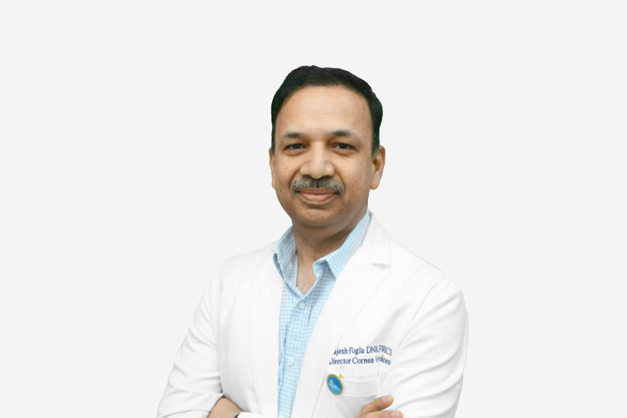
Trusted by 3,500+ International Patients Across 50+ Countries | Speak with a Dedicated Medical Coordinator 24/7 | Personalized Care Every Step of the Way
+91 83682 47758
info@medivisorhealth.com
Medivisor India Treatment Logo
  • Home
    • About Us
    • Our Services
    • Our Team
    • Our Safety Measures
    • Loading treatments...
    • Loading hospitals...
    • Loading doctors...
    • Treatment Cost
    • Treatment Process
    • Visa Process
    • Travel Guide
    • FAQs
    • Why Medivisor
    • Success Stories
    • Patient Activities
    • Social Activities
    • News Coverage
    • Blog
  • Become a Partner

Medivisor India Treatment is a trusted New Delhi–based medical travel partner connecting global patients to advanced, affordable, high-quality healthcare in India.

  • Medivisor House 359, Sector 1, Vaishali, Ghaziabad, (Delhi/NCR) India
  • +91 8368 247 758
  • info@medivisorhealth.com

About Us

  • About Us
  • Our Services
  • Our Team
  • Our Medical Advisors
  • Our Safety Measures
  • Join Our Network
  • Contact Us

India Treatment

  • Hospitals
  • Treatments
  • Treatment Cost
  • Treatment Process
  • Visa Process
  • Travel Guide
  • FAQs
  • Why Medivisor

Gallery

  • Patient Testimonials
  • Patient Activities
  • News Coverage
  • Blog

Get in Touch

© 2026 Medivisor India Treatment. All rights reserved.

Dr Rajesh Fogla

25+ years experience

 Dr Rajesh Fogla
  1. Home
  2. Doctors
  3. Dr Rajesh Fogla

About Dr Rajesh Fogla

Dr. Rajesh Fogla is a highly distinguished specialist in cornea, cataract, and refractive ophthalmology, recognized for his technical mastery in advanced microsurgical interventions, corneal transplantations, and vision correction procedures. He specializes in utilizing precision-guided diagnostic, imaging, and laser platforms to treat complex ocular surface diseases, restore visual acuity, and deliver comprehensive sight-saving treatment pathways.

Mastery in Advanced Cornea and Refractive Sciences

He specializes in the advanced evaluation, micro-mapping, and clinical management of a wide range of acute and chronic conditions affecting the anterior segment of the eye. His clinical practice leverages a quarter-century of refined ophthalmic acumen to perform high-precision therapeutic interventions, ensuring optimal corneal stabilization and long-term visual preservation for his patients.

Innovation in Microsurgical and Vision Correction Pathways

Dr. Fogla possesses profound expertise in implementing state-of-the-art microsurgical techniques and targeted laser therapies within his operative framework. By addressing intricate structural disorders—including corneal dystrophies, advanced cataracts, and complex refractive errors—he executes detail-driven interventions that protect delicate ocular tissues, minimize surgical trauma, and facilitate optimal recovery outcomes.

Elite International Fellowships and Global Acclaim

A primary focus of his distinguished career involves the integration of global standards into localized patient care, built upon a foundation of elite medical training from premier institutions worldwide. His comprehensive credentials include prestigious fellowships from the Royal College of Surgeons of Edinburgh, the Royal College of Ophthalmologists of London, and the American College of Surgeons, complemented by high-profile international gold medals and achievement awards from the AAO and APAO.

Clinical Governance, Multi-Lingual Communication, and Academic Leadership

Throughout his extensive career spanning 25 years, Dr. Fogla has combined modern ophthalmic innovations with rigorous clinical protocols and a deep commitment to medical education. Practicing within premier healthcare networks in Hyderabad, Telangana, his exceptional multi-lingual proficiency in English, Hindi, and Telugu supports a highly diverse patient base, while his 17-year tenure conducting international fellowship courses underscores his role as a trusted authority training the next generation of eye specialists.

Dr. Rajesh Fogla at a Glance

  • Specialist in Cornea, Advanced Cataract Surgery, and Refractive Vision Correction.

  • Extensive clinical experience with 25 years of dedicated service in the medical and surgical field.

  • Elite global foundation holding advanced fellowships from Edinburgh, London, the USA, and Singapore.

  • Honored with prestigious international achievement awards from the AAO, APAO, and global medical bodies.

  • Actively involved for over 17 years in directing advanced instructional training and corneal fellowship courses.

  • Highly proficient in multiple languages, including English, Hindi, and Telugu, to support a diverse patient base.

DNB
FRCS (Edin)
FRCO (London)
FACS (USA)
MMed (Singapore)

No awards & achievements available

Affiliated Hospitals

Apollo Health City, Jubilee Hills

Multi Specialty

Hyderabad, Telangana

1988

Estd.

550+

Beds

200+

Doctors

May we help you?

Enter digits only (6-15 digits). We'll add your country code.

Related Treatments

Corneal Transplant (Keratoplasty)

Corneal Transplant Surgery (Keratoplasty) Corneal transplant surgery, or keratoplasty, is a specialized procedure to replace damaged or diseased corneal tissue with healthy tissue from a human donor. Modern surgical techniques allow for "component surgery," where surgeons replace only the specific damaged layers of the eye, significantly improving success rates and recovery times. When You Should Consider Corneal Transplant Surgery Keratoconus: When progressive thinning causes the cornea to bulge into a cone shape and specialty lenses no longer restore vision. Fuchs’ Dystrophy: If you experience persistent swelling or cloudiness due to the deterioration of the innermost cell layers. Corneal Scarring: To restore vision lost to physical trauma, chemical burns, or severe infections like herpes keratitis. Graft Failure: When a previous donor transplant has been rejected by the body or has stopped functioning. Advanced Thinning: When medical treatments like cross-linking can no longer provide structural integrity or relieve chronic eye pain. How Is Performed Anesthesia: Typically performed as an outpatient procedure under either local or general anesthesia, depending on the complexity. Tissue Removal: The surgeon uses high-precision tools to carefully remove the diseased or damaged layers of the patient's cornea. Methods of Transplant: Penetrating Keratoplasty (PKP): A full-thickness transplant where the entire central cornea is replaced and secured with microscopic sutures. Deep Anterior Lamellar Keratoplasty (DALK): Replaces the front and middle layers while preserving the patient's own healthy inner lining. Endothelial Keratoplasty (DMEK/DSAEK): Minimally invasive procedures to replace only the innermost layer (endothelium). Securing the Graft: In full-thickness surgeries, fine sutures are used. In endothelial transplants, a specialized Air Bubble Tamponade is used to hold the new tissue in place against the eye. Duration: The surgical procedure typically lasts between 1 to 2 hours. Pre-Procedure Preparation Comprehensive Eye Examination: To determine exactly which layers of the cornea need replacement (full-thickness vs. lamellar). Graft Matching: Sourcing high-quality donor tissue from an eye bank and reviewing success rates, which often exceed 90%. Recovery Planning: Preparing for post-operative requirements, such as strict face-up positioning for several days. Medication Review: Discussing the necessity of long-term or life-long steroid eye drops to inhibit immune rejection. Anesthesia Assessment: Evaluating overall health to determine the safest sedation method for the patient. Tests Before Corneal Transplant Surgery Pachymetry: To measure corneal thickness and identify specific areas of extreme thinning or fluid buildup (edema). Specular Microscopy: A detailed evaluation of the health and density of the endothelial cell layer. Corneal Topography: Creating a 3D map of the curvature and surface irregularities of the diseased cornea. Visual Acuity Test: Establishing a clear baseline of current vision impairment to measure post-surgical improvement. Life After Corneal Transplant Surgery Positioning Requirements: For DMEK or DSAEK, patients must maintain a face-up position for several days to allow the air bubble to support the graft. Steroid Regimen: A committed long-term schedule of steroid eye drops is required to prevent the immune system from attacking the new tissue. Rejection Monitoring: Patients must immediately report "RSVP" symptoms: Redness, Sensitivity to light, Vision blurriness, or Pain. Recovery Timeline: Healing varies by procedure—weeks for endothelial transplants (DMEK) and 6 to 12 months for full-thickness (PKP) procedures. Follow-up Care: Regular appointments are necessary to manage the sutures and monitor for potential risks like glaucoma or astigmatism. Why Specialized Treatment Is Highly Effective Restores Transparency: Effectively clears vision by replacing opaque or scarred tissue with crystal-clear donor tissue. Reduced Rejection Risk: Modern "component" surgery (DALK/DMEK) leaves more of the patient's own tissue intact, lowering the chance of an immune response. Last-Resort Solutions: For patients who cannot receive human donor tissue, an Artificial Cornea (Keratoprosthesis) offers a high-tech synthetic alternative. Eliminates Chronic Pain: Resolves the significant discomfort associated with corneal surface diseases and recurring erosions. Proven Success: Keratoplasty remains the most common and successful type of human organ transplant worldwide.

Cross-linking (for Keratoconus)

Corneal Collagen Cross-linking (CXL) Corneal Collagen Cross-linking (CXL) is a specialized medical procedure used to treat Keratoconus, a progressive condition where the cornea thins and bulges into a cone shape. Unlike LASIK or SMILE, which are designed to correct vision, the primary goal of CXL is to strengthen the corneal tissue to stop the disease from worsening and prevent future vision loss. When You Should Consider CXL Progressive Keratoconus: When repeat eye exams show that the cornea is continuing to thin or the "cone" shape is becoming more pronounced. Post-LASIK Ectasia: A rare complication where the cornea becomes unstable and thins following refractive surgery. Pellucid Marginal Degeneration: A similar corneal thinning condition that affects the lower part of the cornea. Fluctuating Vision: When your eyeglass or contact lens prescription is changing rapidly due to corneal instability. Early Diagnosis: It is highly effective when performed early to stabilize the cornea before significant vision loss occurs. How Is Performed Numbing: Anesthetic eye drops are applied so the patient remains comfortable and feels no pain during the process. Epithelium Management: Epi-off Method: The thin outer layer (epithelium) is gently removed to allow the medication to saturate the deeper layers more effectively. Epi-on (Trans-epithelial): The outer layer is left intact, which may reduce post-operative discomfort. Riboflavin Saturation: Vitamin B2 (riboflavin) drops are applied to the eye every few minutes for about 30 minutes until the cornea is fully saturated. UV Light Exposure: The eye is exposed to a controlled amount of Ultraviolet A (UVA) light for several minutes. The Chemical Reaction: The interaction between the Riboflavin and UV light creates new "cross-links" (chemical bonds) between the collagen fibers, making the cornea stiffer and more stable. Bandage Lens: A clear, soft contact lens is placed on the eye to protect the surface while it heals. Duration: The entire treatment typically takes 30 to 60 minutes. Pre-Procedure Preparation Corneal Mapping: Detailed scans (topography) are taken to document the current shape and thickness of the cornea as a baseline. Contact Lens Holiday: Patients must stop wearing hard or gas-permeable contact lenses for several weeks before the procedure to allow the cornea to settle. Medication Review: Discussing any history of slow healing or eye infections with your specialist. Transportation: Arranging for a ride home, as the eye may be sensitive to light and vision may be blurry immediately after the UV exposure. Tests Before CXL Pachymetry: To measure the thinnest point of the cornea; a minimum thickness is often required to safely perform the UV light stage. Corneal Topography/Tomography: To identify the "apex" of the cone and track the rate of disease progression. Slit-Lamp Examination: To check for any pre-existing corneal scarring that might affect the treatment outcome. Visual Acuity: Establishing the current level of corrected and uncorrected vision. Life After CXL Initial Discomfort: Especially in "Epi-off" cases, the eye may feel sore, gritty, or highly light-sensitive for the first 3–5 days. Vision Fluctuations: It is normal for vision to be slightly "foggy" or worse immediately after the procedure before it stabilizes over several weeks. Medication Regimen: Patients must use prescribed antibiotic and steroid drops for several weeks to prevent infection and manage inflammation. Healing Timeline: While the surface heals within a week, the full strengthening effect of the cross-linking takes 3 to 6 months to reach maximum stability. Follow-up Care: Regular checkups are essential to monitor the "haze" (temporary cloudiness) and ensure the epithelium has regrown properly. Why Specialized Treatment Is Highly Effective Prevents Disease Progression: CXL has a success rate of over 90% in stopping Keratoconus from getting worse. Avoids Major Surgery: By stabilizing the cornea early, the vast majority of patients can avoid the need for a full corneal transplant later in life. Long-Term Stability: The new chemical bonds created between collagen fibers provide a permanent increase in corneal rigidity. Preserves Vision: While it does not eliminate the need for glasses, it "locks" the vision in place and prevents further deterioration. Minimally Invasive: It is an outpatient procedure that does not require stitches or a long hospital stay.

Corneal Collagen Cross-linking

Corneal Collagen Cross-linking (CXL) Corneal Collagen Cross-linking (CXL) is a specialized medical treatment designed to stop the progression of Keratoconus and other corneal thinning disorders. Unlike most laser eye surgeries, its goal is not to improve vision, but to strengthen and stabilize the cornea's structure. By creating new chemical bonds between corneal fibers, CXL acts as a support system to prevent the eye from bulging further. When You Should Consider CXL Progressive Keratoconus: When regular eye exams show that the cornea is continuing to thin or the "cone" shape is becoming more pronounced. Post-LASIK Ectasia: A rare condition where the cornea becomes unstable and begins to thin following previous refractive surgery. Pellucid Marginal Degeneration: A specific type of corneal thinning that typically affects the lower portion of the cornea. Rapidly Changing Vision: When your eyeglass or contact lens prescription is shifting frequently due to structural changes in the eye. Early Diagnosis: It is most effective when performed early to "lock" the cornea in place before significant vision loss occurs. How Is Performed Numbing: Anesthetic eye drops are applied so the patient remains comfortable and feels no pain throughout the process. Epithelium Management: Epi-off Method: The thin outer layer (epithelium) is gently removed to allow the riboflavin medicine to penetrate the deeper layers more effectively. Epi-on (Trans-epithelial): The outer layer is left intact, which may lead to less post-operative discomfort but sometimes slower saturation. Riboflavin Loading: Vitamin B2 (riboflavin) drops are applied every few minutes for about 30 minutes until the cornea is fully saturated. UVA Activation: The eye is exposed to a precise, controlled beam of Ultraviolet-A light. The Cross-linking: The UV light reacts with the riboflavin to create new "cross-links" (chemical bonds) between the collagen fibers, making the cornea stiffer. Protection: A clear, soft bandage contact lens is placed on the eye to protect the surface while the epithelium regrows. Duration: The entire treatment typically takes between 30 and 60 minutes. Pre-Procedure Preparation Baseline Mapping: Detailed 3D scans (topography) are taken to document the current shape and thickness of the cornea. Contact Lens Holiday: Patients must stop wearing hard or gas-permeable contact lenses for several weeks before the procedure to ensure the cornea is in its natural state. Medication Audit: Reviewing any history of slow healing, dry eyes, or chronic eye infections with the surgical team. Transportation Planning: Arranging for a ride home, as the eye will be sensitive to light and vision may be blurry immediately following the UV exposure. Tests Before CXL Pachymetry: To measure the thickness of the cornea at its thinnest point; a safe minimum thickness is required for the UV light phase. Corneal Topography/Tomography: To identify the "apex" of the cone and track the exact rate of disease progression over time. Slit-Lamp Examination: A microscopic check for any pre-existing corneal scarring or inflammation that might impact the outcome. Visual Acuity Test: Establishing the current level of corrected vision to monitor stability after the procedure. Life After CXL Initial Discomfort: Especially in "Epi-off" cases, the eye may feel sore, gritty, or highly light-sensitive for the first 3–5 days. Vision Fluctuations: It is normal for vision to be "hazy" or slightly worse than before surgery for a few weeks while the surface heals. Medication Regimen: Patients must strictly follow the schedule for prescribed antibiotic and steroid drops for several weeks. Stabilization Timeline: While the surface heals within a week, the full strengthening effect and final vision stabilization can take 3 to 6 months. Follow-up Care: Regular checkups are essential to monitor "corneal haze" (temporary cloudiness) and confirm the epithelium has fully regrown. Why Specialized Treatment Is Highly Effective Stops Disease Progression: CXL is highly successful at preventing the cornea from bulging further and worsening the condition. Prevents Major Surgery: By stabilizing the cornea early, most patients can avoid the need for a full corneal transplant later in life. Permanent Strengthening: The new chemical bonds provide a long-term increase in corneal rigidity and structural integrity. Preserves Vision Quality: While it may not eliminate the need for glasses, it "locks" the vision in place and prevents further loss of sight. Minimally Invasive: It is an outpatient procedure that requires no incisions into the inner eye and no stitches.

LASIK / Femto-LASIK
LASIK / Femto-LASIK

LASIK & Femto-LASIK LASIK and Femto-LASIK are refractive surgeries designed to correct vision problems like nearsightedness (myopia), farsightedness (hyperopia), and astigmatism by reshaping the cornea. While both follow the same basic restorative principles, the primary difference lies in how the initial corneal flap is created, with Femto-LASIK offering a completely "bladeless" experience. When You Should Consider LASIK Nearsightedness (Myopia): When nearby objects are clear, but distant objects appear blurry. Farsightedness (Hyperopia): When you can see distant objects clearly, but close-up tasks cause eye strain or blur. Astigmatism: When the cornea is shaped more like a football than a basketball, causing overall distorted vision. Dependence on Corrective Lenses: When you wish to eliminate or significantly reduce the need for glasses or contact lenses for daily activities. Stable Vision: When your eye prescription has remained unchanged for at least one to two years. How Is Performed Step 1: Numbing: Anesthetic eye drops are applied so you remain awake and comfortable throughout the procedure. Step 2: Flap Creation: A thin, hinged flap is created on the outer layer of the cornea and folded back to access the underlying tissue. Traditional LASIK: Uses a mechanical handheld blade called a microkeratome. Femto-LASIK: Uses a high-precision femtosecond laser to create the flap, making it "bladeless." Step 3: Reshaping: An excimer laser is used to remove microscopic amounts of tissue from the inner cornea to improve how light focuses on the retina. Step 4: Repositioning: The corneal flap is folded back into its original position, where it bonds naturally and begins healing immediately without the need for stitches. Duration: The entire outpatient process typically takes about 10 to 20 minutes for both eyes. Pre-Procedure Preparation Contact Lens Holiday: You must stop wearing contact lenses (usually 1–3 weeks before) to allow the cornea to return to its natural shape. Corneal Mapping: Detailed 3D scans are taken of the eye's surface to program the laser for your specific correction needs. Medication Audit: Discussing any history of dry eyes or autoimmune conditions with your surgeon that might affect healing. Transportation: Arranging for a ride home after the procedure, as your vision will be foggy and you may experience light sensitivity. Tests Before LASIK Corneal Topography: A specialized map that measures the curvature and thickness of the cornea to ensure it is healthy enough for surgery. Pachymetry: Measuring corneal thickness; Femto-LASIK is often preferred for patients with thinner corneas. Dry Eye Assessment: Checking tear production to determine if post-operative dryness needs to be managed proactively. Wavefront Analysis: A highly detailed "fingerprint" of your vision that detects subtle imperfections to guide a customized laser treatment. Life After LASIK Initial Vision: Most patients see clearly within 24 to 48 hours, though vision may fluctuate slightly for the first few days. Post-Op Symptoms: Temporary grittiness, dryness, or light sensitivity is normal immediately following the surgery. Prescribed Drops: You must strictly follow the schedule for antibiotic and anti-inflammatory eye drops to prevent infection. Eye Protection: Wearing protective sunglasses or shields is recommended, especially while sleeping, to prevent accidental rubbing. Activity Restrictions: For the first week, avoid wearing eye makeup, swimming, or participating in contact sports. Why Specialized Treatment Is Highly Effective Higher Precision: Femto-LASIK allows for customizable flap dimensions, ensuring a safer and more predictable surgical outcome. Expanded Eligibility: Bladeless technology makes refractive surgery an option for patients who were previously told their corneas were too thin for a blade. Reduced Complications: The use of a computer-guided laser minimizes risks such as irregular flap creation or flap dislocation. Rapid Results: Most patients return to work and light daily activities within 24 hours of the procedure. Permanent Vision Correction: By physically reshaping the cornea, the procedure provides a long-term solution for refractive errors.

SMILE Surgery (Small Incision Lenticule Extraction)

SMILE (Small Incision Lenticule Extraction) SMILE (Small Incision Lenticule Extraction) is the latest advancement in laser vision correction. It is a flapless, minimally invasive procedure used to treat nearsightedness (myopia) and astigmatism. Unlike LASIK, which requires creating a large circular flap, SMILE utilizes a single high-precision laser to perform the entire correction through a microscopic opening. When You Should Consider SMILE Active Lifestyles: Ideal for individuals involved in contact sports, professional athletics, or high-intensity professions where a corneal flap displacement risk must be zero. Dry Eye Concerns: For patients prone to dry eyes, as the procedure preserves more corneal nerves than traditional refractive surgeries. Nearsightedness (Myopia): Specifically designed for patients with high degrees of myopia who have adequate corneal thickness. Astigmatism: Effective for correcting blurred vision caused by an irregularly shaped cornea. Structural Preference: For those who prefer a procedure that maintains the maximum structural integrity of the upper corneal layers. How Is Performed Numbing: Anesthetic drops are applied to the eye so the patient remains awake and comfortable, feeling no pain during the process. Lenticule Creation: Using the VisuMax femtosecond laser, the surgeon creates a thin, contact-lens-shaped layer of tissue (a lenticule) inside the intact cornea. Small Incision: The same laser creates a microscopic incision—typically less than 4 mm—on the corneal surface. Extraction: The surgeon manually removes the lenticule through this tiny incision, which reshapes the cornea and allows light to focus correctly on the retina. Quiet & Odourless: The laser operates silently, and unlike other methods, there is no "burning" smell during the correction. Duration: The entire process usually takes about 10–15 minutes for both eyes. Pre-Procedure Preparation Corneal Topography: Detailed mapping of the eye's surface to ensure the curvature is suitable for the lenticule extraction method. Contact Lens Holiday: Patients must stop wearing contact lenses for a specified period (usually 1–2 weeks) to allow the cornea to reach its natural shape. Tear Film Analysis: Assessing the quality of the tear film to establish a baseline for post-operative comfort. Fasting: Following clinical instructions regarding food and water intake on the morning of the procedure. Logistics: Arranging for a ride home, as vision may be "foggy" immediately following the laser application. Tests Before SMILE Pachymetry: Measuring corneal thickness to confirm there is enough tissue to safely create and remove the lenticule. Refraction Test: A precise measurement of your prescription to program the laser for the exact correction needed. Slit-Lamp Exam: A microscopic evaluation to ensure the cornea is clear and free of any underlying structural issues or infections. Pupillometry: Measuring pupil size in different lighting conditions to optimize the treatment zone and prevent night vision issues. Life After SMILE Vision Restoration: Most patients see clearly within 24 hours, though full stabilization of the vision may take a few days. Medication: Use of prescribed antibiotic and lubricating eye drops for several weeks is necessary to ensure smooth healing. Fast Return to Activity: You can typically return to sports, swimming, and wearing makeup much sooner—often within 3–5 days—than with traditional methods. Post-Op Symptoms: Some temporary light sensitivity or a "halo" effect around lights at night may occur during the first few weeks of healing. Follow-up: Scheduled checkups allow the surgeon to monitor the microscopic incision site and confirm the new corneal shape is focusing light correctly. Why Specialized Treatment Is Highly Effective No Flap Complications: Since no flap is created, there is zero risk of flap displacement, "folds," or epithelial ingrowth. Superior Stability: Leaves the strongest, outermost layers of the cornea mostly untouched, preserving more of the eye's natural biomechanical strength. Reduced Dry Eye Risk: Because fewer corneal nerves are severed during the small incision, the risk and severity of post-operative dry eye syndrome are significantly lower. High Precision: The use of a single, highly advanced femtosecond laser reduces the number of steps and specialized tools required for the correction. Minimally Invasive: The tiny incision heals very quickly, often resulting in less discomfort and a faster return to a high-intensity lifestyle.

May we help you?

Enter digits only (6-15 digits). We'll add your country code.