Glaucoma Drainage Device Implant
Glaucoma Drainage Device (GDD) Implant
A Glaucoma Drainage Device (GDD) implant, also known as a tube shunt or aqueous shunt, is a specialized medical device used to lower intraocular pressure (IOP). This procedure is typically reserved for cases where conventional treatments, such as eye drops, laser therapy, or trabeculectomy, have been insufficient or are likely to fail.
When You Should Consider a GDD Implant
Refractory Glaucoma: When previous glaucoma surgeries, like a trabeculectomy, have failed or are high-risk due to scarring.
Neovascular Glaucoma: If abnormal blood vessel growth in the eye is blocking the natural drainage angles.
Uveitic Glaucoma: For managing high pressure caused by chronic internal eye inflammation.
Traumatic Glaucoma: When physical injury to the eye has permanently damaged the natural drainage meshwork.
Congenital Glaucoma: In pediatric cases where the eye's drainage system did not develop correctly.
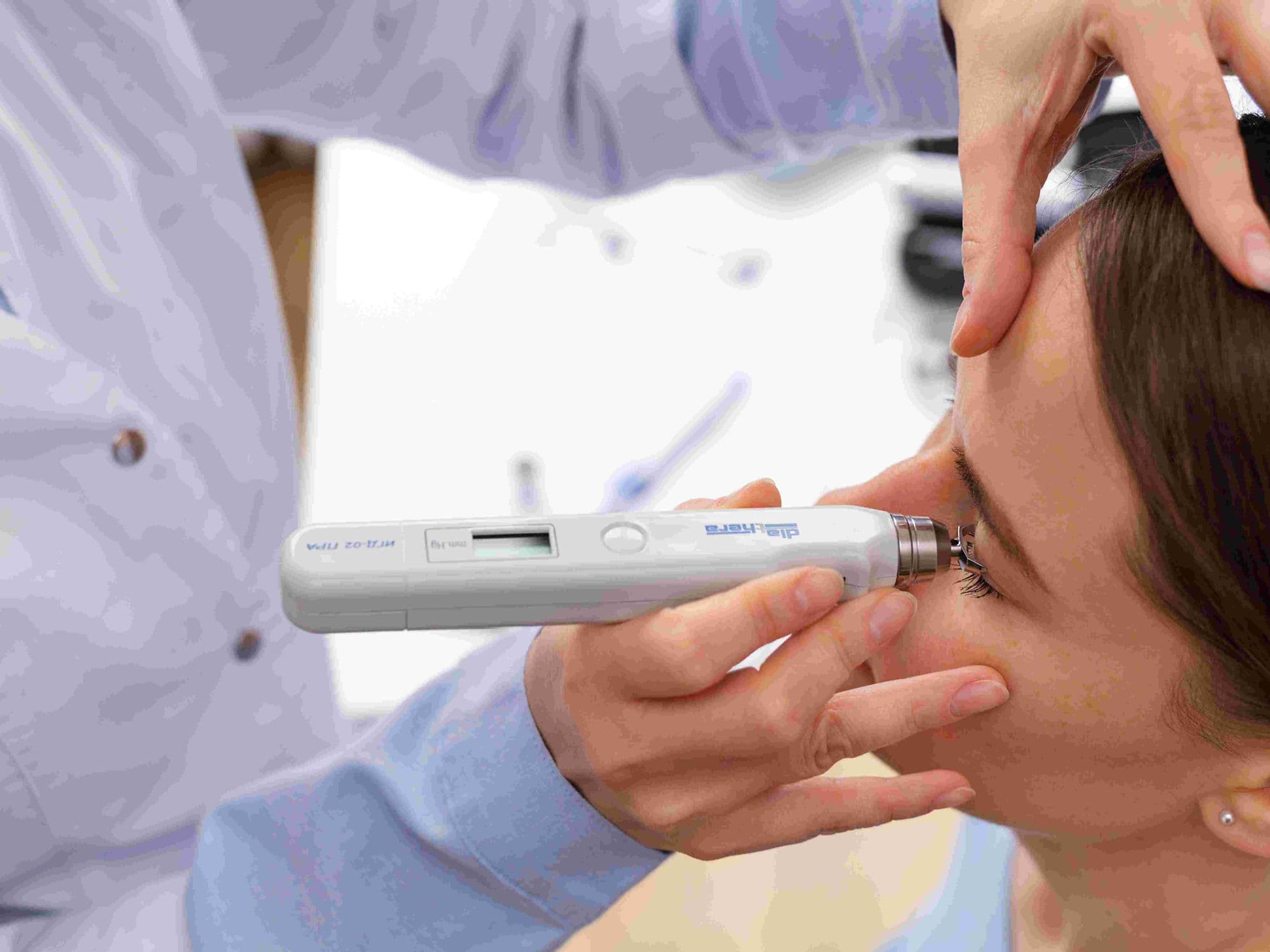
How Is Performed
Anesthesia: The procedure is usually performed as an outpatient surgery under local anesthesia with sedation and takes about one hour.
Incision: A small pocket is created under the conjunctiva (the clear membrane covering the white of the eye).
Plate Fixation: A thin, curved silicone plate is sutured to the sclera (the white part of the eye), usually tucked high under the upper eyelid.
Tube Insertion: A microscopic, flexible tube is trimmed and inserted into the front chamber of the eye to create a new drainage channel.
Patch Graft: A small piece of donor tissue (sclera or cornea) is often placed over the tube to protect it and prevent it from eroding through the eye's surface.
Fluid Flow: Excess fluid (aqueous humor) flows through the tube to the plate, where it forms a reservoir (bleb) and is naturally absorbed by the body.
Common Implant Models
Valved Implants (e.g., Ahmed Valve): These feature a pressure-sensitive valve that limits fluid flow until a specific pressure is reached, helping to prevent the eye pressure from dropping too low (hypotony) immediately after surgery.
Non-Valved Implants (e.g., Baerveldt or Molteno): These lack a valve and offer a larger surface area for drainage. The surgeon often temporarily ties off the tube during surgery to allow a protective capsule to form before drainage begins.
Pre-Procedure Preparation
Surgical Planning: A detailed evaluation to choose between a valved or non-valved device based on your specific pressure goals.
Medication Review: Discussing current glaucoma drops; some may need to be adjusted to manage inflammation before the implant.
Donor Tissue Coordination: Ensuring the necessary patch graft material is available for the day of surgery.
Transportation: Arranging for a companion to drive you home, as the eye will be patched and vision will be temporarily blurry.
Physical Readiness: Confirming you are comfortable lying still for approximately one hour during the micro-surgical steps.
Tests Before GDD Surgery
Gonioscopy: A specialized exam to view the internal drainage angle and determine the safest placement for the tube.
Visual Field Test: Establishing a baseline of peripheral vision to monitor the long-term success of the pressure control.
Endothelial Cell Count: Checking the health of the inner cornea, as the tube's position must not interfere with these delicate cells.
Intraocular Pressure (IOP) Profile: Tracking your pressure fluctuations to confirm the need for a surgical shunt.
Life After a GDD Implant
Initial Vision: Vision may be blurry for several days to a few weeks as the eye adjusts to the new drainage system.
Activity Restrictions: Patients must avoid bending over, straining, or lifting objects heavier than 5 kg for several weeks to prevent pressure spikes or tube movement.
Medication Use: Most patients will still need to continue some glaucoma medications even after the implant is fully functional.
Monitoring: Regular checkups are essential to ensure the tube is in the correct position and the plate is draining fluid effectively.
Safety Watch: Monitor for rare signs of complications, such as double vision (if the plate affects eye muscles) or redness at the patch graft site.
Why Specialized Treatment Is Highly Effective
Robust Pressure Control: Offers a reliable long-term solution for eyes that do not respond to other forms of glaucoma treatment.
Bypasses Scarred Tissue: Because the tube is inserted directly into the eye, it bypasses damaged or scarred natural drainage channels.
Standard of Care for Complex Cases: Successfully manages pressure in high-risk conditions like neovascular or inflammatory glaucoma.
Durable Design: The silicone materials used are highly biocompatible and designed to remain functional inside the eye for many years.
Predictable Outcomes: Modern surgical techniques and a variety of implant sizes allow surgeons to tailor the treatment to the specific volume of the patient's eye.