+91 83682 47758
info@medivisorhealth.com
Medivisor India Treatment Logo
  • Home
    • About Us
    • Our Services
    • Our Team
    • Our Safety Measures
    • Loading treatments...
    • Loading hospitals...
    • Loading doctors...
    • Loading tests & diagnoses...
    • Treatment Cost
    • Treatment Process
    • Visa Process
    • Travel Guide
    • FAQs
    • Why Medivisor
    • Success Stories
    • Patient Activities
    • Social Activities
    • News Coverage
    • Blog
  • Become a Partner

Medivisor India Treatment is a trusted New Delhi-based medical travel company assisting international patients with advanced, affordable, and seamless treatment in India.

  • Medivisor House 359, Sector 1, Vaishali, Ghaziabad, (Delhi/NCR) India
  • +91 8368 247 758
  • info@medivisorhealth.com

About Us

  • About Us
  • Our Services
  • Our Team
  • Our Safety Measures
  • Join Our Network
  • Contact Us

India Treatment

  • Hospitals
  • Treatments
  • Treatment Cost
  • Treatment Process
  • Visa Process
  • Travel Guide
  • FAQs
  • Why Medivisor

Gallery

  • Patient Testimonials
  • Patient Activities
  • News Coverage
  • Blog

Get in Touch

© 2026 Medivisor India Treatment. All rights reserved.

Dr Manoj Jain

Director - Gastrointestinal, Bariatric, Laparoscopic & Robotic Surgery

Nanavati-Max Super Specialty Hospital, Mumbai

25+ years experience

Dr Manoj Jain, Director - Gastrointestinal, Bariatric, Laparoscopic & Robotic Surgery at Nanavati-Max Super Specialty Hospital, Mumbai - Max Healthcare
  1. Home
  2. Doctors
  3. Dr Manoj Jain

About Dr Manoj Jain

Dr. Manoj Jain is a highly distinguished surgeon currently serving as a Director and Consultant Surgeon at Nanavati Max Super Speciality Hospital, Mumbai. With over 25 years of experience in the medical field, he is recognized as a leader in high-precision abdominal surgery. He specializes in a broad spectrum of surgical interventions, including Gastrointestinal, Bariatric, Laparoscopic, and Robotic Surgery, and is noted for his commitment to minimally invasive techniques that prioritize patient recovery.

Clinical Mastery and Procedural Expertise

Dr. Jain’s surgical career is marked by a massive volume of successful interventions, reflecting a deep technical mastery of keyhole and robotic platforms:

  • Laparoscopic Expertise: He has independently performed more than 7,000 laparoscopic surgeries to date. His expertise covers a wide range of abdominal procedures, from routine gallbladder and hernia repairs to complex gastrointestinal resections.

  • Bariatric & Metabolic Surgery: He is a specialist in weight-loss surgeries, including gastric bypass and sleeve gastrectomy, helping patients manage morbid obesity and its associated metabolic disorders like type 2 diabetes.

  • Robotic Surgery: Utilizing the latest robotic-assisted platforms, Dr. Jain performs high-precision surgeries that offer enhanced visualization and dexterity, leading to smaller incisions and faster discharge times.

Specialized Areas of Focus

Dr. Jain provides comprehensive care for a variety of complex conditions using the most advanced surgical modalities:

  • Gastrointestinal Surgery: Expert management of cancers and benign conditions of the stomach, intestines, liver, and pancreas.

  • Complex Hernia Repair: Advanced laparoscopic and robotic techniques for inguinal, incisional, and hiatal hernias.

  • Minimal Access Colorectal Surgery: Treatment for inflammatory bowel diseases and colorectal cancers.

  • General Surgery: Mastery in managing acute surgical emergencies and advanced trauma care.

Leadership and Academic Contributions

At Nanavati Max Super Speciality Hospital, one of Mumbai's premier healthcare institutions, Dr. Jain plays a pivotal role in the leadership of the surgical department.

  • Institutional Leadership: As Director, he oversees the strategic and clinical standards of the surgical units, ensuring the integration of modern technology into daily practice.

  • Mentorship: He is actively involved in training and proctoring young surgeons in the nuances of laparoscopic and robotic techniques.

  • Continuous Innovation: He is a frequent participant and speaker at national surgical conferences, where he shares his insights on improving surgical outcomes through minimal access technology.

Dr. Manoj Jain at a Glance

  • Director and Surgeon at Nanavati Max Super Speciality Hospital, Mumbai.

  • Over 25 years of experience in advanced surgical practice.

  • Performed more than 7,000 laparoscopic surgeries.

  • Specialist in Bariatric (Weight Loss) and Metabolic Surgery.

  • Expert in Robotic-assisted Gastrointestinal and Hernia procedures.

  • Renowned for excellence in minimal access surgery and faster patient recovery.

  • Leading consultant for complex abdominal and colorectal oncological surgeries.

MNAMS – National Board of Examinations, New Delhi (Mar 2004).
MRCS – Royal College of Surgeons, Edinburgh, UK (Apr 2003).
DNB (General Surgery) – National Board of Examinations, New Delhi (Mar 2003).
MS (General Surgery) – Seth G.S. Medical College & KEM Hospital, Mumbai (Mar 2002).

No awards & achievements available
May we help you?

Enter digits only (6-15 digits). We'll add your country code.

Related Treatments

Appendectomy (Open/Laparoscopic)

An Appendectomy is the surgical removal of the appendix, a small, finger-shaped pouch attached to the large intestine. It is the standard treatment for appendicitis, an inflammation of the organ that can lead to a life-threatening rupture if left untreated.

When You Should Consider Appendectomy

  • Pain that starts near the belly button and moves to the lower right abdomen.

  • "Rebound tenderness" or sharp pain in the lower right abdomen when pressure is released.

  • Fever and chills accompanying abdominal discomfort.

  • Nausea, vomiting, or loss of appetite.

  • High white blood cell counts detected during blood testing.

Methods of Appendectomy

  • Laparoscopic Appendectomy: The modern minimally invasive standard using three small incisions and a camera (laparoscope).

  • Open Appendectomy: A traditional approach using a single 2- to 4-inch incision, typically reserved for ruptured cases.

  • Emergency Appendectomy: Immediate surgical intervention performed to prevent or treat an appendix rupture.

  • Interval Appendectomy: A delayed surgery performed after a severe infection has been initially managed with antibiotics.

How Appendectomy Is Performed

  • Insufflation: In laparoscopic cases, carbon dioxide gas is used to inflate the abdomen for better visibility.

  • Appendix Identification: The surgeon locates the appendix and carefully ties off its base to separate it from the large intestine.

  • Irrigation: If the appendix has ruptured, the abdominal cavity is washed with saline to remove bacteria and pus.

  • Drainage: A small tube may be placed in the incision site for a few days to allow excess fluid to escape in complex cases.

  • Closure: Incisions are secured using absorbable sutures, surgical glue, or sterile strips.

Pre-Procedure Preparation

  • Maintaining NPO (nothing by mouth) status for several hours prior to the operation.

  • Starting Intravenous (IV) fluids to ensure proper hydration.

  • Administration of prophylactic antibiotics to reduce the risk of surgical site infection.

  • Undergoing general anesthesia to remain completely unconscious throughout the procedure.

Tests Before Appendectomy

  • Physical Examination: To check for localized pain and abdominal wall tension.

  • Ultrasound or CT Scan: Imaging used to confirm the inflammation or see if the appendix has burst.

  • Complete Blood Count (CBC): To identify signs of infection through elevated white blood cell levels.

  • Urine Test: Often performed to rule out a urinary tract infection or kidney stones which can mimic appendicitis symptoms.

Life After Appendectomy

  • For uncomplicated cases, hospital discharge usually occurs within 24 hours.

  • Recovery for laparoscopic surgery includes avoiding heavy lifting (over 5kg) for 1–2 weeks.

  • Recovery for open surgery requires avoiding strenuous exercise for 4–6 weeks to prevent hernia.

  • Diet begins with clear liquids and progresses as bowel function returns to normal.

  • Patients should monitor for signs of abscess, such as new fevers or persistent bloating.

Benefits of Appendectomy

  • Provides a permanent cure for appendicitis by removing the source of inflammation.

  • Prevents life-threatening complications associated with an appendix rupture or peritonitis.

  • Minimal scarring and faster recovery times when performed using laparoscopic techniques.

  • Rapidly alleviates severe abdominal pain and restores normal digestive health.

Starting Cost: $ 400

Body Contouring (Post Bariatric)

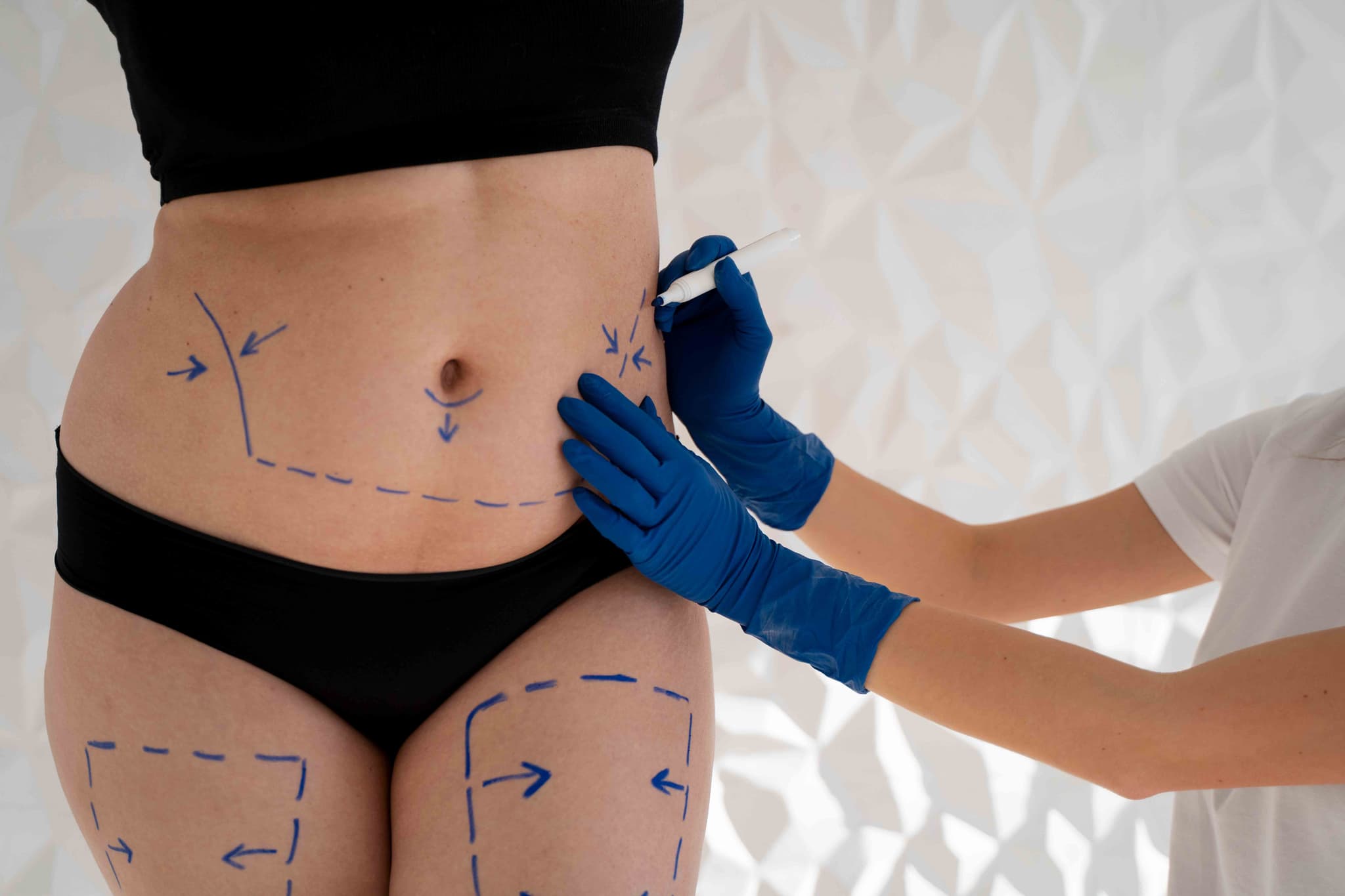
Body Contouring after bariatric surgery is a sequence of reconstructive procedures in 2026 designed to remove massive amounts of redundant skin and residual fat. It is the definitive final step for patients who have lost significant weight (50–100+ lbs), as excess skin can cause persistent hygiene issues, chronic infections, and physical discomfort that exercise alone cannot resolve.

When You Should Consider Body Contouring

This reconstructive journey is recommended for those who have completed their weight-loss phase and are seeking to improve their quality of life. Key indications include:

  • Redundant Skin Folds: Excessive skin on the abdomen, arms, or thighs that interferes with movement or clothing.

  • Chronic Skin Irritation: Recurring rashes or infections (intertrigo) within the skin folds.

  • Functional Limitations: Heavy skin that causes back pain, postural issues, or prevents a return to active fitness.

  • Stabilized Weight: Successful maintenance of a stable weight for at least 6 to 12 months.

  • Psychological Closure: A desire to have one's physical appearance reflect their health and weight-loss achievements.

Common Stages of Body Contouring

  • Phase 1 (The Core): Typically involves a Lower Body Lift or a Fleur-de-lis Abdominoplasty to stabilize the midsection.

  • Phase 2 (The Upper Body): Focuses on a Breast Lift (Mastopexy) and an Arm Lift (Brachioplasty).

  • Phase 3 (The Lower Extremities & Face): Includes a Thigh Lift or a Face and Neck Lift to address remaining laxity.

  • Auto-Augmentation: A 2026 standard where redundant tissue is used to rebuild volume in the breasts or buttocks, avoiding the need for synthetic implants.

  • Fleur-de-lis Technique: A specialized approach using both vertical and horizontal incisions to address massive skin redundancy in both directions.

How Body Contouring Is Performed

  • Staged Planning: Due to the extensive nature of these surgeries, procedures are spaced 4–6 months apart to allow the body to recover and ensure patient safety.

  • Incision & Removal: The surgeon makes precise incisions to remove wide sections of skin and fat, carefully repositioning the remaining tissue for a natural look.

  • Muscle & Tissue Repair: Weakened muscles (especially in the abdomen) are tightened to provide structural support for the new body shape.

  • Refined Contouring: Targeted liposuction is used to smooth the transitions between the areas where skin was removed.

  • Closure: Complex, multi-layered suturing is used to manage tension on the long incisions, and surgical drains are placed to prevent fluid buildup.

Pre-Procedure Preparation for Body Contouring

  • Nutritional Optimization: Surgeons require comprehensive blood panels to ensure protein, iron, and B12 levels are optimized, as post-bariatric patients often have deficiencies.

  • Weight Maintenance: Patients must demonstrate a stable "plateau" weight for at least 6 months prior to the first stage.

  • Mental Health Assessment: In 2026, professional support is often encouraged to help patients adjust to their changing body image.

  • Smoking/Vaping Cessation: Strict abstinence is mandatory for several weeks before and after to ensure tissue survival and wound healing.

Tests Before Body Contouring

  • Comprehensive Metabolic Panel: To check electrolyte balance, kidney function, and liver health.

  • Protein & Micronutrient Testing: To confirm the patient has the nutritional reserves (especially albumin) needed for massive tissue healing.

  • ECG & Cardiac Clearance: Essential due to the multiple, lengthy surgical sessions required.

  • Standard Blood Panel: To evaluate hemoglobin and clotting factors to minimize the risk of post-operative anemia.

Life After Body Contouring (Recovery)

  • High-Intensity Healing: The first week of each stage requires around-the-clock assistance and careful drain management.

  • Compression Support: Heavy-duty medical compression garments are worn for 6–8 weeks to support the tissues and minimize swelling.

  • Total Timeline: A full body transformation typically takes 12 to 18 months to complete all stages and see the final settled results.

  • Scar Management: While scars are extensive and permanent, 2026 protocols include advanced topical treatments and laser therapies to help them fade over time.

Benefits of Body Contouring

  • Elimination of Skin Issues: Directly resolves chronic rashes, infections, and hygiene struggles associated with skin folds.

  • Restored Mobility: Dramatically improves the ability to exercise, run, and move without the hindrance of heavy skin.

  • Permanent Tissue Correction: Once the redundant skin is surgically removed, it does not return if weight remains stable.

  • Complete Transformation: Provides the physical and emotional "finish line" to a significant weight-loss journey.

Starting Cost: $ 1500

Cholecystectomy (Open/Laparoscopic)

A Cholecystectomy is the surgical removal of the gallbladder, a small organ located under the liver that stores bile. It is primarily performed to treat gallstones (cholelithiasis), gallbladder inflammation (cholecystitis), or biliary colic, which is intense pain caused by stones blocking the bile ducts.

When You Should Consider Cholecystectomy

  • Sudden and rapidly intensifying pain in the upper right portion of the abdomen.

  • Nausea or vomiting following high-fat meals.

  • Yellowing of the skin and the whites of the eyes (jaundice).

  • Fever or chills, which may indicate a gallbladder infection.

  • Confirmation of gallstones or a non-functional gallbladder through diagnostic testing.

Methods of Cholecystectomy

  • Laparoscopic Cholecystectomy: The modern "gold standard" minimally invasive approach used in over 90% of cases, involving four small incisions.

  • Open Cholecystectomy: A traditional surgical method using a larger incision, typically reserved for complex cases or severe scarring.

  • Emergency Cholecystectomy: Immediate removal performed when acute inflammation or infection poses a significant health risk.

  • Single-Incision Laparoscopic Surgery (SILS): A specialized technique where the gallbladder is removed through one small incision in the umbilicus.

How Cholecystectomy Is Performed

  • Pneumoperitoneum: During laparoscopic surgery, the abdomen is inflated with carbon dioxide gas to provide a clear workspace for the surgeon.

  • Duct and Artery Clipping: The surgeon identifies the cystic duct and cystic artery and secures them with permanent clips before separation.

  • Gallbladder Detachment: The organ is carefully separated from the liver bed using specialized surgical instruments.

  • Organ Extraction: The gallbladder is placed in a sterile bag and removed through one of the small abdominal incisions.

  • Drainage: In open or complex cases, a Jackson-Pratt (JP) drain may be placed temporarily to remove excess fluid or bile.

Pre-Procedure Preparation

  • Fasting (NPO) for at least 6 to 8 hours prior to the operation.

  • Discontinuing blood-thinning medications several days before surgery as directed by the surgical team.

  • Receiving general anesthesia and the placement of a breathing tube for the duration of the procedure.

  • Undergoing a pre-operative evaluation to check liver enzymes and white blood cell counts.

Tests Before Cholecystectomy

  • Abdominal Ultrasound: The "gold standard" imaging test used to identify gallstones and gallbladder wall thickening.

  • HIDA Scan: A specialized nuclear medicine test used to evaluate the functional emptying of the gallbladder.

  • Blood Panels: To check for elevated liver enzymes or signs of infection (leukocytosis).

  • MRCP or ERCP: Advanced imaging or endoscopic procedures used to check for stones that may have moved into the common bile duct.

Life After Cholecystectomy

  • Most laparoscopic patients return home the same day or after one overnight stay.

  • Recovery for open surgery involves a hospital stay of 3 to 5 days and restricted activity for 4 to 6 weeks.

  • A low-fat diet is recommended for the first few weeks as the body adjusts to bile flowing directly into the small intestine.

  • Patients may experience temporary shoulder pain or bloating due to the gas used during laparoscopic procedures.

  • Monitoring for Post-Cholecystectomy Syndrome (PCS), which affects 10–15% of patients and involves lingering indigestion or nausea.

Benefits of Cholecystectomy

  • Provides permanent relief from the recurring pain of biliary colic and gallstones.

  • Prevents serious complications such as gallbladder rupture, gangrene, or severe pancreatitis.

  • Minimally invasive techniques result in smaller scars, less post-operative pain, and a faster return to daily activities.

  • Eliminates the risk of future gallbladder infections and inflammation.

Starting Cost: $ 400

Laparoscopic Gastric Bypass Surgery

Gastric Sleeve and Gastric Bypass are the primary surgical treatments for severe obesity and its associated metabolic conditions, such as Type 2 diabetes and hypertension. While both procedures aim to facilitate significant weight loss and metabolic health, they utilize different surgical mechanisms—one focusing on reducing the stomach's size and the other involving the rerouting of the digestive tract. By 2026, the use of robotic-assisted platforms has become the standard for these procedures, providing surgeons with the precision needed to ensure optimal outcomes and faster patient recovery.

When You Should Consider Bariatric Surgery

  • Class II or III Obesity: Generally recommended for individuals with a BMI over 35, or over 30 when accompanied by serious health conditions.

  • Uncontrolled Type 2 Diabetes: When blood sugar remains high despite intensive medical management.

  • Severe Obstructive Sleep Apnea: When weight-related airway obstruction impacts cardiovascular health and daily energy.

  • Non-Alcoholic Fatty Liver Disease (NAFLD): To reduce liver fat and prevent the progression toward cirrhosis.

  • Chronic Joint Pain: For patients whose weight has led to severe debilitating osteoarthritis in the knees or hips.

  • Failed Conventional Weight Loss: After multiple documented attempts at diet and exercise have failed to produce sustainable results.

Conditions That Require Specialized Care

  • Severe Acid Reflux (GERD): Patients with significant reflux are often steered toward Gastric Bypass, as the Gastric Sleeve can sometimes exacerbate the condition.

  • Sweet Eaters: Those with a high intake of processed sugars may benefit from the "Dumping Syndrome" feedback of Gastric Bypass to help modify behavior.

  • Previous Abdominal Surgeries: Presence of extensive scar tissue that may require a robotic-assisted approach for safe navigation.

  • Polycystic Ovary Syndrome (PCOS): Requiring careful hormonal monitoring as rapid weight loss begins to restore fertility and metabolic balance.

  • Extreme BMI (>60): Sometimes managed with a "staged" approach—performing a sleeve first and converting to a bypass later if needed.

How These Procedures Are Performed

  • Robotic-Assisted Access: Both surgeries are performed through several tiny "keyhole" incisions, utilizing a 3D high-definition camera and robotic wrists for extreme dexterity.

  • Sleeve Gastrectomy (Gastric Sleeve): Approximately 75–80% of the stomach is permanently removed along the "greater curvature," leaving a narrow, sleeve-like pouch.

  • Gastric Bypass (RYGB): A small stomach pouch (roughly the size of an egg) is created and connected directly to the middle part of the small intestine, bypassing the rest of the stomach.

  • Hormonal Modulation: Both procedures involve the removal or bypassing of the area where "Ghrelin" (the hunger hormone) is produced, significantly reducing appetite.

  • Staple Line Reinforcement: Surgeons use advanced stapling technology, often reinforced with specialized sutures or "buttress" material, to ensure a leak-proof seal.

  • Leak Testing: Before finishing the surgery, a specialized dye or air test is performed to confirm the absolute integrity of all new connections.

Innovations in Bariatric Surgery

  • Robotic Precision PlatformsAdvanced systems that provide surgeons with tremor-free movement and 3D visualization, making the complex suturing of a bypass significantly safer.

  • Digital Health IntegrationModern care involves wearable tech and apps that sync with the bariatric team, tracking protein intake and hydration in real-time.

  • Smart StaplersDevices that automatically sense tissue thickness and adjust the firing pressure to create more consistent and secure staple lines.

  • Endoscopic Revision (TORE)The ability to "tighten" a previous bypass or sleeve using a camera through the mouth, avoiding the need for a second major surgery if weight is regained.

  • Fluorescence-Guided SurgeryUsing injectable dyes that glow under specialized light to ensure a perfect blood supply to the newly created stomach pouch.

  • Vagal Nerve BlockingExperimental neuro-modulators that can be adjusted after surgery to help control hunger signals and improve long-term success.

Pre-Procedure Preparation

  • Psychological Evaluation: To ensure the patient has the mental health support and coping strategies needed for a major lifestyle transition.

  • Nutritional Counseling: Multiple sessions with a bariatric dietitian to learn the "protein-first" rule and the importance of lifelong vitamins.

  • Pre-Op Liquid Diet: A 1-to-2-week specialized diet intended to shrink the liver, making the surgery safer and easier for the surgeon.

  • Cardiac and Sleep Clearance: Ensuring the heart and lungs are strong enough for general anesthesia and the post-operative healing phase.

  • Smoking Cessation: A mandatory requirement for many programs, as nicotine significantly increases the risk of stomach ulcers and poor healing.

Diagnostic and Safety Monitoring

  • Upper GI Series (Swallow Test): An X-ray performed shortly after surgery to verify that liquids are passing correctly through the new anatomy.

  • Vitamin Level Tracking: Regular blood tests to monitor levels of Iron, B12, Vitamin D, and Calcium to prevent long-term deficiencies.

  • Body Composition Analysis: Using advanced scales to ensure that weight loss is coming from fat mass rather than muscle tissue.

  • Gallbladder Ultrasound: Monitoring for gallstones, which can occasionally form during the rapid weight loss phase following surgery.

  • Hemoglobin A1c Monitoring: Tracking the rapid (often near-instant) improvement in blood sugar levels in diabetic patients.

Why This Treatment Is Highly Effective

  • Metabolic Reset: These surgeries do more than "shrink the stomach"; they fundamentally change the body's hormonal response to food and insulin.

  • Sustainable Results: Unlike traditional dieting, bariatric surgery alters the body's "set point," making it easier to maintain weight loss long-term.

  • Resolution of Co-morbidities: Many patients see a complete remission of Type 2 diabetes, sleep apnea, and high blood pressure within months.

  • Increased Longevity: Studies consistently show that bariatric surgery reduces the long-term risk of heart disease, stroke, and certain cancers.

  • Enhanced Quality of Life: Patients often report a dramatic increase in mobility, energy levels, and social confidence.

Recovery and Aftercare

  • Early Mobilization: Walking is required within 4 hours of surgery to promote circulation and help clear the surgical gas from the body.

  • Phased Diet: A strict transition from clear liquids to purees, then soft foods, and finally solid proteins over a 6-to-8-week period.

  • Lifelong Supplementation: Daily bariatric-specific multivitamins are required to compensate for the changes in nutrient absorption.

  • Support Groups: Regular attendance at peer support groups is highly correlated with long-term weight loss success.

  • Follow-up Schedule: Frequent visits with the surgical team during the first year (1, 3, 6, and 12 months) to monitor progress and nutrition.

Life After Bariatric Surgery

  • A transformed relationship with food, where "fullness" is felt quickly and hunger signals are significantly muted.

  • The ability to engage in physical activities that were previously impossible, such as running, hiking, or playing with children.

  • Significant improvement in self-image and mental well-being as health goals are met and surpassed.

  • A requirement for ongoing mindful eating—surgery is a powerful "tool," but long-term success depends on a healthy partnership with that tool.

  • A renewed lease on life, often described by patients as a "second chance" at a healthy, active future.

Starting Cost: $ 1500

Laparoscopic Hernia Repair

Laparoscopic Hernia Repair, also known as minimally invasive or keyhole surgery, uses specialized tools and a camera to repair a weakness in the abdominal wall from the inside out. It is most commonly used for inguinal (groin), umbilical (navel), and ventral hernias.

When You Should Consider Laparoscopic Hernia Repair

  • A visible bulge in the groin or abdomen that may become more prominent when standing or coughing.

  • Pain or pressure at the hernia site during physical activity or lifting.

  • Inguinal hernias that occur on both sides (bilateral) of the groin.

  • Recurrent hernias where a previous open surgical repair has failed.

  • Desire for a faster return to work and daily activities with minimal scarring.

Methods of Laparoscopic Hernia Repair

  • TAPP (Transabdominal Preperitoneal): The surgeon enters the peritoneal cavity where the organs are located to reach and repair the hernia.

  • TEP (Totally Extraperitoneal): The surgeon operates in the space between the muscle and the abdominal lining, avoiding the organ cavity entirely.

  • IPOM (Intraperitoneal Onlay Mesh): Primarily used for ventral hernias, where a specialized mesh is placed on the inside of the abdominal wall.

  • Robotic-Assisted Laparoscopy: Using a robotic interface for enhanced precision and 3D visualization during the repair.

How Laparoscopic Hernia Repair Is Performed

  • Insufflation: The abdominal cavity is inflated with Carbon Dioxide (CO2) gas to create a dome-shaped workspace.

  • Port Insertion: Three to four small incisions (0.5–1 cm) are made to allow the insertion of a laparoscope and long, thin instruments.

  • Reduction: Protruding tissue, such as fat or a loop of intestine, is carefully pulled back into the abdominal cavity from the inside.

  • Mesh Reinforcement: A lightweight, flexible synthetic mesh is unfurled over the defect to reinforce the weakened wall.

  • Fixation: The mesh is secured in place using surgical tacks, staples, or specialized surgical glue to prevent shifting.

Pre-Procedure Preparation

  • Fasting (NPO) for 6–8 hours prior to the surgery to ensure safety during anesthesia.

  • Pausing blood-thinning medications several days in advance as coordinated with the surgical team.

  • Medical clearance to ensure the patient can tolerate general anesthesia and abdominal CO2 inflation.

  • Discussing the specific approach (TAPP vs. TEP) based on the hernia's location and surgical history.

Tests Before Laparoscopic Hernia Repair

  • Physical Examination: The primary method to determine if the hernia is "reducible" or "incarcerated."

  • Ultrasound or CT Scan: Imaging used to confirm the diagnosis and map the size of the abdominal wall defect.

  • Electrocardiogram (ECG): Often required for patients over a certain age to ensure heart health for general anesthesia.

  • Blood Panels: To check for infection markers and ensure proper kidney and liver function.

Life After Laparoscopic Hernia Repair

  • Most patients return home the same day as the procedure (day-care surgery).

  • Walking is encouraged immediately following surgery to prevent blood clots.

  • Desk-based work can typically be resumed within 3 to 7 days.

  • Strict lifting restrictions—usually no more than 5–10 kg—must be followed for 4–6 weeks.

  • Monitoring for temporary shoulder pain, which is a common side effect of the CO2 gas used during surgery.

Benefits of Laparoscopic Hernia Repair

  • Results in significantly smaller scars and a lower risk of wound infection compared to open surgery.

  • Offers a faster recovery timeline and a quicker return to professional and athletic activities.

  • Provides a superior view for the surgeon to identify and repair multiple hernia defects through the same incisions.

  • Utilizes a "tension-free" mesh technique that lowers the risk of the hernia returning.

Starting Cost: $ 500

Laparoscopic Polypectomy

Laparoscopic Polypectomy generally refers to the minimally invasive removal of polyps from the gallbladder or, in specific gynecological cases, subserosal uterine polyps. While most uterine polyps are removed via hysteroscopy (through the cervix), the laparoscopic approach is reserved for polyps or small growths located on the outer surface of an organ or within the abdominal cavity.

When You Should Consider Laparoscopic Polypectomy

  • Symptomatic Polyps: Growths causing localized pain or persistent digestive issues.

  • Suspect Features: Polyps showing irregular shapes or rapid growth during an ultrasound.

  • Size-Dependent Risks: Gallbladder polyps exceeding 1 cm in size, which is a standard surgical indication in 2026 guidelines.

  • Outer Organ Growth: When a polypoid growth is located on the exterior of the uterus or attached to pelvic ligaments.

  • Cancer Mitigation: To prevent the risk of malignancy in polyps that show signs of growth.

Methods of Removal and Techniques

  • Laparoscopic Cholecystectomy: Removal of the entire gallbladder for polyps larger than 10mm, as they cannot be safely scraped off the wall.

  • Laparoscopic Excision: The use of precision shears or lasers to excise a growth at its base from the exterior of an organ.

  • High-Definition Visualization: Utilizing zoomed-in views to identify a polyp’s blood supply and seal vessels before cutting.

  • Oncological Safety Protocols: The use of specimen bags during removal to ensure tissue does not contact the abdominal wall.

  • Hysteroscopy (Alternative): The standard approach for internal uterine polyps, distinct from the laparoscopic method used for external growths.

How Laparoscopic Polypectomy Is Performed

  • Anesthesia: The surgery is performed under general anesthesia to ensure the patient is comfortable and still.

  • Access Incisions: The surgeon makes 1 to 3 tiny incisions, ranging from 5–10 mm, in the abdominal wall.

  • Instrument Insertion: A laparoscope and specialized micro-instruments are inserted through the small incisions.

  • Vessel Sealing: Surgeons use high-definition cameras to seal blood vessels before removal, resulting in near-zero blood loss.

  • Abdominal Inflation: Carbon dioxide gas is used to create space, though this can lead to temporary post-operative bloating.

Pre-Procedure Preparation

  • Fasting Requirements: Patients must fast for 8-12 hours before the surgery or catheterization.

  • Medical Assessments: Blood tests, ECG, and chest X-rays are conducted to assess overall health.

  • Medication Adjustments: Current medications must be adjusted as directed by the medical team.

  • Allergy Discussion: Patients must discuss any allergies, particularly to materials used in surgical devices or dyes.

  • Post-Op Planning: Arranging for post-operative care and a support person for the recovery period.

Tests Before Laparoscopic Polypectomy

  • Ultrasound Imaging: Used to monitor the size and shape of the polyp and detect rapid growth.

  • Blood Tests: To assess overall health and check for markers of inflammation or infection.

  • ECG: To monitor the heart's electrical rhythm and check for conduction issues.

  • Pulse Oximetry: Used to evaluate oxygen saturation levels in the blood before anesthesia.

  • CT Scan: For detailed mapping of the abdominal cavity and complex growths.

Life After Laparoscopic Polypectomy

  • Hospital Stay: Usually a same-day discharge or a short 23-hour observation stay.

  • Immediate Recovery: Walking is encouraged immediately to aid digestion and prevent blood clots.

  • Pain Management: Discomfort is typically managed with over-the-counter anti-inflammatories.

  • Return to Work: Patients can generally return to work within 5 to 7 days post-procedure.

  • Activity Restrictions: Heavy lifting must be avoided for approximately 4 weeks to ensure proper healing.

Benefits of Laparoscopic Polypectomy

  • Minimal Tissue Disruption: Rapid recovery due to the use of tiny incisions rather than large open cuts.

  • Precise Visualization: High-definition cameras allow for the zoom-in on blood supplies for safer removal.

  • Near-Zero Blood Loss: Advanced sealing techniques minimize bleeding during the excision.

  • Strict Safety: The use of specialized bags prevents tissue fragments from remaining in the pelvic cavity.

  • Long-Term Cure: Provides a definitive treatment with high technical success rates for symptomatic growths.

Starting Cost: $ 300

Myomectomy (Open/Laparoscopic/Robotic)

A myomectomy is a surgical procedure to remove uterine fibroids (benign growths) while keeping the uterus intact. Unlike a hysterectomy, it preserves fertility and is the preferred option for women who wish to become pregnant or maintain their uterine health. This procedure specifically targets the fibroids while surgically repairing the uterine wall.

When You Should Consider a Myomectomy

  • Fertility Preservation: For women who plan to have children in the future and need to keep the uterus functional.

  • Heavy Menstrual Bleeding: When fibroids cause anemia, flooding, or prolonged periods that disrupt daily life.

  • Pelvic Pain or Pressure: Large fibroids pressing on the bladder (causing frequent urination) or the rectum.

  • Infertility or Miscarriage: When fibroids distort the uterine cavity and interfere with embryo implantation or maintenance of pregnancy.

  • Rapid Growth: If ultrasound monitoring shows fibroids are growing quickly, even if they are currently asymptomatic.

Surgical Approaches

  • Abdominal Myomectomy (Open): A traditional "bikini cut" horizontal incision is made in the lower abdomen. This is used for very large fibroids (typically over 10 cm), numerous growths, or those buried deep in the uterine wall.

  • Laparoscopic Myomectomy: Performed through 3–4 tiny "keyhole" incisions. The surgeon uses a camera and specialized tools to cut the fibroids into smaller pieces (morcellation) for removal through the small ports.

  • Robotic-Assisted Myomectomy: Similar to the laparoscopic approach, but the surgeon operates via a robotic console. This provides superior 3D visualization and greater precision, which is helpful for complex or hard-to-reach fibroids.

  • Hysteroscopic Myomectomy: No abdominal incisions are made. A specialized scope is inserted through the cervix to remove fibroids that are bulging into the uterine cavity (submucosal fibroids).

How Is Performed

  • Anesthesia: The surgery is performed under general anesthesia and typically takes between 1 to 3 hours depending on the number of fibroids.

  • Incision and Access: Depending on the approach, the surgeon accesses the uterus either through the abdomen or the vaginal canal/cervix.

  • Fibroid Removal: Each fibroid is carefully "shelled out" from the surrounding healthy uterine muscle.

  • Uterine Reconstruction: The surgeon meticulously sutures the layers of the uterine muscle back together to ensure the wall is strong enough to support a future pregnancy.

  • Closure: Abdominal incisions are closed with stitches or surgical glue. For hysteroscopic cases, there are no external wounds to close.

Pre-Procedure Preparation

  • Pelvic Imaging: An MRI or detailed ultrasound is mandatory to map the exact size, number, and depth of all fibroids.

  • Hormonal Therapy (Optional): In some cases, medication (like GnRH agonists) may be prescribed for 2–3 months before surgery to shrink the fibroids and reduce blood loss.

  • Iron Supplementation: If you are anemic due to heavy bleeding, you may need to take iron to build up your blood count before surgery.

  • Fasting: Adhering to strict "nothing by mouth" instructions for at least 8 hours prior to your general anesthesia.

  • Medication Audit: Stopping blood thinners, aspirin, or anti-inflammatory drugs 7–10 days before the procedure.

Tests Before Myomectomy

  • Saline Infusion Sonogram (SIS): Injecting sterile water into the uterus during an ultrasound to get a clear view of the uterine lining.

  • Hysteroscopy (Diagnostic): A quick office procedure to look inside the uterus and confirm if fibroids are affecting the cavity.

  • Blood Panels: Checking hemoglobin and hematocrit levels to assess for anemia.

  • ECG: A standard heart check to ensure you are healthy enough for the administration of anesthesia.

Life After Myomectomy

  • Hospital Stay: Usually 1–2 nights for open surgery; often same-day discharge or one night for laparoscopic and robotic approaches.

  • Physical Activity: Walking is encouraged immediately to prevent blood clots. Avoid heavy lifting and strenuous exercise for 4 weeks (laparoscopic) to 6 weeks (open).

  • Pregnancy Timing: Surgeons usually recommend waiting 3 to 6 months before trying to conceive to allow the uterine wall to heal completely.

  • Delivery Method: If the surgery involved deep incisions into the uterine wall, a C-section is often required for future deliveries to prevent uterine rupture during labor.

  • Follow-up Imaging: A repeat ultrasound is often done 3–6 months post-op to ensure the uterus has healed well and no small fibroids were left behind.

Why Specialized Treatment Is Highly Effective

  • Preserves Reproductive Potential: Unlike a hysterectomy, this procedure keeps the option of biological motherhood open.

  • Targeted Symptom Relief: Successfully removing fibroids eliminates the source of heavy bleeding and pelvic pressure while keeping the organ intact.

  • High Success Rates: Modern robotic and laparoscopic techniques allow for the removal of complex fibroids with minimal blood loss and faster recovery.

  • Improved Fertility Outcomes: For many women, removing cavity-distorting fibroids significantly increases the chances of successful natural conception or IVF.

  • Maintains Pelvic Integrity: Keeping the uterus helps maintain the natural support of the pelvic floor and avoids the early onset of surgical menopause.

Starting Cost: $ 800

Ovarian Cyst Removal (Laparoscopic Cystectomy)

Laparoscopic ovarian cystectomy is a minimally invasive surgery to remove an ovarian cyst while preserving the healthy part of the ovary and the fallopian tube. It is the preferred method for treating benign (non-cancerous) cysts that are causing pain, growing rapidly, or at risk of twisting (torsion). This technique allows for precise removal of the cyst wall with minimal impact on future fertility.

When You Should Consider an Ovarian Cystectomy

  • Persistent Cysts: Functional cysts that do not disappear naturally after several menstrual cycles.

  • Endometriomas: Also known as "chocolate cysts," these are caused by endometriosis and can significantly affect fertility if left untreated.

  • Dermoid Cysts: Complex cysts containing different types of tissue (such as skin or hair) that will not resolve on their own and carry a risk of rupture.

  • Chronic Pelvic Pain: Large cysts that create constant pressure on the bladder or rectum, or cause sharp pain during physical activity.

  • Rapid Growth: If ultrasound monitoring shows a cyst is increasing in size, even if it is currently asymptomatic.

How Is Performed

  • Anesthesia: The surgery is performed under general anesthesia and typically takes between 45 to 90 minutes.

  • Incision: The surgeon makes 3–4 tiny "keyhole" incisions (5–10 mm) in the abdomen, including one inside the belly button for the camera (laparoscope).

  • Visualization: Carbon dioxide gas is used to gently inflate the abdomen, providing the surgical team with a clear, magnified view of the pelvic organs.

  • Cyst Removal: Using specialized micro-instruments, the surgeon carefully peels the cyst wall away from the healthy ovarian tissue, ensuring the ovary remains functional.

  • Extraction: The cyst is placed in a small surgical bag and removed through one of the incisions. Large cysts may be drained first to fit through the small opening.

  • Closure: The small incisions are closed with dissolvable stitches or surgical glue, resulting in minimal scarring.

Pre-Procedure Preparation

  • Pelvic Ultrasound or MRI: To determine the exact size, location, and internal structure of the cyst before surgery.

  • Blood Testing: Checking "tumor markers" (such as CA-125) to confirm the cyst appears benign and to assess overall health.

  • Fasting: Adhering to strict "nothing by mouth" instructions for at least 8 hours prior to your general anesthesia.

  • Medication Audit: Stopping blood thinners, aspirin, or certain supplements 7–10 days before the procedure as directed.

  • Bowel Prep (Optional): In some cases, a mild laxative may be recommended the day before to clear the pelvic area for better visualization.

Tests Before Ovarian Cystectomy

  • Transvaginal Ultrasound: The primary tool used to distinguish between fluid-filled simple cysts and complex solid masses.

  • Pregnancy Test: To rule out ectopic pregnancy or ensure the safety of the developing fetus if pregnancy is known.

  • Coagulation Profile: To ensure your blood's ability to clot is optimal for a safe, minimally invasive recovery.

  • ECG: A standard heart check to confirm you are healthy enough for the administration of anesthesia.

Life After Ovarian Cystectomy

  • Hospital Stay: Most patients are discharged the same day or stay for one night of observation.

  • Shoulder Pain: It is common to feel temporary pain in the shoulders for 24–48 hours; this is caused by the gas used during surgery irritating the diaphragm.

  • Activity Restrictions: You can usually return to light daily activities within 3 to 5 days. However, avoid heavy lifting and strenuous exercise for 2 to 3 weeks.

  • Wound Care: Keep the small incisions clean and dry. Most stitches are internal and will dissolve on their own within 10–14 days.

  • Sexual Activity: Most doctors recommend waiting at least 2 weeks before resuming sexual intercourse to allow internal healing.

Why Specialized Treatment Is Highly Effective

  • Preserves Fertility: By removing only the cyst wall and saving the healthy ovarian tissue, the "ovarian reserve" and hormone production are protected.

  • Minimal Scarring: The use of tiny incisions results in superior cosmetic outcomes compared to traditional open surgery.

  • Rapid Recovery: The minimally invasive approach allows patients to return to their jobs and families much faster than with a laparotomy.

  • High Precision: The magnified view provided by the laparoscope allows surgeons to identify and protect the delicate fallopian tubes and surrounding nerves.

  • Lower Complication Rates: Laparoscopy is associated with less post-operative pain, lower risk of infection, and a reduced chance of developing internal scar tissue (adhesions).

Starting Cost: $ 500

Radical Prostatectomy (Open/Laparoscopic/Robotic)

Radical Prostatectomy is the surgical removal of the entire prostate gland and surrounding tissues, typically performed to treat Prostate Cancer. Unlike treatments for an enlarged prostate (BPH) which only remove the "core," this treatment removes the entire organ to ensure the cancer is fully eliminated.

When You Should Consider Radical Prostatectomy

  • Localized Prostate Cancer: When imaging confirms the cancer is confined within the prostate capsule (Stages T1 or T2).

  • Aggressive Disease: For patients with a higher Gleason Score who require definitive surgical removal rather than "active surveillance."

  • Long Life Expectancy: Typically recommended for patients expected to live 10+ years, where surgical removal offers the best long-term cure rate.

  • Failure of Radiation: As a "salvage" treatment if the cancer returns after previous radiation therapy.

  • Patient Preference: For individuals who prefer the psychological certainty of having the cancerous organ physically removed.

Methods of Radical Prostatectomy

  • Robotic-Assisted (Da Vinci): The modern gold standard. The surgeon sits at a console controlling robotic arms with 3D magnification and 360-degree "wristed" instruments for extreme precision.

  • Laparoscopic Surgery: A minimally invasive approach using 5–6 "keyhole" incisions, a camera, and long instruments to operate while viewing a 2D screen.

  • Open Surgery (Retropubic): The traditional method involving a single 4–5 inch incision in the lower abdomen to remove the gland.

  • Nerve-Sparing Technique: A meticulous process where the surgeon peels the delicate "neurovascular bundles" away from the prostate to preserve sexual function.

  • Vesicourethral Anastomosis: The reconstructive step where the bladder is stitched directly back to the urethra to restore the urinary path after the gland is removed.

How the Procedure Is Performed

  • Anesthesia: Performed under General Anesthesia to ensure the patient is completely asleep and the abdominal muscles are relaxed.

  • Dissection: The surgeon carefully separates the prostate from the bladder above it and the urethra below it.

  • Lymph Node Removal: Depending on the cancer's aggressiveness, nearby pelvic lymph nodes are often removed to check for microscopic spread.

  • Gland Extraction: The entire prostate and the attached seminal vesicles are removed as a single unit.

  • Catheterization: A Foley catheter is inserted through the penis into the bladder to act as a "splint" while the new connection (anastomosis) heals.

  • Drain Placement: A small suction tube may be left in the abdomen for 24–48 hours to remove excess surgical fluid.

Pre-Procedure Preparation

  • Cancer Staging: Includes a Multiparametric MRI (mpMRI) and often a PSMA PET-CT scan to ensure the cancer has not spread.

  • Biopsy Review: The surgical team reviews the Gleason Score and genomic markers to plan the extent of the surgery.

  • Pelvic Floor Training: Patients are taught Kegel exercises weeks before surgery to strengthen the muscles responsible for urinary control.

  • Medication Audit: Patients must stop blood thinners (like Aspirin or Warfarin) 7–10 days prior to the operation.

  • Fasting: Maintaining a "nil per oral" status for 8 hours before the procedure for anesthesia safety.

Pre-Surgery Tests

  • PSA Blood Test: To establish the final pre-operative baseline for monitoring future "undetectable" levels.

  • EKG and Chest X-ray: Standard tests to ensure heart and lung fitness for a multi-hour surgical procedure.

  • Basic Metabolic Panel: Checking kidney function (Creatinine) and electrolytes to manage IV fluids during surgery.

  • Coagulation Profile: Testing PT/INR and Platelet counts to ensure safe surgical healing and minimal blood loss.

  • Complete Blood Count (CBC): To check baseline hemoglobin levels in case a blood transfusion is required (rare in robotic cases).

Life After Radical Prostatectomy (Recovery & Risks)

  • Hospital Stay: Usually 1–2 days for Robotic/Laparoscopic surgery, or 3–4 days for the Open approach.

  • Catheter Management: The Foley catheter must remain in place for 7 to 14 days to allow the bladder-urethra connection to heal water-tight.

  • Trial of Void: After 1–2 weeks, the catheter is removed in the clinic to ensure the patient can urinate independently.

  • Activity Restrictions: No heavy lifting (over 5kg) or driving for 4 weeks; early walking is encouraged to prevent blood clots.

  • Urinary Incontinence: Most patients experience leaking initially; this typically improves over 3–6 months with consistent pelvic floor exercises.

  • Erectile Dysfunction (ED): It can take 6–18 months for erections to return; doctors often start "Penile Rehabilitation" medication shortly after surgery.

Why Specialized Treatment Is Highly Effective

  • Definitive Cure: Offers the highest probability of completely eliminating localized prostate cancer in a single treatment.

  • Pathological Certainty: Removing the gland allows for a total biopsy, giving the most accurate information on the cancer's stage and grade.

  • Robotic Precision: The 3D-high-definition view in 2026 allows surgeons to see nerves and vessels that are nearly invisible to the naked eye.

  • Predictable Monitoring: Post-surgery, the PSA level should drop to "undetectable," making it very easy to monitor for any future recurrence.

  • Limb-Sparing Mindset: Modern techniques focus heavily on "quality of life" preservation, aiming for the "Trifecta" of cancer control, continence, and potency.

Starting Cost: $ 2500

Revisional Bariatric Surgery

Revisional bariatric surgery is a specialized procedure performed on patients who have already undergone a primary weight-loss surgery but require a second operation. This may be due to medical complications, insufficient weight loss, or significant weight regain. Because of existing scar tissue and altered anatomy, these procedures are more complex and carry higher risks than initial weight-loss surgeries.

When You Should Consider Revisional Surgery

  • Inadequate Weight Loss or Regain: If the initial stomach pouch has stretched over time or the body has adapted to the metabolic changes of the first surgery.

  • Severe Acid Reflux (GERD): Persistent or worsening reflux that does not respond to medication, frequently occurring after a gastric sleeve procedure.

  • Mechanical Complications: Issues such as a "slipped" gastric band, a staple line leak, or a narrowing (stricture) of a previous surgical connection.

  • Chronic Ulcers or "Dumping Syndrome": Persistent digestive issues following a bypass that cannot be managed through dietary changes alone.

  • Nutritional Malabsorption: Severe deficiencies in vitamins or protein that remain uncontrolled despite high-dose supplementation.

Common Revisional Procedures

  • Sleeve to Bypass Conversion: Converting a gastric sleeve into a Roux-en-Y gastric bypass. This is highly effective for treating severe reflux and triggering further weight loss.

  • Gastric Band Conversion: Removing a "Lap-Band" and converting the anatomy into either a sleeve or a bypass, often performed in a single or two-stage process.

  • Bypass Revision (Stoma Tightening): For a stretched gastric bypass, a surgeon may "re-tighten" the connection (stoma) to restore the feeling of fullness.

  • Intestinal Lengthening: Adjusting the "Roux" limb to increase the bypassed section of the intestine, thereby increasing calorie malabsorption.

  • Sleeve Re-Sleeving: Trimming a previously stretched gastric sleeve to restore its original narrow, restrictive shape.

How Is Performed

  • Anesthesia: The surgery is performed under general anesthesia and typically takes between 2 to 4 hours due to the precision required.

  • Surgical Approach: Almost always performed laparoscopically or robotically to navigate through previous scar tissue with high-definition visualization and accuracy.

  • Managing Adhesions: The surgeon must carefully separate internal scarring (adhesions) from previous operations before the new revision can begin.

  • Stapling and Joins: New surgical joins are created using specialized stapling instruments designed for thicker or scarred tissue.

  • Leak Testing: Extensive intra-operative testing is performed to ensure all new connections are watertight and airtight before finishing.

Pre-Procedure Preparation

  • Anatomy Mapping: Extensive imaging is required to visualize the current state of the stomach and intestines before entering the operating room.

  • Nutritional Optimization: Correcting any existing vitamin deficiencies through intensive supplementation prior to surgery.

  • Psychological Assessment: A thorough review to ensure the patient is prepared for the "reset" in dietary habits and lifestyle expectations.

  • Smoking Cessation: Patients must stop smoking at least 6 to 8 weeks before surgery, as nicotine significantly increases the risk of staple line leaks in scarred tissue.

  • Weight Management: Following a specific pre-operative diet to reduce liver size and improve surgical access.

Tests Before Revisional Surgery

  • Upper Endoscopy (EGD): Inserting a camera into the stomach to inspect the pouch and check for ulcers, inflammation, or stretching.

  • Barium Swallow (UGI Series): A specialized X-ray where the patient swallows contrast liquid to show the exact size and flow of the current anatomy.

  • Abdominal CT Scan: To provide a 3D view of the internal organs and identify any hidden hernias or mechanical issues.

  • Comprehensive Blood Panels: Detailed analysis of iron, B12, Vitamin D, and protein levels to establish a baseline.

Life After Revisional Surgery

  • Hospital Stay: Usually involves 2 to 3 nights of close monitoring to ensure the new surgical joins are healing correctly.

  • Dietary Reset: Patients must return to a "Phase 1" clear liquid diet and progress slowly through pureed and soft foods over a 4–6 week period.

  • Slower Weight Loss: It is important to note that weight loss after a revision is typically slower and less dramatic than after the first surgery.

  • Lifelong Monitoring: Regular follow-up appointments and blood tests are essential to monitor for malabsorption and nutritional health.

  • Activity Restrictions: Light walking is encouraged immediately, but heavy lifting and core-straining exercises are restricted for 6 weeks.

Why Specialized Treatment Is Highly Effective

  • Resolves Chronic Pain and Reflux: Provides immediate relief for patients suffering from the mechanical failures or side effects of a primary surgery.

  • Second Chance for Health: Allows patients who did not reach their goals to restart their weight-loss journey with a more powerful metabolic tool.

  • Precision Technology: The use of robotic platforms allows surgeons to operate safely in "hostile" abdomens filled with previous scar tissue.

  • Customized Anatomy: Every revision is tailored specifically to the patient’s existing anatomy, ensuring the most effective solution for their unique situation.

  • Prevents Long-Term Damage: Addressing issues like chronic ulcers or severe reflux prevents permanent damage to the esophagus and digestive tract.

Starting Cost: $ 2000

Vaginal Hysterectomy (Open/Laparoscopic/Robotic)

A vaginal hysterectomy is a surgical procedure to remove the uterus through the vaginal canal. Unlike an abdominal hysterectomy, this approach requires no external incisions on the abdomen, which typically results in a shorter hospital stay, lower costs, and a significantly faster recovery. It is a preferred method for treating various non-cancerous gynecological conditions.

When You Should Consider a Vaginal Hysterectomy

  • Uterine Fibroids: Benign growths in the uterine wall that cause heavy menstrual bleeding, pelvic pressure, or persistent pain.

  • Uterine Prolapse: When the pelvic floor muscles and ligaments weaken, causing the uterus to slip down into or even out of the vaginal canal.

  • Adenomyosis: A condition where the uterine lining grows into the muscular wall of the uterus, causing severe cramping and heavy periods.

  • Endometriosis: When tissue similar to the uterine lining grows outside the uterus, leading to chronic pain and scarring.

  • Abnormal Uterine Bleeding: When heavy or irregular bleeding cannot be managed with medication or less invasive procedures.

Surgical Approaches

  • Traditional Vaginal Hysterectomy: The entire procedure is performed through an incision inside the vagina. This is the least invasive method and leaves no visible scars.

  • Laparoscopically Assisted Vaginal Hysterectomy (LAVH): A thin camera (laparoscope) and micro-instruments are inserted through 3–4 tiny "keyhole" incisions in the belly to detach the upper uterus. The uterus is then removed through the vagina.

  • Robotic-Assisted Hysterectomy: Similar to the laparoscopic approach, but the surgeon uses a robotic console for superior 3D visualization and enhanced precision, which is ideal for complex cases like extensive endometriosis.

  • Vaginal Vault Creation: After the uterus is removed, the surgeon sews the top of the vagina closed, creating what is known as a "vaginal cuff."

How Is Performed

  • Anesthesia: The surgery is performed under general anesthesia or regional anesthesia (spinal/epidural) and typically takes 1 to 2 hours.

  • Access: The surgeon makes an incision inside the vagina to reach the uterus.

  • Detachment: Using specialized tools, the surgeon clamps and cuts the blood vessels and ligaments that support the uterus.

  • Removal: The uterus (and sometimes the fallopian tubes or ovaries) is withdrawn through the vaginal opening.

  • Closure: The internal incisions are closed with dissolvable stitches. Because there are no abdominal cuts in the traditional approach, there is no external scarring.

Pre-Procedure Preparation

  • Pelvic Examination: To assess the size of the uterus and ensure it can be safely removed through the vaginal canal.

  • Fasting: Adhering to strict "nothing by mouth" instructions for at least 8 hours prior to your general anesthesia.

  • Medication Audit: You may need to stop taking blood thinners, aspirin, or certain herbal supplements 7–10 days before the procedure.

  • Bowel Prep: In some cases, your surgeon may recommend a mild laxative or a specific diet the day before surgery.

  • Smoking Cessation: Stopping smoking at least 4 weeks before surgery is vital to promote healthy tissue healing and reduce the risk of infection.

Tests Before Vaginal Hysterectomy

  • Pelvic Ultrasound: To map the size and location of fibroids and check the health of the ovaries.

  • Endometrial Biopsy: To rule out any cancerous or precancerous cells within the uterine lining.

  • Pap Smear: To ensure there are no cervical abnormalities before the uterus and cervix are removed.

  • Blood Panels: Checking hemoglobin levels and blood type to prepare for the rare possibility of a transfusion.

  • ECG: A standard heart check to ensure you are healthy enough for the administration of anesthesia.

Life After Vaginal Hysterectomy

  • Hospital Stay: Most patients are discharged the same day or after one night of observation.

  • Physical Activity: Walking is encouraged immediately to prevent blood clots, but you must avoid heavy lifting (over 5 kg) and strenuous exercise for 4 to 6 weeks.

  • Vaginal Health: You must not use tampons or have sexual intercourse for 6 to 8 weeks to allow the vaginal cuff to heal completely.

  • Hormonal Changes: If the ovaries are left intact, you will not enter menopause prematurely, though you will no longer have periods. If ovaries are removed, menopause begins immediately.

  • Follow-up Care: A post-operative checkup is typically scheduled for 2 to 6 weeks after surgery to ensure the internal stitches are dissolving correctly.

Why Specialized Treatment Is Highly Effective

  • Faster Recovery: Patients generally return to normal activities much sooner than those undergoing traditional abdominal surgery.

  • No Visible Scarring: Because the entry point is internal, there are no cosmetic changes to the abdomen.

  • Reduced Complication Rates: Vaginal approaches are associated with lower risks of wound infection and less post-operative pain.

  • Cost-Efficient: Shorter hospital stays and quicker operating times often result in lower overall medical costs.

  • High Patient Satisfaction: Most women report a significant improvement in quality of life once the symptoms of fibroids or prolapse are resolved.

Starting Cost: $ 500

Related Specialists

Dr Suresh Advani
Dr Suresh Advani
Consultant - Medical Oncology
Nanavati-Max Super Specialty Hospital, Mumbai
46+years experience
View Profile
Dr Pradeep B Bhosale
Chairman - Robotic Joint Replacement, Hip and Knee Surgery
Nanavati-Max Super Specialty Hospital, Mumbai
40+years experience
View Profile
Dr Niraj Vora
Dr Niraj Vora
Senior Director - Orthopedics Joint Replacement and Robotic Joint Replacement
Nanavati-Max Super Specialty Hospital, Mumbai
35+years experience
View Profile
Dr Rajan Shah
Dr Rajan Shah
Senior Director- Neuro Surgery
Nanavati-Max Super Specialty Hospital, Mumbai
32+years experience
View Profile
Dr Neeta Shah
Dr Neeta Shah
Consultant
Nanavati-Max Super Specialty Hospital, Mumbai
43+years experience
View Profile
Dr Pradyumna J Oak
Dr Pradyumna J Oak
Principal Director - Neurology
Nanavati-Max Super Specialty Hospital, Mumbai
22+years experience
View Profile
Dr Mihir Bapat
Dr Mihir Bapat
Vice Chairman - Orthopedics & Spinal Surgery
Nanavati-Max Super Specialty Hospital, Mumbai
25+years experience
View Profile
Dr Sanjiv Badhwar
Dr Sanjiv Badhwar
Senior Director – ENT, Program Head - Cochlear Implants & ENT Robotic Surgery
Nanavati-Max Super Specialty Hospital, Mumbai
34+years experience
View Profile
Dr Gayatri Deshpande
Director - Obstetrics & Gynecology
Nanavati-Max Super Specialty Hospital, Mumbai
32+years experience
View Profile
Dr Sanjay S Nabar
Dr Sanjay S Nabar
Consultant
Nanavati-Max Super Specialty Hospital, Mumbai
36+years experience
View Profile
Dr Avanish Arora
Dr Avanish Arora
Principal Director - Urology & Uro-Oncology
Nanavati-Max Super Specialty Hospital, Mumbai
26+years experience
View Profile
Dr Vishal Khullar
Dr Vishal Khullar
Director - CTVS and Heart and Lung Transplant
Nanavati-Max Super Specialty Hospital, Mumbai
32+years experience
View Profile
Dr Chandrashekhar Kulkarni
Visiting Consultant - Cardiothoracic & Vascular Surgery, Heart & Lung Transplant
Nanavati-Max Super Specialty Hospital, Mumbai
24+years experience
View Profile
Dr Jatin Kothari
Dr Jatin Kothari
Principal Director, Nephrology & Chief Consultant, Renal Transplant Medicine
Nanavati-Max Super Specialty Hospital, Mumbai
30+years experience
View Profile

May we help you?

Enter digits only (6-15 digits). We'll add your country code.