
+91 83682 47758
info@medivisorhealth.com
Medivisor India Treatment Logo
  • Home
    • About Us
    • Our Services
    • Our Team
    • Our Safety Measures
    • Loading treatments...
    • Loading hospitals...
    • Loading doctors...
    • Treatment Cost
    • Treatment Process
    • Visa Process
    • Travel Guide
    • FAQs
    • Why Medivisor
    • Success Stories
    • Patient Activities
    • Social Activities
    • News Coverage
    • Blog
  • Become a Partner

Medivisor India Treatment is a trusted New Delhi-based medical travel company assisting international patients with advanced, affordable, and seamless treatment in India.

  • Medivisor House 359, Sector 1, Vaishali, Ghaziabad, (Delhi/NCR) India
  • +91 8368 247 758
  • info@medivisorhealth.com

About Us

  • About Us
  • Our Services
  • Our Team
  • Our Medical Advisors
  • Our Safety Measures
  • Join Our Network
  • Contact Us

India Treatment

  • Hospitals
  • Treatments
  • Treatment Cost
  • Treatment Process
  • Visa Process
  • Travel Guide
  • FAQs
  • Why Medivisor

Gallery

  • Patient Testimonials
  • Patient Activities
  • News Coverage
  • Blog

Get in Touch

© 2026 Medivisor India Treatment. All rights reserved.

Bombay Hospital & Medical Research Centre

Bombay Hospital & Medical Research Centre

Bombay Hospital, 12, Vitthaldas Thackersey Marg, near Liberty cinema, New Marine Lines, Marine Lines, Mumbai, Maharashtra 400020

  1. Home
  2. Hospitals
  3. Bombay Hospital & Medical Research Centre

Quick Overview

1952

Established

Multi

Speciality

830

Beds

440

Doctors

Bombay Hospital - Leading Healthcare Services in Mumbai, India

Bombay Hospital and Medical Research Centre is one of India’s oldest and most prestigious multi-specialty tertiary hospitals. Founded in 1952 by the visionary philanthropist Shri Rameshwardas Birla, it was built on the radical principle of providing the same quality of medical service to the poor that the wealthy receive. Located in the heart of South Mumbai at New Marine Lines, it has evolved from a 440-bed facility into a massive healthcare complex that treats over 200,000 patients annually.

Clinical Specializations and Centres of Excellence

The hospital is divided into broad specialties and super-specialty services, manned by a panel of over 240 eminent consultants and 200 resident doctors.

  • R D Birla International Cardiac Centre: One of the most advanced cardiac centers in India, conducting over 1,200 surgeries and 4,000 angioplasties/angiographies annually. It features two state-of-the-art Cath Labs and dedicated cardiac operation theatres.

  • Institute of Neurosciences: Renowned for treating complex brain tumors and spinal disorders. It is equipped with advanced Neuro-navigation systems and specialized operating microscopes.

  • Orthopaedics & Joint Replacement: A leading center for total hip, knee, and shoulder replacements. It utilizes specialized Ortho-navigation systems and features five dedicated operation theatres for orthopedic procedures.

  • Nephrology & Urology: Home to a high-capacity artificial dialysis unit and a comprehensive Kidney Transplant Centre supported by a dedicated tissue-typing laboratory.

  • Cancer Care: Offers holistic oncology services including surgical oncology, radiation therapy (Linear Accelerator), and medical oncology (chemotherapy).

Advanced Infrastructure and Technology

The hospital comprises four buildings with a combined built-up area of 600,000 sq. ft. and is fully NABH accredited.

  • Diagnostic Prowess: Features cutting-edge technology including a 3 Tesla Cardiac MRI, 128-Slice CT Scanner, and a PET-CT for precise metabolic imaging.

  • Surgical Suites: Operates 22 to 24 modular Operation Theatres designed with the latest infection control systems.

  • Taparia Institute of Ophthalmology (TIO): A specialized wing for advanced eye care, from cataract surgery to complex retinal procedures.

Academics and Research in Bombay Hospital

The Bombay Hospital Institute of Medical Sciences (BHIMS) is a premier postgraduate training institute affiliated with the Maharashtra University of Health Sciences (MUHS).

  • Postgraduate Training: Offers MD, MS, DM, and M.Ch. courses across 22 specialties, making it one of the few private sector hospitals to provide such extensive medical education.

  • Nursing Education: The hospital runs its own Nursing Training Institute, producing highly skilled nursing professionals through B.Sc. and M.Sc. Nursing programs.

  • Research: The M P Birla Medical Centre serves as a hub for clinical trials and medical research, ensuring that treatment protocols remain at the cutting edge of global standards.

The "Bedrock of Charity"

True to its founder's vision, Bombay Hospital maintains a unique social balance:

  • Subsidized Care: Approximately 33% to 40% of the hospital's capacity is dedicated to the general ward, where patients pay only for medicines and consumables.

  • Free OPD: Treats over 100,000 patients for free every year.

  • Affordable Surgery: About 60% of all operations performed at the hospital are either free or conducted at nominal charges.

May we help you?

Enter digits only (6-15 digits). We'll add your country code.

Bombay Hospital & Medical Research Centre

Myringotomy with Grommet Insertion

Myringotomy with grommet insertion is a specialized minor surgical procedure used to treat persistent middle ear fluid (commonly known as "glue ear") or recurrent ear infections. The procedure involves making a microscopic incision in the eardrum to drain trapped fluid and inserting a small ventilation tube, or grommet, to maintain continuous airflow and pressure equalization.

  • Persistent Glue Ear: When thick fluid remains trapped behind the eardrum for more than three months, causing muffled hearing.

  • Recurrent Ear Infections: If a child or adult experiences frequent, painful middle ear infections (otitis media) that do not respond well to antibiotics.

  • Speech and Language Delays: In children, when fluid-related hearing loss interferes with their ability to develop clear speech or follow instructions.

  • Ear Pressure or Fullness: A constant, uncomfortable sensation of "clogged" ears or pressure that affects balance or daily comfort.

  • Retracted Eardrum: When a chronic vacuum in the middle ear causes the eardrum to be sucked inward, potentially damaging the delicate hearing bones.

  • Anesthesia: Children typically undergo a brief general anesthesia for safety, while the procedure can often be performed under local anesthesia for adults.

  • Microscopic Incision (Myringotomy): Using a high-powered operating microscope, the surgeon makes a microscopic cut in the eardrum (tympanic membrane).

  • Aspiration: Any trapped fluid, mucus, or pus is gently suctioned out from the middle ear space to clear the visual and auditory path.

  • Grommet Insertion: A tiny, bobbin-shaped tube called a grommet is placed into the incision. This tube acts as a pressure equalization valve, allowing air to enter the middle ear.

  • No Stitches: The eardrum naturally grips the tube to hold it in place; no sutures or bandages are required inside the ear.

  • Duration: The entire clinical process typically takes between 15 and 30 minutes.

  • Audiometry (Hearing Test): A baseline test to document the degree of hearing loss caused by the trapped fluid.

  • Tympanometry: A specialized test that measures the movement of the eardrum and confirms the presence of fluid or a vacuum.

  • Infection Screen: Ensuring the ear is not actively draining or severely inflamed on the day of the procedure.

  • Fasting: Following "nothing by mouth" instructions if general anesthesia is required for a child.

  • Anesthesia Consultation: Briefly reviewing the health history to ensure the safest level of sedation is used.

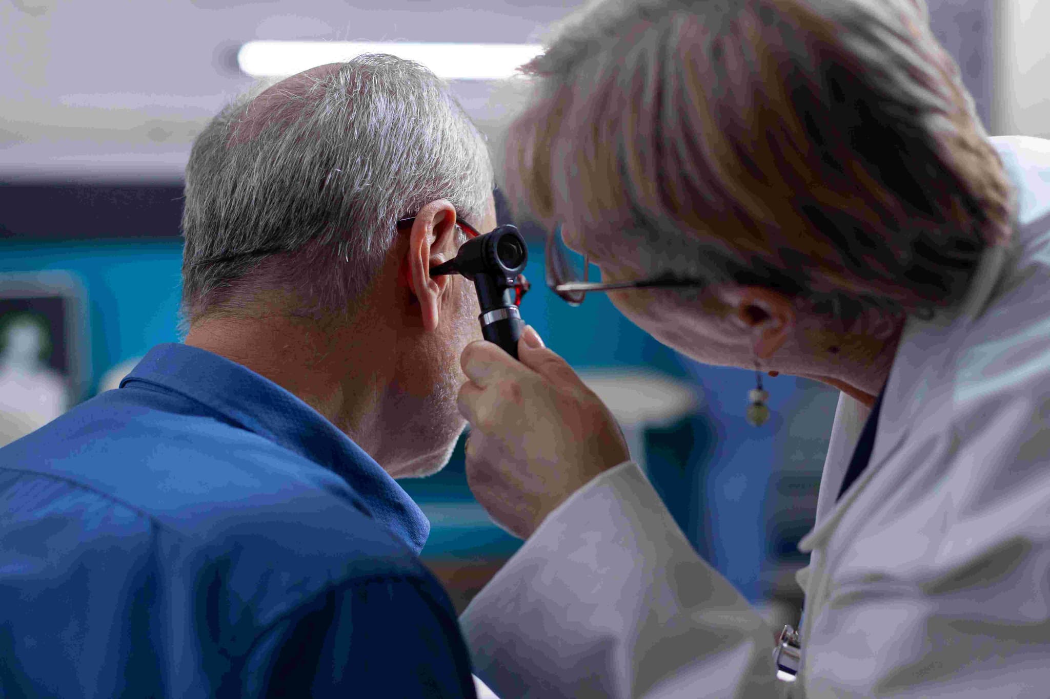
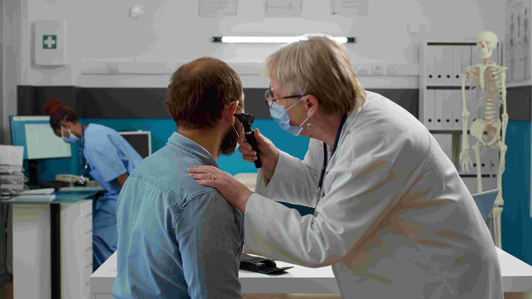
  • Otoscopy: A high-magnification view of the eardrum to check for redness, bulging, or scarring.

  • Acoustic Reflex Testing: To assess how the middle ear muscles respond to sound, which is often dampened by fluid.

  • Speech Reception Threshold (SRT): Especially in children, to determine the lowest volume at which they can recognize words.

  • Medical Review: Confirming that the fluid has not cleared on its own during the "watchful waiting" period.

  • Immediate Results: Most patients notice a dramatic improvement in hearing and a relief of internal pressure as soon as they wake up.

  • Activity: Recovery is rapid; most children can return to school or daycare the very next day.

  • Water Precautions: It is important to keep the ears dry while the tubes are in place. Your doctor may recommend earplugs or cotton wool with petroleum jelly during hair washing or swimming.

  • Natural Extrusion: Grommets are temporary. As the eardrum heals, it naturally pushes the tube out, usually after 6 to 18 months.

  • Monitoring: Regular follow-up appointments are necessary to ensure the tubes remain open and to check the eardrum's healing once they fall out.

  • Instant Hearing Restoration: Physically removing the "glue" immediately eliminates the conductive hearing loss associated with fluid buildup.

  • Break the Infection Cycle: By keeping the middle ear ventilated, grommets prevent the stagnant environment where bacteria and viruses thrive.

  • Supports Development: Resolving hearing issues early is critical for a child's educational and social progress.

  • Minimally Invasive: The procedure is performed entirely through the ear canal, leaving no external scars or visible changes to the ear.

  • Self-Resolving: Because the body eventually rejects the tubes and the eardrum heals itself, it is a low-impact solution for a common childhood condition.

Tympanoplasty (Eardrum Repair)

Tympanoplasty is a specialized surgical procedure performed to repair a perforated (torn) eardrum or to reconstruct the tiny bones (ossicles) of the middle ear. The primary goals are to prevent recurrent ear infections and to improve hearing by restoring the natural vibration of the eardrum and sound conduction through the middle ear.

  • Chronic Perforations: When a hole in the eardrum fails to heal on its own after 3 to 6 months, leaving the middle ear exposed.

  • Recurrent Ear Infections: If you experience frequent bouts of chronic otitis media caused by water or bacteria entering through the perforation.

  • Conductive Hearing Loss: When a ruptured eardrum or damaged middle ear bones prevent sound from reaching the inner ear effectively.

  • Cholesteatoma: If a skin cyst begins to grow behind the eardrum, requiring removal and subsequent reconstruction of the ear structure.

  • Water Sensitivity: When you must constantly protect your ear from any moisture to avoid immediate pain or infection.

  • Anesthesia: The surgery is typically performed as an outpatient procedure under general or local anesthesia and takes between 1 and 2 hours.

  • Surgical Approach: The surgeon accesses the eardrum through a precise incision made either inside the ear canal (transcanal) or behind the ear (post-auricular).

  • Grafting: A small piece of the patient's own tissue—usually fascia (connective tissue from a muscle) or tragus cartilage (from the outer ear)—is harvested to act as a permanent "patch" for the hole.

  • Placement: The graft is carefully tucked over or under the perforation and supported by dissolvable packing material (gel foam) to keep it in place during the initial healing phase.

  • Ossiculoplasty (If Needed): If the middle ear bones are damaged, the surgeon may repair them using synthetic implants or the patient's own bone to restore sound conduction.

  • Closure: Any external incisions are closed with fine sutures, and the ear canal is protected with a sterile dressing.

  • Audiometry (Hearing Test): A comprehensive baseline test to document the degree of hearing loss before the reconstruction.

  • Infection Clearance: Ensuring the ear is "dry" and free of active infection for several weeks leading up to the surgery.

  • Imaging: In complex cases involving the middle ear bones, a CT scan of the temporal bone may be used to map the internal anatomy.

  • Fasting: Following strict "nothing by mouth" instructions if general anesthesia is planned.

  • Transportation: Arranging for a ride home, as you may feel dizzy or "off-balance" immediately following the procedure.

  • Otoscopy: A high-magnification view of the eardrum to determine the size and location of the perforation.

  • Tympanometry: To assess the pressure and mobility of the eardrum and the status of the middle ear space.

  • Speech Discrimination Test: To evaluate how well you can understand words, which helps predict the potential for hearing improvement.

  • Microscopic Clearance: Cleaning the ear canal of any debris or wax to provide the surgeon with a clear surgical field.

  • Initial Recovery: You may feel dizzy or have a sense of "fullness" in the ear for a few days due to the internal packing material.

  • Water Protection: It is critical to keep the ear bone-dry for at least 4 to 6 weeks. Use a cotton ball coated in petroleum jelly during showers to prevent any moisture entry.

  • Pressure Restrictions: Avoid blowing your nose, flying, or strenuous lifting for 2 to 4 weeks, as internal pressure can dislodge the delicate graft.

  • Healing Timeline: The internal packing dissolves or is removed after 2 to 3 weeks. A follow-up hearing test is usually performed 6 to 12 weeks later to confirm success.

  • Sensation Changes: Some patients notice a temporary metallic taste on one side of the tongue if a small nerve near the eardrum is stretched during surgery.

  • High Success Rates: Modern grafting techniques result in successful eardrum closure in approximately 85% to 90% of cases.

  • Restores Natural Barrier: Successfully sealing the eardrum prevents bacteria from entering the middle ear, ending the cycle of chronic infections.

  • Improves Quality of Life: Restoring hearing and eliminating the need for strict water precautions allows for a return to swimming and other daily activities.

  • Prevents Bone Damage: Closing the perforation protects the delicate ossicles from erosion caused by chronic inflammation.

  • Permanent Repair: Using the patient's own tissue (fascia or cartilage) provides a highly biocompatible and durable repair that lasts a lifetime.

Mastoidectomy (Chronic Infection/Cholesteatoma)

Mastoidectomy is a specialized surgical procedure performed to remove diseased air cells from the mastoid bone—the honeycomb-like bone located directly behind the ear. It is primarily used to treat a cholesteatoma, a non-cancerous skin cyst that can erode ear structures, or to clear severe, chronic infections that have not responded to intensive antibiotic therapy.

  • Cholesteatoma: When a skin cyst forms in the middle ear and begins to expand into the mastoid bone, risking damage to the hearing bones or facial nerve.

  • Chronic Mastoiditis: If a persistent ear infection spreads into the bone, causing pain, swelling, and a risk of deeper abscesses.

  • Failed Antibiotic Therapy: When chronic drainage (otorrhea) continues despite multiple rounds of specialized ear drops and medications.

  • Complicated Ear Infections: If an infection threatens to spread toward the brain (meningitis) or the inner ear (labyrinthitis).

  • Aural Polyps: When chronic inflammation causes fleshy growths to form in the ear canal, often masking underlying bone disease.

  • Anesthesia: The surgery is performed under general anesthesia and typically takes between 2 to 4 hours due to the delicate nature of the structures involved.

  • Incision: The surgeon makes a precise incision either behind the ear (post-auricular) or inside the ear canal to gain access to the bone.

  • Bone Removal: Using a high-speed surgical drill and a high-powered operating microscope, the surgeon carefully opens the mastoid bone to reach the diseased air cells.

  • Cleaning: All infected tissue or the cholesteatoma cyst is meticulously removed to create a "dry, safe ear" and prevent recurrence.

  • Surgical Variations:
    Simple (Cortical) Mastoidectomy: Only the mastoid air cells are removed; the ear canal and middle ear structures remain intact.
    Modified Radical Mastoidectomy: The most common approach for cholesteatoma, where some middle ear structures are preserved to allow for future hearing reconstruction.
    Radical Mastoidectomy: The eardrum and most middle ear structures are removed to create a single "common cavity" for the ear canal and mastoid.

  • High-Resolution CT Scan: A vital "roadmap" used by the surgeon to visualize the exact anatomy of your mastoid bone and the location of the facial nerve.

  • Audiometry (Hearing Test): Establishing a detailed baseline of your current hearing levels to plan for potential reconstruction.

  • Infection Management: Using specialized ear drops in the weeks leading up to surgery to reduce inflammation and "quiet" the ear.

  • Fasting: Following strict "nothing by mouth" instructions for the hours preceding general anesthesia.

  • Transportation: Arranging for a ride home and a caregiver for the first 24 hours, as dizziness is common immediately following the procedure.

  • Ear Swab/Culture: To identify the specific bacteria causing the infection and ensure the most effective antibiotics are used.

  • Microscopic Ear Exam: A thorough cleaning and inspection of the ear canal by the specialist to assess the extent of the disease.

  • Tympanometry: Measuring the pressure in the middle ear and the mobility of the eardrum.

  • Neurological Screening: Checking the function of the facial nerve and balance system before the bone is opened.

  • Hospital Stay: Most patients go home the same day or remain for one night of observation. A large "head bandage" is often worn for the first 24 hours.

  • Immediate Symptoms: It is normal to feel dizzy, have a "stuffy" sensation, or hear popping and crackling sounds as internal packing dissolves.

  • Water Protection: The ear must be kept completely dry for 4 to 6 weeks. Use a cotton ball with petroleum jelly during showers to prevent any moisture entry.

  • Pressure Restrictions: Avoid blowing your nose, heavy lifting, or flying for at least 3 to 4 weeks to prevent internal pressure from damaging the healing area.

  • Wound Care: External stitches are usually removed after 7 to 10 days. Follow-up appointments are critical to clean the "mastoid cavity" and monitor for regrowth.

  • Prevents Life-Threatening Complications: By removing deep-seated disease, the procedure stops infections from spreading to the brain or spinal fluid.

  • Eliminates Chronic Drainage: Successfully clearing the mastoid air cells finally ends the cycle of foul-smelling ear discharge and pain.

  • Safety-First Approach: Modern microsurgical techniques allow for the meticulous removal of cysts while protecting the delicate facial nerve and inner ear.

  • Hearing Potential: While the primary goal is safety, a "safe ear" provides the necessary foundation for future hearing bone reconstruction (ossiculoplasty).

  • Long-Term Stability: For many patients with chronic disease, a mastoidectomy is the only way to achieve a permanently dry and healthy ear.

Cochlear Implant Surgery (Hearing Restoration)

Cochlear implant surgery is a specialized procedure that restores a sense of sound by bypassing damaged parts of the inner ear and directly stimulating the auditory nerve. Unlike hearing aids, which amplify sound, cochlear implants convert sound into electrical signals that the brain interprets as speech and environmental noise.

  • Severe-to-Profound Hearing Loss: When high-power hearing aids no longer provide enough clarity for speech understanding in one or both ears.

  • Poor Speech Recognition: For adults who score poorly on speech recognition tests (typically less than 50%) even with well-fitted hearing aids.

  • Early Childhood Intervention: Children as young as 9 to 12 months who are born with profound hearing loss to support near-normal speech and language development.

  • Hybrid Candidacy: For individuals who have some residual low-frequency hearing but suffer from profound high-frequency hearing loss.

  • Sensorineural Loss: When the tiny hair cells in the cochlea are damaged or missing, but the auditory nerve remains functional.

  • Anesthesia: The surgery is an outpatient procedure performed under general anesthesia and typically lasts between 1.5 and 2.5 hours.

  • Incision and Access: The surgeon makes a small, discreet incision behind the ear and creates a tiny opening in the mastoid bone to reach the inner ear.

  • Electrode Insertion: A thin, flexible electrode array is carefully threaded into the cochlea—the spiral-shaped hearing organ.

  • Internal Component Placement: The receiver-stimulator is secured under the skin against the skull, where it will communicate with the external processor.

  • Verification: Surgeons use intraoperative tests or X-rays to confirm the electrodes are correctly positioned before closing the incision with sutures.

  • Same-Day Discharge: Most patients are monitored for a few hours and then allowed to return home the same day.

  • Comprehensive Audiological Evaluation: Detailed testing to ensure hearing aids are no longer providing sufficient benefit.

  • CT or MRI Imaging: High-resolution scans to confirm the cochlea is shaped correctly and the auditory nerve is present.

  • Vaccination Review: Recipients are often recommended to receive meningitis vaccinations prior to surgery as a standard safety precaution.

  • Expectation Setting: Discussing the "rehabilitation" phase, as the brain requires time and practice to interpret the new electrical signals.

  • Transportation: Arranging for a ride home, as you will be drowsy from the anesthesia and may experience temporary dizziness.

  • Speech-in-Noise Testing: Measuring your ability to understand sentences in a noisy environment while wearing hearing aids.

  • Promontory Stimulation: In some cases, a small test to confirm the auditory nerve responds to electrical pulses.

  • Vestibular Testing: Assessing your balance system, as the inner ear is responsible for both hearing and equilibrium.

  • Otoacoustic Emissions (OAEs): To check the function of the outer hair cells in the cochlea.

  • Initial Healing (1–2 weeks): Stitches are typically removed after 7–14 days. You may experience mild swelling, temporary changes in taste, or a "full" feeling in the ear.

  • Activation (2–4 weeks post-op): Hearing is not immediate. Once the site has healed, an audiologist fits the external processor and "switches on" the device for the first time.

  • Mapping and Rehabilitation: Initial sounds often seem "robotic" or "mechanical." You will undergo multiple "mapping" sessions to fine-tune the device settings.

  • Brain Training: Consistent participation in auditory therapy is essential to help the brain learn to interpret these new signals into recognizable speech.

  • Activity Restrictions: Avoid heavy lifting or strenuous exercise for the first 1–2 weeks to allow the internal components to settle.

  • Direct Nerve Stimulation: By bypassing damaged hair cells, the implant provides a clear signal directly to the brain, overcoming severe biological barriers.

  • Proven Success in Children: Early implantation allows children to attend mainstream schools and develop complex language skills alongside their peers.

  • Restores Social Connection: Significantly improves the ability to communicate in groups, use the telephone, and enjoy music.

  • Advanced Technology: Modern implants are "MRI-safe" and offer water-resistant processors, allowing for a highly active lifestyle.

  • Lifelong Support: Once implanted, the internal device is designed to last for decades, while the external processor can be upgraded as technology improves.

Stapedectomy (Otosclerosis)

Stapedectomy is a specialized microsurgical procedure used to treat otosclerosis, a condition where the smallest bone in the human body—the stapes (stirrup)—becomes stuck in place by abnormal bone growth. This prevents the bone from vibrating, leading to progressive conductive hearing loss. By replacing the immobile bone with a medical-grade prosthesis, the surgery restores the natural transmission of sound waves to the inner ear.

  • Otosclerosis Diagnosis: When clinical exams confirm that abnormal bone remodeling is "fixing" the stapes bone in place.

  • Conductive Hearing Loss: If you have significant difficulty hearing faint sounds or identifying speech, particularly in the lower frequencies.

  • Air-Bone Gap: When hearing tests show a large difference between how well you hear through the air versus through the bone.

  • Desire to Eliminate Hearing Aids: For patients who prefer a permanent surgical solution over the daily use of external amplification devices.

  • Good Inner Ear Health: When the hearing nerve (cochlea) is still healthy and capable of processing sound once the mechanical blockage is removed.

  • Anesthesia: The surgery is typically performed as an outpatient procedure under local anesthesia with sedation or general anesthesia and takes about 45 to 90 minutes.

  • Access: The surgeon reaches the middle ear directly through the ear canal using a high-powered microscope or endoscope, usually leaving no external incision.

  • Removal: The immobile stapes bone is carefully removed, or a microscopic hole is made in its base (a variation known as a Stapedotomy).

  • Prosthesis Placement: A tiny artificial piston, usually made of biocompatible materials like Teflon, titanium, or stainless steel, is inserted into the middle ear.

  • Restoration: The prosthesis is securely attached to the second ear bone (the incus) and the inner ear, allowing sound vibrations to bypass the diseased bone and reach the hearing nerve.

  • Closure: Small pieces of tissue or dissolvable packing are placed to support the new prosthesis while it heals.

  • Audiometry (Hearing Test): A detailed baseline test to measure the exact degree of conductive hearing loss and ensure the inner ear is healthy.

  • Tympanometry: To confirm that the eardrum is moving correctly and that the hearing loss is caused by bone stiffness rather than fluid.

  • CT Scan of the Temporal Bone: In some cases, to visualize the thickness of the otosclerotic bone and plan the surgical approach.

  • Medication Review: Discussing the management of blood thinners and supplements that could interfere with delicate microsurgery.

  • Transportation: Arranging for a ride home, as it is very common to feel "off-balance" or dizzy immediately following the procedure.

  • Rinne and Weber Tests: Simple tuning fork tests used by the specialist to differentiate between conductive and sensorineural hearing loss.

  • Speech Discrimination Score: To determine how clearly you can understand words once sound is loud enough, which helps predict surgical success.

  • Acoustic Reflex Testing: Measuring the involuntary muscle contraction in the middle ear, which is typically absent in cases of otosclerosis.

  • Physical Examination: Ensuring the ear canal and eardrum are free of infection or inflammation before the sterile procedure.

  • Initial Recovery: It is very common to feel dizzy or experience mild vertigo for the first 24–48 hours. The ear will feel "blocked" due to the internal packing material.

  • The "Pop": Hearing often improves dramatically once the packing is removed (usually 1–3 weeks later), though some patients notice an immediate difference.

  • Pressure Restrictions: For 4 weeks, you must strictly avoid blowing your nose hard, flying, high-altitude travel, or heavy lifting, as internal pressure can dislodge the prosthesis.

  • Water Protection: Keep the ear completely dry for at least 2–3 weeks. Use a cotton ball coated in petroleum jelly during showers to prevent any moisture entry.

  • Taste Disturbance: A metallic taste on the side of the tongue is common because the taste nerve runs right next to the surgical site; this usually resolves within a few weeks.

  • Superior Success Rates: Significant and lasting improvement in hearing is achieved in approximately 90% to 95% of cases.

  • Permanent Mechanical Fix: Unlike hearing aids that require batteries and maintenance, the prosthesis provides a lifelong mechanical restoration of sound.

  • No External Scars: Because the procedure is performed through the ear canal, there are typically no visible signs of surgery once the area has healed.

  • Rapid Return to Normalcy: Most patients return to light daily activities within a few days, with a significant boost in their quality of life and social communication.

  • Standard of Care: Stapedectomy is the global gold standard for treating conductive hearing loss caused by otosclerosis, offering a highly predictable outcome.

Functional Endoscopic Sinus Surgery (FESS)

Functional Endoscopic Sinus Surgery (FESS) is a minimally invasive procedure used to treat chronic sinus conditions, such as persistent sinusitis and nasal polyps. By restoring natural drainage and airflow, FESS eliminates the root cause of chronic inflammation. The procedure is performed entirely through the nostrils using a specialized nasal endoscope (a thin tube with a magnified camera), meaning there are no external incisions or visible scars.

  • Chronic Sinusitis: If you suffer from sinus inflammation lasting more than 12 weeks that hasn't responded to medical therapy.

  • Nasal Polyps: Benign growths in the nasal passages that physically obstruct your breathing and block natural fluid drainage.

  • Recurrent Acute Infections: When you experience four or more severe sinus infections per year, significantly impacting your quality of life.

  • Anatomical Obstructions: To correct issues such as a deviated septum or enlarged turbinates that contribute to chronic blockages.

  • Failed Medication: When antibiotics, steroid sprays, and saline rinses are no longer effective in managing your symptoms.

  • Anesthesia: The surgery is usually performed as an outpatient (day-care) procedure under general anesthesia and typically lasts 2 to 3 hours.

  • Visualisation: The surgeon inserts the endoscope into the nostrils to obtain a high-definition, magnified 3D view of the internal sinus passages.

  • Tissue Removal: Using precise micro-instruments, the surgeon removes obstructions such as nasal polyps, diseased tissue, or small pieces of bone blocking the sinus openings.

  • Opening the Passageways: The natural drainage pathways (ostia) are carefully widened to improve long-term ventilation and allow future nasal rinses to reach deep into the sinus cavities.

  • Completion: To control minor bleeding, the surgeon may place dissolvable packing or temporary splints inside the nose, which do not require manual removal.

  • CT Scan of the Sinuses: A mandatory high-resolution "map" used by the surgeon to navigate the complex anatomy near the eyes and brain.

  • Medication Review: You may be prescribed a short course of oral steroids or antibiotics before surgery to reduce inflammation and bleeding.

  • Smoking Cessation: Patients are strongly advised to stop smoking several weeks before surgery, as tobacco use significantly hinders the healing of sinus tissue.

  • Fasting: Following strict "nothing by mouth" instructions for the hours preceding your general anesthesia.

  • Transportation: Arranging for a ride home, as you will be drowsy and may have temporary nasal congestion affecting your comfort.

  • Nasal Endoscopy: An in-office diagnostic check to visualize the extent of polyps or structural blockages.

  • Allergy Testing: To determine if underlying allergies need to be managed alongside surgery for the best long-term outcome.

  • Blood Tests: Standard pre-operative panels to ensure healthy clotting factors and general surgical readiness.

  • Olfactory Testing: Assessing your baseline sense of smell to monitor improvements after the airways are cleared.

  • Immediate Symptoms: It is normal to experience stuffiness, mild facial pressure, and blood-stained drainage for the first 3 to 5 days.

  • Nasal Irrigation: Using a saline nasal wash (usually 4+ times daily) is essential to flush out debris, prevent crusting, and ensure the new openings stay clear.

  • Crucial Restrictions: You must strictly avoid blowing your nose for at least 7 to 14 days and avoid strenuous activity or heavy lifting for 2 to 3 weeks to prevent bleeding.

  • Debridement: Follow-up visits are vital for your surgeon to professionally clean out dried mucus and ensure the sinuses are healing without excessive scar tissue.

  • Healing Timeline: While most patients return to work or school within one week, full internal healing and the stabilization of the sinus lining can take several months.

  • Targeted Precision: FESS focuses only on the diseased areas, preserving as much healthy nasal tissue and mucous membrane as possible.

  • No External Scarring: Because the surgery is entirely internal, there is no change to your physical appearance and no external stitches to remove.

  • High Safety Profile: With major complication rates of less than 0.5%, FESS is considered one of the safest and most effective ways to treat chronic sinus disease.

  • Long-Term Relief: By widening the natural ostia, the surgery provides a permanent structural improvement that allows for better drainage for years to come.

  • Improved Medication Delivery: Post-operative nasal sprays and rinses can finally reach the deep sinus cavities, making ongoing maintenance far more effective.

Septoplasty (Deviated Septum Correction)

Septoplasty is a specialized surgical procedure performed to straighten a deviated septum—the internal wall of bone and cartilage that separates your two nostrils. When the septum is crooked or "deviated," it can block one side of the nose and significantly reduce airflow. By repositioning the septal midline, this procedure restores balanced breathing and improves overall nasal health.

  • Chronic Nasal Obstruction: If one or both nostrils feel constantly blocked, making it difficult to breathe through your nose.

  • Recurrent Sinus Infections: When a deviated septum prevents proper drainage, leading to frequent sinus pressure and infections.

  • Chronic Snoring or Sleep Apnea: If the nasal blockage contributes to disrupted breathing patterns during sleep.

  • Frequent Nosebleeds: When the air flowing over a crooked septum dries out the nasal lining, causing recurrent bleeding.

  • Unilateral Congestion: A persistent "stuffy" feeling that is always worse on the same side of the nose.

  • Anesthesia: The surgery is typically an outpatient procedure performed under general or local anesthesia and takes about 30 to 90 minutes.

  • Incision: The surgeon makes a precise incision entirely inside the nose. In most cases, there are no external cuts or visible scars.

  • Mucosal Elevation: The protective lining of the septum (mucosa) is gently lifted away from the underlying cartilage and bone.

  • Reshaping: The crooked portions of the cartilage or bone are trimmed, repositioned, or removed to straighten the midline.

  • Repositioning: The mucosal lining is laid back over the newly straightened structural framework.

  • Stabilization: The surgeon may place soft silicone splints or dissolvable packing inside the nostrils to support the septum as it heals in its new position.

  • Nasal Examination: A thorough evaluation of the internal nasal structure to ensure the breathing issues are caused by the septum and not just enlarged turbinates.

  • Medication Audit: You must stop taking aspirin, ibuprofen, and certain herbal supplements for 1–2 weeks prior to surgery to minimize the risk of bleeding.

  • Smoking Cessation: Stopping smoking is highly recommended, as nicotine restricts blood flow and can slow the healing of the nasal mucosa.

  • Fasting: Following strict "nothing by mouth" instructions for the hours preceding your anesthesia.

  • Transportation: Arranging for a ride home, as you will be drowsy and your nose will feel quite congested immediately after the procedure.

  • Nasal Endoscopy: Using a thin camera to view the back of the nasal passage and confirm the extent of the deviation.

  • Acoustic Rhinometry: A non-invasive test that uses sound waves to measure the width of the nasal cavity and the location of the narrowest point.

  • Peak Nasal Inspiratory Flow (PNIF): Measuring the maximum amount of air you can inhale through your nose to establish a functional baseline.

  • CT Scan (Optional): Sometimes used if the surgeon needs to evaluate the sinuses simultaneously for chronic sinusitis.

  • Immediate Symptoms: Expect some internal swelling, "stuffiness," and mild blood-stained drainage for the first 3 to 5 days.

  • Nasal Splints: If splints were used, they are typically removed by the doctor during a follow-up visit approximately one week after surgery.

  • Crucial Rule: You must not blow your nose for at least 7 to 10 days post-surgery to prevent bleeding or displacement of the healing tissue.

  • Nasal Care: Using saline nasal sprays or rinses is essential to keep the passages clean, moist, and free of crusting.

  • Activity Restrictions: Avoid strenuous exercise, heavy lifting, and contact sports for 2 to 4 weeks to prevent injury or pressure-related nosebleeds.

  • Restores Natural Airflow: Directly addresses the structural cause of nasal blockage, providing a permanent improvement in breathing quality.

  • No External Changes: Unlike a rhinoplasty, a standard septoplasty focuses on the internal structure and does not change the outward shape of your nose.

  • Reduces Sinus Pressure: By opening the nasal airway, the procedure allows for better ventilation and drainage of the paranasal sinuses.

  • Rapid Recovery: Most patients return to work or sedentary activities within a few days, with a significant boost in energy levels due to better oxygen intake.

  • High Patient Satisfaction: Septoplasty is one of the most successful functional surgeries, with the majority of patients reporting a dramatic improvement in their quality of life.

Turbinate Reduction
Turbinate Reduction

Turbinate reduction (or turbinoplasty) is a specialized surgical procedure performed to shrink enlarged bony and soft tissue structures inside the nose. These structures, known as turbinates, are responsible for cleansing, warming, and humidifying the air you breathe. When they become chronically swollen—often due to allergies or irritants—they can severely block nasal airflow.

  • Chronic Nasal Congestion: If you experience a persistent "stuffy" feeling that does not improve with steroid sprays or antihistamines.

  • Difficulty Breathing Through the Nose: When enlarged tissue physically obstructs the airway, forcing you to breathe through your mouth.

  • Disrupted Sleep: If nasal blockages contribute to heavy snoring or interfere with CPAP therapy for sleep apnea.

  • Persistent Post-Nasal Drip: When swollen turbinates lead to excessive mucus production and a constant "dripping" sensation in the throat.

  • Failed Medical Management: When you have exhausted non-surgical options like nasal rinses, allergy shots, or decongestants.

  • Anesthesia: The procedure is typically performed as an outpatient surgery under either local or general anesthesia and takes approximately 15 to 30 minutes.

  • Surgical Techniques:
    Radiofrequency Ablation (RFA): A needle-like probe uses controlled heat energy to shrink the soft tissue beneath the surface. This is the least invasive method.
    Coblation: Similar to RFA, but utilizes lower temperatures to dissolve excess tissue while preserving the surrounding area.
    Microdebrider Submucosal Resection: A tiny, rotating device removes a small amount of bone or deep tissue through a microscopic incision, keeping the outer lining (mucosa) intact.
    Partial Resection: A small portion of the turbinate bone and tissue is physically trimmed away to create a wider airway.

  • Access: All work is performed entirely through the nostrils; there are no external incisions or visible scars.

  • Completion: Dissolvable packing or light gauze may be placed in the nose to control minor bleeding and support the tissues as they heal.

  • Nasal Endoscopy: A diagnostic check to confirm that the obstruction is caused by turbinate hypertrophy rather than polyps or a deviated septum.

  • Medication Audit: You must stop taking blood-thinning medications, including aspirin and certain herbal supplements, at least one week before surgery.

  • Allergy Assessment: Identifying underlying triggers to ensure long-term success, as unmanaged allergies can cause tissue to regrow over time.

  • Fasting: Following strict "nothing by mouth" instructions for the hours preceding your anesthesia.

  • Transportation: Arranging for a ride home, as you will feel congested and may be drowsy from the sedation or anesthesia.

  • Acoustic Rhinometry: To measure the cross-sectional area of the nasal cavity and identify the narrowest points of resistance.

  • Rhinomanometry: A functional test that evaluates the pressure and flow of air during active breathing through the nose.

  • Decongestant Challenge: Applying a temporary spray to see how much the tissue is capable of shrinking, which helps predict the surgical outcome.

  • CT Scan (Optional): Usually only required if the surgeon is planning to address the sinuses or a deviated septum during the same session.

  • Immediate Symptoms: You will feel very "stuffy" for the first 1 to 2 weeks due to internal swelling and natural crusting during the healing phase.

  • Mandatory Nasal Rinses: Using saline nasal sprays or neti pots (4–6 times daily) is essential to wash away crusts and prevent infection.

  • Crucial Rule: You must not blow your nose for at least 7 to 10 days post-surgery to prevent a sudden nosebleed.

  • Activity Restrictions: Most people return to work within 2 to 3 days, but you must avoid heavy lifting or intense exercise for at least 2 weeks.

  • Full Recovery: While breathing improves as swelling subsides, the final stabilized result is typically achieved 3 to 6 weeks after the procedure.

  • Permanent Airway Opening: Provides a long-term structural solution for patients who have become dependent on temporary nasal sprays.

  • Preserves Nasal Function: Modern "submucosal" techniques shrink the bulk of the turbinate while leaving the protective lining healthy and functional.

  • Minimally Invasive: Most methods involve zero external cutting and a very rapid return to daily routines with minimal discomfort.

  • Improved Sleep Quality: Clearing the nasal passage often leads to significantly reduced snoring and better oxygen intake during rest.

  • High Success Rates: When combined with proper allergy management, turbinate reduction offers a highly predictable and transformative improvement in breathing.

Nasal Polyp Removal
Nasal Polyp Removal

Nasal polyp removal, medically known as a polypectomy, is a surgical procedure to extract non-cancerous, teardrop-shaped growths from the lining of the nasal passages or sinuses. When these polyps become large enough to block breathing, reduce the sense of smell, or cause chronic sinus infections, surgical intervention is the most effective way to clear the airway and restore health.

  • Persistent Nasal Obstruction: If large polyps physically block your nostrils, making it difficult to breathe through your nose.

  • Loss of Smell (Anosmia): When polyps block scent molecules from reaching the olfactory nerves at the top of the nasal cavity.

  • Chronic Sinusitis: If you experience ongoing sinus pressure, pain, and infections that do not respond to antibiotics or steroid sprays.

  • Sleep Disruption: When nasal blockages lead to heavy snoring or contribute to obstructive sleep apnea.

  • Post-Nasal Drip: Persistent mucus drainage down the back of the throat caused by polyps interfering with natural sinus clearance.

  • Anesthesia: The surgery is typically an outpatient procedure performed under general or local anesthesia and takes about 45 to 90 minutes.

  • Endoscopic Approach: The surgeon inserts a thin, flexible tube with a high-definition camera (endoscope) into the nostrils. There are no external incisions or visible scars.

  • Removal Techniques: Using small precision instruments or a microdebrider (a tiny, rotating suction tool), the surgeon carefully shaves away or snips the polyps from the mucosal lining.

  • Sinus Opening: The surgeon often opens the natural drainage pathways of the sinuses to prevent future polyps from forming and to allow medicated sprays to reach deeper into the cavity.

  • Packing: Dissolvable packing or soft splints may be placed in the nose to control minor bleeding and support the tissues during the initial healing phase.

  • CT Scan of the Sinuses: A detailed "roadmap" used by the surgeon to visualize the extent of the polyps and their proximity to the eyes and brain.

  • Steroid Pre-Treatment: You may be prescribed a short course of oral steroids before surgery to shrink the polyps and reduce the risk of bleeding.

  • Medication Audit: You must stop taking aspirin, ibuprofen, and certain herbal supplements for 1–2 weeks prior to surgery to minimize bleeding risks.

  • Smoking Cessation: Patients are strongly advised to stop smoking, as it irritates the nasal lining and can cause polyps to return more quickly.

  • Transportation: Arranging for a ride home, as you will feel congested and may be drowsy from the anesthesia.

  • Nasal Endoscopy: An in-office diagnostic check to confirm the size, location, and color of the polyps.

  • Allergy Testing: To identify triggers that may be causing the chronic inflammation responsible for polyp growth.

  • Aspirin Sensitivity Test: Particularly for patients with asthma, to check for "Samter’s Triad," which indicates a higher risk of polyp recurrence.

  • Olfactory Testing: Assessing your baseline sense of smell to track improvement after the obstruction is removed.

  • Immediate Symptoms: You will feel very "stuffed up" for the first 1 to 2 weeks due to internal swelling and natural crusting. Mild blood-stained drainage is normal for a few days.

  • Mandatory Saline Rinses: Using a Neti pot or saline spray (4–6 times daily) is essential to wash away blood and crusts and to speed up the healing process.

  • Long-Term Medication: You will likely need to continue using nasal steroid sprays long-term to manage underlying inflammation and prevent the polyps from growing back.

  • Activity Restrictions: Most patients return to work in 1 week. You must avoid heavy lifting, bending over, or blowing your nose for at least 2 weeks.

  • Follow-up Debridement: Your surgeon will perform minor office cleanings to remove any remaining crusts or early scar tissue (adhesions).

  • Restores Clear Breathing: Removing the physical mass of the polyps provides immediate and dramatic relief from nasal congestion.

  • Revives the Sense of Taste and Smell: Clearing the olfactory pathway often allows patients to enjoy food and scents that were previously blocked.

  • Reduces Infection Frequency: By restoring natural sinus drainage, the surgery eliminates the stagnant environment where bacteria thrive.

  • Minimally Invasive: The endoscopic technique allows for thorough removal with zero external cutting and a relatively fast recovery.

  • Synergistic Care: When combined with ongoing allergy or asthma management, a polypectomy offers the best chance for long-term respiratory health and comfort.

CSF Leak Repair (Endoscopic)

Endoscopic CSF (Cerebrospinal Fluid) leak repair is a specialized, minimally invasive surgery performed to seal a hole in the skull base—the critical bone barrier separating the brain from the nasal cavity. This procedure is essential for stopping the drainage of brain fluid and, more importantly, preventing bacteria from entering the central nervous system and causing life-threatening infections such as meningitis.

  • Clear Nasal Drainage (Rhinorrhea): If you experience a persistent, watery discharge from one side of the nose that often increases when leaning forward or straining.

  • Salty or Metallic Taste: A distinct sensation in the back of the throat caused by spinal fluid draining down from the skull base.

  • Recurrent Meningitis: If you have suffered from unexplained or repeat bacterial infections of the brain lining.

  • Post-Traumatic Leaks: Following a head injury or facial fracture that has compromised the integrity of the skull base.

  • Iatrogenic Leaks: As a follow-up to previous sinus or neurosurgical procedures where a small opening in the dura was created.

  • Spontaneous Leaks: Often associated with high intracranial pressure, frequently seen in patients with idiopathic intracranial hypertension.

  • Anesthesia: The surgery is performed under general anesthesia and typically takes between 2 to 4 hours due to the microscopic precision required.

  • Endoscopic Approach: The surgeon operates entirely through the nostrils using a high-definition endoscope. There are no external incisions and no visible facial scarring.

  • Localization: Surgeons use the endoscope and sometimes a fluorescent dye (fluorescein) injected into the spinal fluid to pinpoint the exact site of the leak with total accuracy.

  • The Multi-Layer Repair: A "sandwich" technique is often used to ensure a watertight seal:
    Grafts: Small pieces of the patient’s own tissue (fat, muscle lining, or nasal mucosa) are placed over or into the defect.
    Vascularized Flap: For larger leaks, a Nasoseptal Flap—tissue with its own blood supply—is rotated from the septum to provide a robust, living cover for the repair.
    Sealants: Medical-grade "glue" or fibrin sealant is applied to bond the grafts to the bone.

  • Packing: The nose is packed with specialized sponge-like material (either dissolvable or removable) to maintain constant pressure on the repair site during the first stage of healing.

  • High-Resolution Imaging: Combining CT scans (to see bone) and MRI (to see fluid) to create a precise surgical roadmap.

  • Beta-2 Transferrin Test: A laboratory confirmation that the fluid draining from the nose is indeed cerebrospinal fluid.

  • Pneumococcal Vaccination: Ensuring you are protected against the most common bacteria that cause meningitis before the skull base is manipulated.

  • Intracranial Pressure Assessment: Evaluating if you have high brain pressure that might have caused the leak and could potentially threaten the new repair.

  • Fasting: Following strict "nothing by mouth" instructions for the hours preceding your general anesthesia.

  • Pledget Study: Placing small cotton sponges in the nose to absorb drainage, which are then tested for the presence of spinal fluid markers.

  • CT Cisternography: A specialized scan where contrast dye is injected into the spinal fluid to visualize the exact point of the "leakage."

  • Neurological Consultation: A full assessment of brain function and cranial nerves to establish a baseline before surgery.

  • Blood Clotting Panel: Ensuring your blood's ability to clot is optimal for a deep, vascularized repair.

  • Hospital Monitoring: Patients typically remain in the hospital for 1 to 3 days. You may need to keep your head elevated at a 30–45 degree angle to reduce pressure on the repair.

  • Lumbar Drain: Occasionally, a small tube is placed in the lower back for 24–48 hours to divert spinal fluid and allow the nasal "patch" to bond without pressure.

  • The "No" Rules (4–6 Weeks): To protect the seal, you must strictly avoid:
    Blowing your nose: The most critical restriction to prevent air from being forced into the brain.
    Straining: Stool softeners are often prescribed to prevent "valsalva" maneuvers during bowel movements.
    Physical Exertion: No heavy lifting or intense exercise that could dislodge the graft.
    Closed-Mouth Sneezing: You must always sneeze with your mouth open to release internal pressure.

  • Sensation Changes: Headaches and a temporary decrease in the sense of smell (anosmia) are common as the nasal passages heal.

  • Prevents Life-Threatening Infection: By restoring the watertight barrier, the procedure eliminates the direct "highway" for bacteria to reach the brain.

  • High Success Rates: Modern endoscopic techniques have a success rate of 90% to 95% on the first attempt for most skull base defects.

  • Avoids Traditional Brain Surgery: This internal approach replaces the need for an "open" craniotomy, which involves removing a portion of the skull and retracting the brain.

  • Rapid Recovery: Because no external bone is cut and the brain is not manipulated, patients recover much faster and with fewer neurological side effects.

  • Preserves Nasal Function: Using localized grafts and flaps allows the surgeon to repair the defect while maintaining the health of the surrounding nasal lining.

Tonsillectomy
Tonsillectomy

A tonsillectomy is the surgical removal of the two oval-shaped pads of tissue at the back of the throat, known as the tonsils. While historically considered a childhood procedure, it is now frequently performed on adults to treat chronic infections, breathing obstructions, or persistent dental and throat issues.

  • Chronic Tonsillitis: If you experience recurrent, severe sore throats—typically defined as 7 episodes in one year or 5 episodes per year for two consecutive years.

  • Sleep Apnea: When enlarged tonsils physically block the airway during sleep, causing heavy snoring, gasping, or dangerous pauses in breathing.

  • Persistent Tonsil Stones (Tonsilloliths): When debris becomes trapped in the tonsil crypts, causing chronic bad breath and localized pain that does not respond to cleaning.

  • Peritonsillar Abscess: Following a severe infection where a collection of pus forms behind the tonsil, often making it difficult to swallow or open the mouth.

  • Asymmetric Tonsils: If one tonsil is significantly larger than the other, surgery may be recommended to rule out underlying pathology.

  • Anesthesia: The surgery is an outpatient procedure performed under general anesthesia and typically takes between 20 to 45 minutes.

  • Method: The surgeon accesses and removes the tonsils entirely through the mouth. There are no external incisions or visible scars.

  • Surgical Techniques:
    Cold Knife Dissection: The tonsils are precisely removed using a scalpel, a traditional and highly effective method.
    Electrocautery: Thermal energy (heat) is used to remove the tissue while simultaneously sealing blood vessels to minimize bleeding.
    Coblation (Radiofrequency): This advanced technique uses low-temperature energy to dissolve the tissue, which often results in less post-operative pain for the patient.

  • Closure: The surgical sites are usually left to heal naturally as "scabs" form over the area; stitches are rarely required.

  • Medical History Review: Discussing any history of bleeding disorders or adverse reactions to anesthesia.

  • Medication Audit: You must strictly avoid aspirin, ibuprofen, and certain herbal supplements for at least two weeks before surgery to reduce the risk of bleeding.

  • Infection Management: Ensuring you do not have an active, acute tonsillitis infection on the day of the procedure.

  • Fasting: Following "nothing by mouth" instructions for the hours preceding your general anesthesia.

  • Transportation: Arranging for a ride home and a caregiver to stay with you for the first 24 hours, especially for adult patients.

  • Polysomnography (Sleep Study): Often required if the primary reason for surgery is suspected obstructive sleep apnea.

  • Blood Clotting Profile: A standard panel to ensure your blood coagulates properly, which is vital for a safe recovery.

  • Throat Culture: To identify specific bacteria if the surgery is being performed during a period of recurrent infection.

  • Physical Exam: A thorough inspection of the throat and neck to assess the size and position of the tonsils.

  • Healing Timeline: Full recovery and the return to a normal diet typically take 10 to 14 days. Recovery is generally more intense for adults than for children.

  • Pain Management: Severe throat pain is common and often radiates to the ears. Staying on a strict, around-the-clock schedule of prescribed medication is vital for comfort.

  • Hydration: This is the most important part of recovery. Drinking plenty of cool fluids prevents the surgical "scabs" from becoming dry, which can lead to premature falling off and bleeding.

  • Dietary Transition: Start with soft, cool foods (ice cream, jelly, pudding). You must strictly avoid "scratchy," spicy, or acidic foods (chips, citrus, toast) for a full 2 weeks.

  • Activity Restrictions: Most patients return to work or school after 10 days. However, you must avoid heavy lifting or vigorous exercise for 14 days to prevent post-operative bleeding.

  • Eliminates Chronic Pain: For those with recurrent tonsillitis, removing the source of infection provides a permanent end to the cycle of illness and antibiotic use.

  • Restores Restful Sleep: By clearing the airway, the procedure can immediately resolve snoring and significantly improve oxygen levels during sleep.

  • Improves Oral Health: Removing tonsil crypts eliminates the site where tonsil stones form, resolving chronic halitosis (bad breath).

  • Rapid Surgical Process: As a short, outpatient procedure, patients can begin their recovery in the comfort of their own home on the same day.

  • Long-Term Health: Once the tonsils are removed, they do not grow back, providing a lifelong solution to the mechanical and infectious issues they caused.

Adenoidectomy
Adenoidectomy

Adenoidectomy is a specialized surgical procedure performed to remove the adenoids—small pads of immune tissue located high in the back of the throat, directly behind the nose. While adenoids help infants fight infections, they can become chronically enlarged or infected in childhood. This enlargement can obstruct breathing, interfere with sleep, and block the drainage of the ears and sinuses.

  • Chronic Mouth Breathing: When enlarged adenoids physically block the nasal passage, forcing a child to breathe through their mouth constantly.

  • Obstructive Sleep Apnea (OSA): If a child experiences heavy snoring, gasping, or pauses in breathing during sleep due to airway obstruction.

  • Recurrent Ear Infections: When adenoids block the Eustachian tubes, leading to persistent fluid buildup ("glue ear") or frequent, painful infections.

  • Chronic Sinusitis: If a child suffers from persistent nasal congestion and sinus infections that do not respond to standard antibiotic therapy.

  • Dental or Facial Changes: Long-term mouth breathing (often called "adenoid face") can sometimes affect the development of the jaw and alignment of the teeth.

  • Anesthesia: The surgery is a quick, outpatient procedure performed under general anesthesia, usually taking between 15 to 30 minutes.

  • Access: The surgeon reaches the adenoids entirely through the open mouth. There are no external incisions and no visible facial scars.

  • Removal Techniques:
    Curette: A traditional method using a small, spoon-shaped instrument to precisely scrape the tissue away.
    Electrocautery: Using thermal energy (heat) to remove the tissue while simultaneously sealing blood vessels to stop any bleeding.
    Coblation: An advanced method using low-temperature radiofrequency energy to dissolve the tissue, which often results in less post-operative discomfort.
    Microdebrider: A tiny, rotating suction tool used for highly precise removal of the adenoid tissue.

  • Hemostasis: The surgeon ensures all bleeding has stopped completely before the patient is woken up. Stitches are almost never required for this procedure.

  • Medical History Review: Discussing any family history of bleeding disorders or previous reactions to anesthesia.

  • Medication Audit: You must stop giving your child aspirin or certain herbal supplements for at least 1–2 weeks prior to surgery to minimize bleeding risks.

  • Infection Screen: Ensuring the child does not have a high fever or an active, acute respiratory infection on the day of surgery.

  • Fasting: Following strict "nothing by mouth" instructions for the hours preceding the general anesthesia.

  • Comfort Planning: Preparing soft, cool foods and cold drinks at home to encourage hydration immediately after the procedure.

  • Lateral Neck X-ray: A simple imaging test to visualize the size of the adenoids and the degree of airway narrowing.

  • Nasal Endoscopy: Using a small, flexible camera in the office to get a direct view of the adenoid tissue behind the nose.

  • Tympanometry: To check for fluid behind the eardrums, which often occurs alongside enlarged adenoids.

  • Sleep Study (Polysomnography): May be recommended if the primary concern is severe snoring or suspected sleep apnea.

  • Healing Timeline: Most children recover fully within 7 to 10 days. A nasal-sounding voice and "stinky" bad breath are normal for 1–2 weeks during the healing process.

  • Hydration: This is the most critical part of recovery. Encourage plenty of cool fluids to keep the throat moist and prevent dehydration.

  • Dietary Transition: Start with soft, cool foods (jelly, yogurt, ice cream). You must strictly avoid "scratchy," spicy, or very hot foods (chips, citrus, toast) for at least one week.

  • Pain Management: Mild to moderate throat or ear pain is common. Regular doses of paracetamol or ibuprofen, as directed by your doctor, are usually sufficient.

  • Activity Restrictions: Children should stay home from school or daycare for about one week. Vigorous exercise or swimming should be avoided for 14 days to prevent bleeding.

  • Restores Natural Breathing: Removing the physical obstruction allows the child to return to healthy nasal breathing almost immediately.

  • Improves Sleep Quality: Clearing the airway resolves snoring and sleep apnea, leading to better rest and improved daytime behavior and focus.

  • Protects Ear Health: By unblocking the Eustachian tubes, the surgery allows the ears to drain naturally, often ending the cycle of chronic ear infections.

  • Rapid Recovery: As a short, outpatient procedure with no external cuts, children typically bounce back to their normal energy levels very quickly.

  • Long-Term Solution: While there is a very small chance of partial regrowth in children under age 3, an adenoidectomy generally provides a lifelong solution to upper airway obstruction.

Microlaryngeal Surgery (Vocal Cord Nodules/Polyps)

Microlaryngeal surgery is a precision microsurgical procedure used to remove benign growths—such as nodules, polyps, or cysts—from the vocal cords. By utilizing a high-powered operating microscope and delicate long-reach instruments, surgeons can operate through the mouth to treat the voice box (larynx). This approach ensures the microscopic layers of the vocal folds are preserved, which is essential for maintaining or restoring high-quality voice function.

  • Vocal Cord Polyps: Typically fluid-filled growths often caused by a single instance of vocal trauma (like screaming or clearing the throat forcefully).

  • Vocal Cord Nodules: Often referred to as "singer's nodules," these are callous-like growths resulting from long-term vocal abuse or strain.

  • Vocal Cord Cysts: Firm, encapsulated lumps embedded deeper within the vocal fold tissue that do not resolve with voice therapy.

  • Persistent Hoarseness: When a raspy or breathy voice quality persists for more than three weeks despite resting the voice.

  • Diagnostic Biopsy: To definitively rule out malignancy or specialized inflammatory conditions in suspicious-looking lesions.

  • Anesthesia: The surgery is an outpatient procedure performed under general anesthesia and typically takes between 30 to 60 minutes.

  • Access: A hollow metal tube called a laryngoscope is inserted through the mouth to provide a direct, stable view of the larynx. There are no external incisions and no visible scars.

  • Visualization: The surgeon uses an operating microscope or a high-definition endoscope to obtain a highly magnified, 3D view of the delicate vocal fold architecture.

  • Removal Techniques:
    Cold Instruments: Micro-scissors and fine forceps are used to precisely "shave" or snip the growth away from the healthy tissue.
    Laser (CO2 or KTP): In specific cases, a laser is used to vaporize tissue or seal blood vessels, which is particularly effective for vascular (bloody) polyps.

  • Preservation: The surgeon takes extreme care to protect the "vocal ligament" and the "lamina propria," the specific layers required for the vocal folds to vibrate correctly.

  • Videostroboscopy: A specialized diagnostic test using a flashing light to observe the vocal folds vibrating in "slow motion" to plan the exact site of surgery.

  • Reflux Management: You may be started on anti-reflux medication (PPIs) before surgery, as stomach acid can irritate the vocal cords and slow down healing.

  • Smoking Cessation: It is mandatory to stop smoking several weeks before and after surgery, as tobacco smoke causes significant inflammation that can lead to permanent scarring.

  • Fasting: Following strict "nothing by mouth" instructions for the hours preceding your general anesthesia.

  • Dental Assessment: Informing your surgeon of any loose teeth, crowns, or veneers, as the laryngoscope rests near the upper teeth during the procedure.

  • Acoustic Voice Analysis: A computer-based recording to measure the pitch, range, and stability of your voice before intervention.

  • Laryngeal Electromyography (LEMG): In some cases, to check the nerve function of the vocal muscles if weakness is suspected alongside a growth.

  • Transnasal Esophagoscopy (TNE): To check the health of the esophagus if chronic acid reflux is the suspected cause of the vocal cord irritation.

  • General Fitness for Anesthesia: Standard blood panels and an EKG to ensure you are healthy for a short period of general anesthesia.

  • Total Voice Rest: The most critical phase of recovery. You will likely be ordered absolute silence (no speaking, whispering, or throat clearing) for 3 to 7 days to allow the vocal folds to heal without friction.

  • Hydration and Humidity: Drinking significant amounts of water and using a room humidifier are essential to keep the vocal folds lubricated as they heal.

  • Dietary Restrictions: Avoiding caffeine, alcohol, and spicy foods for 2 weeks to prevent acid reflux from reaching the healing surgical site.

  • Voice Therapy: Most patients are referred to a Speech-Language Pathologist (SLP) after the initial healing phase to learn "vocal pacing" and healthy speaking techniques to prevent the growths from returning.

  • Activity Limits: Avoid heavy lifting or strenuous exercise for at least one week, as "bearing down" puts significant pressure on the vocal folds.

  • Restores Voice Clarity: Removing the physical obstruction allows the vocal folds to close completely and vibrate symmetrically, eliminating hoarseness.

  • Microscopic Precision: Using a microscope allows the surgeon to remove only the diseased tissue while leaving the surrounding healthy vocal fold completely untouched.

  • No External Scarring: Because the entire procedure is performed through the mouth, there is no change to your physical appearance and a very rapid recovery.

  • Long-Term Vocal Health: When combined with professional voice therapy, the surgery addresses both the physical growth and the behavioral causes of the vocal injury.

  • Definitive Diagnosis: Provides an immediate tissue sample for pathology, offering peace of mind regarding the nature of the vocal cord lesion.

Vocal Cord Surgery
Vocal Cord Surgery

Vocal cord surgery (laryngeal surgery) encompasses a range of specialized procedures to treat conditions affecting the voice box (larynx). These surgeries aim to restore voice quality, improve breathing, or protect the airway by addressing structural abnormalities, growths, or nerve issues within the delicate vocal folds.

  • Benign Growths: For persistent nodules, polyps, or cysts that cause chronic hoarseness and do not respond to voice therapy.

  • Vocal Cord Paralysis: When one or both vocal folds do not move correctly, leading to a breathy voice, difficulty projecting, or frequent choking on liquids.

  • Spasmodic Dysphonia: If neurological spasms cause the voice to sound "tight" or "strangled," requiring surgical intervention or specialized injections.

  • Laryngeal Cancer: For the removal of malignant tumors or precancerous lesions (dysplasia) while attempting to preserve as much vocal function as possible.

  • Airway Stenosis: When scarring or narrowing of the larynx makes it difficult to breathe, necessitating widening procedures.

  • Anesthesia: Most vocal cord surgeries are performed under general anesthesia as outpatient procedures, typically lasting between 30 and 90 minutes.

  • Surgical Approaches:
    Endoscopic (Microlaryngeal): The most common approach, performed entirely through the mouth using a hollow tube (laryngoscope) and a high-powered microscope. No external incisions are made.
    Laser Surgery: A CO2 or KTP laser is used to precisely vaporize lesions or seal blood vessels with minimal bleeding.
    Framework Surgery (Thyroplasty): An external incision is made in the neck to adjust the position of the vocal cord, often by placing a small implant to help a paralyzed cord close more effectively.
    Injection Laryngoplasty: "Filler" materials (like collagen or hydroxyapatite) are injected into a weakened vocal cord to add bulk and improve closure.

  • Precision: Surgeons use micro-instruments to operate on the "lamina propria," the microscopic layers of the vocal fold that are essential for vibration.

  • Videostroboscopy: A high-speed camera test that uses synchronized light to view the vocal folds vibrating in slow motion to identify the exact source of the voice issue.

  • Reflux Management: You may be prescribed anti-reflux medication (PPIs) before surgery to ensure the throat is not inflamed by stomach acid.

  • Smoking Cessation: It is mandatory to stop smoking several weeks before surgery, as tobacco smoke causes severe inflammation and permanent scarring of the vocal folds.

  • Vocal Rest Planning: Preparing for a period of absolute silence following the procedure to allow the surgical site to heal without friction.

  • Fasting: Following strict "nothing by mouth" instructions for the hours preceding your general anesthesia.

  • Acoustic Analysis: A recording of your voice to measure pitch, volume stability, and "shimmer," providing a baseline for post-operative comparison.

  • Laryngeal Electromyography (LEMG): Testing the electrical activity in the laryngeal muscles to determine if a voice issue is caused by nerve damage.

  • Aerodynamic Testing: Measuring the airflow and pressure required to produce sound, which helps assess how efficiently the vocal cords are closing.

  • Biopsy: Taking a small tissue sample if there is any suspicion of malignancy or unusual inflammatory disease.

  • Strict Voice Rest: The most vital part of recovery. You may be required to remain completely silent (no speaking, whispering, or throat clearing) for 3 to 7 days.

  • Hydration: Drinking significant amounts of water and using a humidifier is necessary to keep the healing vocal membranes lubricated.

  • Dietary Adjustments: Avoiding spicy foods, caffeine, and alcohol for 2 weeks to prevent acid reflux from irritating the healing tissue.

  • Voice Therapy: Almost all patients participate in post-operative therapy with a Speech-Language Pathologist (SLP) to learn healthy vocal habits and prevent recurrence.

  • Activity Restrictions: Avoid heavy lifting or "straining" for 1 to 2 weeks, as these actions put significant pressure on the larynx.

  • Restores Communication: Successfully removing a growth or correcting paralysis can return a patient’s ability to speak clearly and confidently.

  • Protects the Airway: Correcting vocal cord position helps prevent "aspiration," where food or liquid accidentally enters the lungs.

  • Microscopic Accuracy: Modern microsurgical tools allow for the removal of lesions as small as 1mm while sparing the surrounding healthy tissue.

  • No Visible Scarring: For endoscopic procedures, there are no external cuts, leading to a faster cosmetic and physical recovery.

  • Tailored Solutions: Whether the issue is a mechanical growth or a nerve problem, modern laryngology offers specific, highly successful interventions for each condition.

Tracheostomy
Tracheostomy

A tracheostomy is a specialized surgical procedure to create a functional opening (stoma) in the front of the neck directly into the trachea (windpipe). A tracheostomy tube is inserted into this opening to provide an alternative airway for breathing. This procedure is typically performed when the upper airway is obstructed or when a patient requires long-term mechanical ventilation to support lung function.

  • Acute Airway Obstruction: Due to tumors, severe facial trauma, vocal cord paralysis, or life-threatening swelling from allergic reactions.

  • Long-term Ventilation: For patients who cannot be "weaned" off a breathing machine (ventilator) through a standard tube in the mouth or nose.

  • Inability to Clear Secretions: To assist in suctioning thick mucus from the lungs in patients with weak cough reflexes, often seen in neuromuscular diseases.

  • Emergency Airway Access: When traditional intubation through the mouth is impossible due to physical blockages or extensive injury.

  • Bypassing Upper Airway Narrowing: To provide a stable airway for patients with chronic conditions like subglottic stenosis or laryngeal cancer.

  • Anesthesia: The procedure is usually performed under general anesthesia in an operating room, though it can be done under local anesthesia at the bedside in emergency situations. It typically takes 20 to 45 minutes.

  • Incision: A precise horizontal or vertical cut is made in the lower neck, positioned between the thyroid cartilage and the sternum.

  • Access: The surgeon carefully moves aside the neck muscles and the thyroid gland to expose the underlying tracheal rings.

  • Tracheotomy: A small, controlled hole is created in the trachea.

  • Tube Insertion: A specialized tracheostomy tube is inserted into the opening. This tube often features an inflatable "cuff" that creates a seal to ensure air from a ventilator reaches the lungs.

  • Securing: The tube is held in place by a secure neck strap or temporary sutures to prevent accidental dislodgement.

  • Neck Anatomy Assessment: Evaluating the physical structure of the neck to identify the best placement for the incision, especially in patients with a "short" neck or enlarged thyroid.

  • Coagulation Profile: A standard blood test to ensure the patient's blood clots correctly, minimizing the risk of internal bleeding.

  • Imaging (Optional): In complex cases, a CT scan or ultrasound of the neck may be used to map the blood vessels and thyroid position.

  • Consent and Communication: Discussing the temporary loss of voice and the specialized care required for the stoma after surgery.

  • Fasting: Adhering to strict "nothing by mouth" instructions if the procedure is a planned surgery under general anesthesia.

  • Arterial Blood Gas (ABG): To measure oxygen and carbon dioxide levels in the blood, establishing a baseline for respiratory function.

  • Chest X-ray: To evaluate the current state of the lungs and the position of any existing breathing tubes.

  • Bronchoscopy: Using a thin camera to inspect the internal airway and confirm the level of obstruction.

  • Electrocardiogram (EKG): To ensure cardiac stability prior to the administration of anesthesia.

  • Hospital Stay: Most patients remain in the hospital for several days to weeks to receive specialized nursing care and learn how to manage the stoma.

  • Communication: Initially, patients cannot speak because air exits through the tube instead of the vocal cords. As healing progresses, specialized speaking valves (such as a Passy-Muir valve) can be used.

  • Suctioning and Cleaning: The tube must be suctioned regularly to keep it clear of mucus. The "inner cannula" of the tube is also removed and cleaned daily.

  • Humidification: Because the nose (which naturally warms and moistens air) is bypassed, patients must breathe humidified air to prevent the windpipe from drying or becoming irritated.

  • Stoma Care: The skin around the tube must be kept clean and dry to prevent irritation and infection.

  • Emergency Awareness: Patients and caregivers are trained to recognize signs of a "mucus plug," which can block the tube and requires immediate clearing.

  • Stable Long-term Airway: Provides a much more comfortable and secure breathing path than a tube through the mouth for patients requiring weeks or months of support.

  • Facilitates Oral Care: Unlike oral intubation, a tracheostomy allows for better mouth hygiene and, eventually, the possibility of eating by mouth.

  • Easier Weaning: A tracheostomy reduces the "work of breathing," making it easier for patients to gradually transition off a ventilator.

  • Improved Patient Comfort: Patients with a tracheostomy often require less sedation and are able to be more mobile and interactive during their recovery.

  • Direct Access for Suctioning: Allows for immediate removal of secretions from the lower airway, significantly reducing the risk of pneumonia in vulnerable patients.a

Thyroidectomy (Non-Cancerous)

Non-cancerous thyroidectomy is a surgical procedure to remove all or part of the thyroid gland to treat benign conditions such as a large goitre, symptomatic nodules, or an overactive thyroid (hyperthyroidism). The primary goals are to relieve physical symptoms—like difficulty breathing or swallowing—and to normalize hormone production when medications are ineffective.

  • Symptomatic Goitre: When an enlarged thyroid compresses the trachea (windpipe) or esophagus, causing shortness of breath or difficulty swallowing.

  • Large Benign Nodules: Growths, typically 4 cm or larger, that are visible as a lump in the neck or cause physical discomfort.

  • Hyperthyroidism: Conditions like Graves' disease or toxic multinodular goitre where the gland produces excess hormone and other treatments (like radioactive iodine) are not preferred or feasible.

  • Cosmetic Concerns: Large, visible goitres that the patient finds unsightly or that cause significant self-consciousness.

  • Suspicious Growths: When a biopsy is inconclusive but the risk of future problems warrants the removal of the affected lobe.

  • Anesthesia: The surgery is performed under general anesthesia and typically takes between 1 and 2 hours.

  • Incision: The surgeon makes a small horizontal incision in the lower neck, often placed within a natural skin crease to minimise scarring and ensure a discreet cosmetic result.

  • Surgical Types:
    Hemithyroidectomy (Lobectomy): Removal of only one lobe (half) of the thyroid. This often preserves enough function that lifelong hormone medication may not be needed.
    Total Thyroidectomy: Removal of the entire gland. This is a definitive cure for multinodular goitre and hyperthyroidism.

  • Nerve Monitoring: Surgeons often use specialized equipment during the procedure to identify and protect the nerves that control the vocal cords.

  • Parathyroid Preservation: Great care is taken to identify and save the tiny parathyroid glands, which sit behind the thyroid and regulate the body's calcium levels.

  • Ultrasound of the Neck: To provide a detailed map of the thyroid size and the exact location of any nodules.

  • Thyroid Function Tests: Blood tests (TSH, T3, T4) to determine if the gland is overactive or underactive before surgery.

  • Vocal Cord Assessment: A baseline check of vocal cord movement, often done via a quick endoscopic exam, to ensure the nerves are functioning correctly.

  • Medication Audit: You may need to stop taking certain blood thinners or supplements 7–10 days prior to the procedure.

  • Fasting: Adhering to strict "nothing by mouth" instructions for the hours preceding your general anesthesia.

  • Fine Needle Aspiration (FNA): A biopsy performed before surgery to confirm that the nodules being removed are indeed benign.

  • Calcium Level Check: To establish a baseline for your calcium levels, which helps in monitoring parathyroid function after surgery.

  • Chest X-ray or CT Scan: In cases of very large goitres, imaging may be used to see how far the thyroid extends into the chest (substernal goitre).

  • Electrocardiogram (EKG): A standard heart check to ensure you are healthy enough for the administration of anesthesia.

  • Hospital Stay: Most patients stay for one night for observation, though some partial removals may be discharged the same day.

  • Hormone Replacement: If the entire gland is removed, you will require lifelong daily thyroid hormone medication (e.g., levothyroxine) to replace what the gland used to produce.

  • Initial Symptoms: You may experience a mild sore throat, neck stiffness, or temporary voice hoarseness for a few days after the procedure.

  • Calcium Monitoring: You may be asked to take temporary calcium supplements if the parathyroid glands experience "stunning" during surgery, which can cause tingling in the fingers or lips.

  • Activity Restrictions: Light activities can resume within a day, but you must avoid strenuous exercise and heavy lifting for 10 to 14 days to prevent internal bleeding.

  • Permanent Resolution: For patients with hyperthyroidism or large goitres, surgery provides an immediate and definitive cure for their symptoms.

  • Relieves Pressure: Removing an enlarged gland instantly clears the airway and esophagus, restoring the ability to breathe and swallow comfortably.

  • Minimal Cosmetic Impact: Using modern surgical techniques and skin-crease incisions, the resulting scar is typically very thin and fades significantly over time.

  • High Safety Standards: When performed by a specialist, the risk of serious complications is extremely low, making it a reliable option for long-term health.

  • Personalized Approach: By choosing between a partial or total removal, the surgeon can tailor the procedure to your specific needs while maximizing your remaining thyroid function.

Obstructive Sleep Apnea (OSA) Surgery

Treatment for Obstructive Sleep Apnea (OSA) focuses on keeping the airway open during sleep to prevent breathing interruptions. Options range from lifestyle changes and wearable devices to advanced surgical interventions, depending on the severity of the condition and the patient’s anatomy.

  • Chronic Snoring: Loud, persistent snoring that is often interrupted by gasps or choking sounds.

  • Daytime Fatigue: Feeling excessively tired or falling asleep during the day despite a full night's rest.

  • Observed Apneas: When a partner notices you stop breathing or struggle for air during sleep.

  • Morning Headaches: Caused by fluctuations in oxygen levels and poor sleep quality throughout the night.

  • High AHI Score: If a sleep study confirms a high Apnea-Hypopnea Index (number of breathing pauses per hour).

  • How it works: Continuous Positive Airway Pressure (CPAP) is the most effective non-surgical treatment. A machine delivers a steady stream of pressurized air through a mask, acting as an "air splint" to keep the throat from collapsing.

  • Variations:
    BiPAP: Uses two different pressures for inhaling and exhaling, often easier for those who struggle to breathe out against a constant flow.
    Auto-CPAP: Automatically adjusts the air pressure throughout the night based on your specific breathing patterns and sleep stage.

  • Mandibular Advancement Devices (MAD): Custom-fitted dental trays that gently pull the lower jaw forward. This creates more physical space at the back of the throat by moving the base of the tongue away from the airway.

  • Tongue Retaining Devices: These hold the tongue in a forward position to prevent it from falling back and blocking the throat during deep sleep.

  • Positional Therapy: Using "sleep bumpers" or specialized pillows to prevent sleeping on your back (the supine position), which is when the tongue and soft tissues are most likely to collapse.

  • UPPP (Uvulopalatopharyngoplasty): The surgeon trims and tightens excess tissue in the soft palate and throat. This is often combined with a Tonsillectomy to maximize the size of the airway.

  • Hypoglossal Nerve Stimulation (Inspire): An implanted "pacemaker for the tongue". The device detects your breathing and sends a mild electrical pulse to the hypoglossal nerve, causing the tongue muscle to move forward during sleep.

  • MMA (Maxillomandibular Advancement): A major procedure that surgically moves both the upper and lower jaws forward to permanently enlarge the entire respiratory pathway.

  • Pillar Procedure: Small polyester strips are inserted into the soft palate to stiffen the tissue, reducing the likelihood of vibration (snoring) and collapse.

  • Polysomnography (Sleep Study): A mandatory diagnostic step to determine your AHI and categorize your OSA as mild, moderate, or severe.

  • Drug-Induced Sleep Endoscopy (DISE): A brief procedure where the surgeon views your airway while you are sedated to see exactly where the collapse is occurring.

  • Weight Management: Patients are often encouraged to lose weight before surgery, as reducing neck fat decreases external pressure on the airway and improves outcomes.

  • CPAP Trial: Most insurance providers require a documented trial of CPAP therapy before approving surgical interventions.

  • Dental Clearance: Ensuring your teeth and gums are healthy enough to support an oral appliance if that is the chosen treatment path.

  • Immediate Energy Boost: Many patients report feeling significantly more alert and focused within the first few days of successful CPAP or oral appliance use.

  • Health Monitoring: Successful treatment reduces the long-term risk of high blood pressure, heart disease, and stroke associated with untreated OSA.

  • Follow-up Sleep Studies: A repeat study is often performed after surgery or device fitting to confirm that the AHI has dropped to a safe level.

  • Maintenance: CPAP masks and oral appliances require regular cleaning and periodic replacement to remain effective and hygienic.

  • Long-Term Habits: Continuing to avoid alcohol and sedatives before bed is vital, as these substances over-relax the throat muscles and can worsen apnea symptoms.

  • Personalized Anatomy: Modern treatments are tailored to whether your obstruction is caused by the tongue, the palate, or the jaw structure.

  • Prevents Serious Complications: Effective management of OSA protects the heart and brain from the strain of repeated oxygen deprivation.

  • Restores Partner Harmony: Resolving loud snoring and gasping often leads to better sleep quality for partners as well as the patient.

  • Cutting-Edge Technology: Innovations like hypoglossal nerve stimulation offer a high-success alternative for those who cannot tolerate traditional masks.

  • Comprehensive Care: Combining lifestyle changes with medical or surgical intervention offers the most robust and lasting solution for a healthy night’s sleep.

Uvulopalatopharyngoplasty (UPPP)

Uvulopalatopharyngoplasty (UPPP) is a surgical procedure used to treat Obstructive Sleep Apnea (OSA) and severe snoring. It involves reshaping the back of the throat by removing or tightening excess tissue to widen the airway and reduce vibrations that cause snoring.

  • Severe Snoring: When the vibration of excess throat tissue causes disruptive snoring that impacts sleep quality.

  • Obstructive Sleep Apnea (OSA): If you have been diagnosed with OSA and the primary blockage is located at the level of the soft palate.

  • Failed Non-Surgical Treatment: When CPAP therapy or oral appliances have been unsuccessful or are not tolerated.

  • Airway Crowding: If large tonsils or an elongated uvula are significantly obstructing the back of the throat.

  • Moderate-to-Severe Apnea: For patients seeking a surgical reduction in the number of apnea events experienced per hour.

  • Anesthesia: The surgery is performed under general anesthesia and typically takes between 45 to 90 minutes.

  • Tissue Removal: The surgeon removes the uvula (the small tissue hanging at the back of the throat) and portions of the soft palate.

  • Tonsillectomy: If the tonsils are still present, they are almost always removed during UPPP to further expand the airway.

  • Repositioning: The remaining tissues and muscles at the side of the throat are pulled tight and sutured to increase the diameter of the airway.

  • Surgical Techniques:
    Traditional Scalpel: Precise manual removal of excess tissue.
    Laser (LAUP): Using laser energy to vaporize or trim the palate.
    Coblation: Utilizing radiofrequency energy to dissolve tissue, which may reduce thermal damage to surrounding areas.

  • Sleep Study: A mandatory polysomnography to confirm the severity of OSA and identify the site of obstruction.

  • Nasal Examination: To ensure the nasal passages are clear, as UPPP is more effective when nasal breathing is optimal.

  • Medication Audit: You must stop taking aspirin, ibuprofen, or certain herbal supplements for 7–10 days prior to surgery to minimize bleeding.

  • Fasting: Following strict "nothing by mouth" instructions for the hours preceding your general anesthesia.

  • Home Recovery Setup: Preparing a "cold and soft" diet and arranging for a support person to assist during the initial recovery period.

  • Polysomnography (Sleep Study): To determine the Apnea-Hypopnea Index (AHI) and categorize the severity of the condition.

  • Drug-Induced Sleep Endoscopy (DISE): A brief procedure to visualize exactly where the throat collapses during sleep.

  • Electrocardiogram (EKG): A standard heart check to ensure you are healthy enough for general anesthesia.

  • Blood Clotting Panel: Ensuring your blood's ability to clot is optimal for a safe recovery.

  • Pain Management: Throat pain is severe and often radiates to the ears; a strict schedule of strong pain medication is required for the first 7 to 10 days.

  • Dietary Restrictions: Start with a "cold and soft" diet (ice cream, jelly, cold soup) and avoid spicy, acidic, or "scratchy" foods for at least 14 days.

  • Hydration: Constant sipping of water is vital to keep the surgical area moist and prevent painful scabbing.

  • Activity Limits: Most patients return to work after 2 weeks, but heavy lifting and vigorous exercise are restricted for 21 days to prevent bleeding.

  • Healing Symptoms: A temporary change where voice may sound more "nasal" or fluids may go up into the nose while swallowing is common during initial healing.

  • Airway Expansion: Directly prevents the throat from collapsing during sleep, reducing or eliminating apnea events.

  • Significant Snoring Relief: By removing the vibrating palate and uvula, the intensity and frequency of snoring are significantly lowered.

  • Permanent Tissue Reshaping: Provides a long-term structural change to the airway that does not require the use of nightly masks or devices.

  • Targeted Solution: Most effective when the primary blockage is identified at the palate level rather than the base of the tongue.

  • Improved Quality of Life: Successful reduction of apnea leads to increased daytime energy, improved focus, and better overall cardiovascular health.

Nasal Airway Correction

Nasal airway correction refers to a group of procedures aimed at fixing physical blockages inside the nose to improve breathing. Depending on the location of the obstruction, a surgeon may perform a single procedure or a combination of treatments to restore clear airflow.

  • Chronic Nasal Obstruction: If you consistently struggle to breathe through one or both nostrils.

  • Persistent Congestion: A "stuffy" feeling that does not resolve with allergy medications or nasal sprays.

  • Sleep Disturbances: If nasal blockages lead to heavy mouth-breathing, snoring, or poor sleep quality.

  • Recurrent Sinus Infections: When physical blockages prevent proper sinus drainage, leading to frequent infections.

  • Reduced Exercise Tolerance: If you cannot get enough oxygen through your nose during physical activity.

  • Septoplasty: The most frequent correction, which involves straightening a deviated septum—the internal wall of cartilage and bone separating the nostrils—to open a blocked airway.

  • Turbinate Reduction: Shrinking enlarged turbinates (small bony structures lined with soft tissue) that often swell due to allergies or chronic inflammation.

  • Nasal Valve Repair: Strengthening the narrowest part of the nasal passage; if these side walls are weak, they can collapse inward during inhalation. This is often fixed using small cartilage grafts or synthetic supports like Latera.

  • Nasal Polyp Removal (Polypectomy): Clearing out non-cancerous, teardrop-shaped growths that physically block airflow and nasal drainage.

  • Technique: Most corrections are minimally invasive and performed entirely through the nostrils using an endoscope.

  • External Appearance: Because the work is internal, there are usually no external scars or changes to the shape of the nose unless the procedure is combined with a rhinoplasty.

  • Anesthesia: These are usually performed under general anesthesia as an outpatient procedure, allowing you to return home the same day.

  • Duration: The surgery typically takes between 30 to 90 minutes, depending on how many areas require correction.

  • Nasal Endoscopy: A diagnostic check where the surgeon uses a small camera to identify the exact site of the obstruction.

  • Medication Audit: You may be asked to stop taking aspirin or other blood thinners for 7–10 days prior to surgery to minimize the risk of bleeding.

  • Imaging: In some cases, a CT scan may be used to provide a detailed map of the internal nasal and sinus structures.

  • Fasting: Following strict "nothing by mouth" instructions for the hours preceding your anesthesia.

  • Home Setup: Preparing saline rinse supplies and arranging for a ride home after the procedure.

  • Acoustic Rhinometry: To measure the internal dimensions of the nasal cavity and locate the narrowest points.

  • Rhinomanometry: A functional test that evaluates the pressure and flow of air during active breathing.

  • Peak Nasal Inspiratory Flow (PNIF): Measuring the maximum amount of air you can inhale through your nose to establish a baseline.

  • Allergy Testing: To determine if chronic swelling is caused by treatable environmental triggers.

  • Immediate Symptoms: You will feel very "stuffy" or congested for the first 1–2 weeks due to internal swelling and the presence of medical packing or splints.

  • Mandatory Nasal Rinses: Using a saline squeeze bottle or Neti pot (usually 4+ times a day) is required to wash away crusting and old blood.

  • Crucial Rule: You must not blow your nose for at least 7–10 days post-surgery to prevent bleeding.

  • Activity Restrictions: Most people return to work in 3–5 days, but heavy lifting and vigorous exercise are restricted for a full 2 weeks.

  • Healing Timeline: While initial breathing improvements are felt quickly, full stabilization of the nasal tissues usually takes several weeks.

  • Easier Breathing: Provides a significant and often permanent increase in airflow through both nostrils.

  • Better Sleep: Reduces the need for mouth-breathing and can lead to a noticeable decrease in snoring.

  • Sinus Relief: Improved drainage helps prevent the bacterial buildup that causes recurrent sinus infections.

  • High Success Rates: Modern endoscopic techniques allow for precise correction with minimal tissue trauma and a rapid recovery.

  • Comprehensive Care: Addressing the specific structural cause—whether it is the septum, valves, or turbinates—ensures a tailored solution for long-term respiratory health.

Rhinoplasty Nose Surgery

Rhinoplasty, or nose reshaping, is a highly individualized surgical procedure designed to enhance facial harmony or improve nasal function. In 2026, modern techniques like preservation rhinoplasty—which maintains the nose's natural framework rather than removing it—are the standard for achieving authentic, long-lasting results.

This procedure is recommended for both aesthetic refinement and the correction of functional breathing issues. Key indications include:

  • Facial Imbalance: A nose that is disproportionately large, small, or wide in relation to other facial features.

  • Nasal Humps or Depressions: Visible bumps or "scoops" on the bridge of the nose.

  • Deviated Septum: Internal structural issues that cause chronic nasal congestion or difficulty breathing.

  • Tip Deformity: A nasal tip that is bulbous, drooping, or upturned.

  • Post-Traumatic Repair: Correcting nasal deformities or fractures caused by previous injuries.

  • Open Rhinoplasty: Involves a small incision across the columella for maximum visibility during complex structural changes.

  • Closed Rhinoplasty: All incisions are made inside the nostrils, leaving no visible external scars.

  • Functional Rhinoplasty (Septorhinoplasty): Focuses on repairing internal structures to improve breathing while simultaneously refining the shape.

  • Preservation Rhinoplasty: A modern approach that reshapes the nose by adjusting existing tissue rather than removing significant amounts of bone and cartilage.

  • Non-Surgical Rhinoplasty: A temporary option using dermal fillers to smooth bumps or improve symmetry without surgery.

  • Surgical Access: Depending on the method, incisions are placed either inside the nostrils or across the narrow strip of skin between them.

  • Reshaping: The surgeon carefully adjusts the underlying bone and cartilage to achieve the desired shape or to clear obstructed airways.

  • Structural Support: In 2026 practice, cartilage grafts (often from the septum) may be used to reinforce the nasal tip or bridge for long-term stability.

  • Closure: The skin is redraped over the new framework, and incisions are closed with fine sutures.

  • Splinting: An external splint is typically applied to protect and support the new nasal structure during the first week of healing.

  • Fasting: Patients are required to fast for 8–12 hours before the surgery.

  • Medical Clearances: Comprehensive blood tests and a physical exam are conducted to ensure the patient is fit for general anesthesia.

  • Smoking Cessation: Patients must quit smoking and vaping several weeks before and after surgery to prevent tissue necrosis and ensure proper blood flow.

  • Medication Review: Avoiding aspirin, ibuprofen, and certain herbal supplements that can increase bleeding risks.

  • Nasal Endoscopy: A detailed internal examination to check for a deviated septum, polyps, or other obstructions.

  • 3D Facial Mapping: High-resolution digital imaging used to plan the surgery and simulate potential aesthetic outcomes.

  • Standard Blood Panel: To confirm healthy hemoglobin levels and clotting function.

  • ECG: To monitor heart health and ensure safety during the procedure.

  • Hospital Stay: Usually performed as an outpatient procedure, allowing the patient to return home the same day.

  • Initial Milestones: The external splint is removed after 1 week. Most patients return to work or light activities by the end of week 2.

  • Activity Restrictions: No nose blowing for 2–3 weeks and no heavy eyewear resting on the bridge for 4–6 weeks.

  • Long-Term Evolution: While 80% of swelling subsides within 3 months, the final refined shape of the nasal tip may take up to a year to fully emerge.

  • Enhanced Facial Symmetry: Provides a balanced appearance by aligning the nose with the forehead, chin, and cheeks.

  • Improved Respiratory Function: Corrects internal blockages, leading to better sleep and easier breathing during physical activity.

  • Boosted Self-Confidence: Resolves long-standing insecurities regarding nasal shape or size.

  • Permanent Results: Surgical rhinoplasty provides a lifelong correction of both aesthetic and structural concerns.

Tympanoplasty Surgery

Tympanostomy, commonly known as ear tube placement, is a surgical procedure where a tiny hollow cylinder is inserted into the eardrum (tympanic membrane). This procedure is the most frequent surgery performed on children but is also used for adults to treat persistent middle ear issues. By creating a reliable path for ventilation and drainage, tympanostomy effectively prevents the buildup of fluid, restores hearing, and significantly reduces the frequency of painful ear infections.

  • Recurrent acute otitis media (typically 3 infections in 6 months or 4 in a year).

  • Chronic Otitis Media with Effusion (fluid that persists for more than 3 months).

  • Conductive hearing loss caused by the presence of trapped fluid behind the eardrum.

  • Significant speech or language delays in children linked to poor hearing.

  • Severe eardrum retraction caused by negative pressure in the middle ear.

  • Chronic Eustachian tube dysfunction that has not responded to medical management.

  • Cleft palate or Down syndrome, where ear anatomy is more susceptible to fluid buildup.

  • Barotrauma in adults, often caused by rapid air pressure changes (flying or diving).

  • Prior ear surgeries or existing eardrum perforations that require careful tube selection.

  • Chronic allergies or sinus issues that exacerbate middle ear inflammation.

  • Cases where standard tubes have repeatedly fallen out too early or stayed in too long.

  • The surgeon uses a high-powered operating microscope or endoscope for maximum precision.

  • A tiny incision (myringotomy) is made in the eardrum to access the middle ear.

  • Any trapped fluid or "glue" behind the eardrum is gently suctioned out.

  • A small tube (made of silicone, fluoroplastic, or metal) is inserted into the incision.

  • The procedure is an outpatient surgery, typically taking between 8 to 15 minutes.

  • Children usually receive brief general anesthesia via mask, while adults may receive local anesthesia.

  • Bio-Compatible "Smart" TubesAdvanced materials that resist bacterial biofilm formation, significantly reducing the risk of post-operative ear discharge.

  • In-Office Pediatric Tube PlacementNewer delivery systems that allow for tube placement in some children without the need for general anesthesia.

  • Laser-Assisted MyringotomyThe use of precision lasers to create the incision, which can sometimes provide temporary ventilation without a tube.

  • Long-Acting "T-Tubes"Specially shaped tubes designed to remain in the eardrum for several years for patients with chronic long-term issues.

  • Endoscopic Visual ConfirmationHigh-definition endoscopes that allow surgeons to view the middle ear space more comprehensively than traditional microscopes.

  • Automated Tube Delivery SystemsAll-in-one devices that perform the incision and tube placement in a single, rapid step for increased safety.

  • Audiology (hearing) test to establish the baseline level of hearing loss.

  • Fasting (NPO) instructions for children undergoing general anesthesia to ensure safety.

  • A brief physical exam to ensure the patient is free of an active, high-fever infection.

  • Discussion with the surgeon regarding the choice between short-term and long-term tubes.

  • Coordination of a 1-to-2-hour hospital stay for the procedure and initial recovery.

  • Tympanometry: A test to measure the movement of the eardrum and confirm the presence of fluid.

  • Audiogram: A comprehensive hearing test to determine the degree of conductive hearing loss.

  • Pneumatic Otoscopy: A visual exam to assess eardrum mobility under gentle air pressure.

  • Standard Pre-Anesthetic Screening: For pediatric patients to ensure fitness for brief sedation.

  • Nasopharyngeal Exam: In some adults to rule out other causes of Eustachian tube blockage.

  • Immediate Hearing Restoration: Hearing often returns to normal levels as soon as the fluid is drained.

  • Infection Prevention: Allows the middle ear to "breathe," preventing the stagnant environment where bacteria grow.

  • Pain Reduction: Eliminates the painful pressure buildup associated with ear infections and fluid.

  • Improved Development: Restored hearing allows children to reach speech and language milestones more effectively.

  • Non-Invasive Nature: The eardrum heals naturally around the tube and usually pushes it out on its own.

  • Most patients go home within 1 to 2 hours after the procedure and resume normal activity the next day.

  • Ear drops are often prescribed for a few days to prevent the tube from clogging and reduce inflammation.

  • Post-operative hearing is usually checked within 4 to 6 weeks to confirm success.

  • Most tubes fall out naturally within 6 to 18 months as the eardrum heals.

  • Regular check-ups every 6 months are recommended until the tubes have successfully extruded.

  • Significant reduction in the need for systemic antibiotics and emergency clinic visits.

  • Improved academic and social performance in children due to clear, consistent hearing.

  • Easy management of "swimmer's ear" or discharge using targeted antibiotic ear drops.

  • Peace of mind for parents as the frequency of painful, sleepless nights from earaches decreases.

  • Potential for a "permanent" fix for middle ear issues as the child's ear anatomy matures.

Aortic Valve Replacement (AVR)

Aortic Valve Replacement (AVR) is an advanced cardiac procedure that replaces a damaged, stiff, or leaking aortic valve with a new mechanical or tissue valve. This restores healthy blood flow, improves heart pumping capacity, reduces symptoms, and prevents long-term heart failure or life-threatening complications.

  • Severe or persistent shortness of breath that limits walking, climbing stairs, or daily activity.

  • Chest pain, pressure, or heaviness due to the heart struggling to push blood through a narrowed valve.

  • Extreme tiredness or low energy even during simple tasks.

  • Dizziness or fainting episodes, especially during exertion.

  • Irregular heartbeat or noticeable palpitations, indicating the heart is under stress.

  • Swelling in the feet, legs, or ankles, a sign of poor blood circulation or early heart failure.

  • Severe Aortic Stenosis – the valve becomes narrowed and heavily calcified, restricting blood flow.

  • Severe Aortic Regurgitation – the valve leaks and allows blood to flow backward into the heart.

  • Congenital valve abnormalities, including bicuspid valves.

  • Infection-related valve damage (endocarditis) that weakens or destroys the valve.

  • Aged, stiff, or heavily calcified aortic valve due to long-term wear and tear.

  • General anesthesia is given to ensure a pain-free and comfortable procedure.

  • The surgeon makes either a full chest incision or a minimally invasive cut depending on your case.

  • The damaged aortic valve is carefully removed.

  • A new mechanical or biological valve is implanted to restore proper blood flow.

  • The heart is restarted, and valve function is tested to ensure smooth operation.

  • You are shifted to the ICU for continuous monitoring and early recovery.

  • Mechanical Valve Replacement
    Long-lasting artificial valve; ideal for younger patients. Requires lifelong blood thinners to prevent clots.

  • Biological (Tissue) Valve Replacement
    Made from natural tissue. Offers natural blood flow and usually requires minimal blood thinner use.

  • Minimally Invasive AVR
    Smaller incisions, less pain, reduced blood loss, and faster healing.

  • Robotic AVR
    Performed using robotic precision tools for high accuracy, minimal scars, and quicker recovery.

  • TAVR (Transcatheter Aortic Valve Replacement)
    A non-surgical, catheter-based procedure performed through the groin. Ideal for elderly or high-risk patients.

  • Quit smoking at least 2–3 weeks before surgery for better lung function.

  • Keep blood pressure, diabetes, and heart rate well controlled.

  • Follow fasting instructions before the procedure.

  • Stop blood thinners only if your cardiologist advises.

  • Complete all required heart and blood tests before the surgery date.

  • ECG to check heart rhythm.

  • 2D Echocardiography to evaluate valve structure and pumping strength.

  • CT scan or MRI for detailed imaging when needed.

  • Coronary Angiography to detect any artery blockages.

  • Chest X-ray to assess lung health.

  • Routine blood tests including CBC, kidney/liver function, and clotting profile.

  • Restores normal forward blood flow from the heart.

  • Reduces breathlessness and chest discomfort.

  • Prevents the heart from becoming enlarged or weak.

  • Improves daily stamina, energy levels, and activity tolerance.

  • Provides long-lasting results with modern valve technology.

  • ICU stay: Usually 1–2 days for close monitoring.

  • Early walking begins within 24 hours.

  • Tubes and drains are removed in 48–72 hours.

  • Home recovery: Typically 4–8 weeks depending on the surgery type.

  • Return to work: Usually within 6–10 weeks.

  • Avoid smoking permanently to protect the new valve.

  • Follow a heart-healthy, low-salt diet for lifelong cardiac wellness.

  • Exercise daily with light walking, avoid heavy lifting initially.

  • Take medications regularly, especially blood thinners if you have a mechanical valve.

  • Join a cardiac rehabilitation program for guided recovery and long-term heart strength.

Mitral Valve Replacement (MVR)

Mitral Valve Replacement (MVR) is a specialized heart procedure that restores healthy blood flow by replacing a diseased mitral valve with a mechanical or biological valve. This improves heart efficiency, reduces symptoms like breathlessness and fatigue, and prevents long-term complications such as heart failure.

  • Shortness of breath during daily activities or while lying down.

  • Chest discomfort or pressure caused by poor blood flow through the heart.

  • Fatigue or low energy during simple tasks.

  • Irregular heartbeat or palpitations due to valve dysfunction.

  • Swelling in feet, legs, or ankles from fluid retention.

  • Fainting or dizziness, especially during physical activity.

  • Severe Mitral Stenosis – narrowing of the mitral valve restricting blood flow.

  • Severe Mitral Regurgitation – leaking mitral valve causing backward blood flow.

  • Congenital mitral valve defects present from birth.

  • Valve damage from infection (endocarditis).

  • Calcified or thickened mitral valve leading to poor heart function.

  • General anesthesia is administered for a safe, painless procedure.

  • A chest or minimally invasive incision is made based on patient suitability.

  • The damaged mitral valve is carefully removed.

  • A mechanical or biological replacement valve is implanted.

  • Heart function is tested before closing the incision.

  • Patient is moved to the ICU for monitored recovery.

  • Mechanical Valve Replacement
    Long-lasting artificial valve; requires lifelong blood thinners.

  • Biological (Tissue) Valve Replacement
    Natural tissue valve; usually requires minimal blood thinner use.

  • Minimally Invasive MVR
    Smaller incisions, less pain, quicker healing, and reduced scarring.

  • Robotic MVR
    Uses robotic precision for high accuracy, minimal scarring, and faster recovery.

  • Transcatheter Mitral Valve Replacement (TMVR)
    Non-surgical, catheter-based procedure for high-risk or elderly patients.

  • Stop smoking 2–3 weeks before surgery.

  • Maintain blood pressure, diabetes, and heart rate within target range.

  • Follow fasting instructions as advised.

  • Pause blood thinners only if instructed by your cardiologist.

  • Complete all cardiac and routine blood tests prior to surgery.

  • ECG to check heart rhythm.

  • Echocardiography (2D/3D) to evaluate mitral valve function.

  • CT or MRI scans for detailed imaging if required.

  • Coronary angiography to detect any blocked arteries.

  • Chest X-ray to assess lung and heart health.

  • Routine blood tests including CBC, kidney/liver function, and clotting profile.

  • Restores normal blood flow through the heart.

  • Reduces shortness of breath, fatigue, and chest discomfort.

  • Prevents heart enlargement and failure.

  • Improves daily activity tolerance and quality of life.

  • Provides long-lasting results with modern valve options.

  • ICU stay: 1–2 days for close monitoring.

  • Walking usually begins within 24 hours.

  • Tubes and drains are removed in 48–72 hours.

  • Home recovery: 4–8 weeks depending on the procedure type.

  • Return to work: Typically 6–10 weeks, gradually increasing activity.

  • Avoid smoking permanently.

  • Follow a heart-healthy, low-salt diet.

  • Engage in daily light exercise; avoid heavy lifting initially.

  • Take prescribed medications regularly, especially blood thinners for mechanical valves.

  • Join a cardiac rehabilitation program for optimal long-term recovery.

Heart Bypass Surgery (CABG)

Coronary Artery Bypass Grafting (CABG), commonly called "heart bypass surgery," is a major surgical procedure used to treat severe coronary artery disease. It creates new pathways for blood to flow to the heart muscle by bypassing clogged or narrowed sections of the coronary arteries. By using healthy blood vessels from elsewhere in the body to "reroute" blood, CABG restores vital oxygen supply to the heart muscle and reduces the risk of a heart attack.

  • Left Main Disease: A severe blockage in the main artery supplying the left side of the heart, which is considered high-risk.

  • Triple Vessel Disease: Significant blockages in all three major coronary arteries.

  • Diabetes: Patients with diabetes and multi-vessel disease often have better long-term outcomes with surgery than with stenting.

  • Complex Anatomy: Blockages that are too long, heavily calcified (hardened), or located in areas where a stent cannot be safely placed.

  • Failed Angioplasty: When previous attempts to open arteries with balloons or stents have not been successful or the artery has narrowed again.

  • On-Pump CABG: The traditional method where a heart-lung bypass machine takes over the work of the heart and lungs, allowing the surgeon to operate on a still, non-beating heart.

  • Off-Pump (Beating Heart) CABG: The surgeon uses specialized stabilizers to operate while the heart continues to beat, avoiding the bypass machine. This is often preferred for patients at high risk for stroke.

  • Minimally Invasive (MIDCAB): Small incisions are made between the ribs rather than through the breastbone. This is typically used for bypassing one or two arteries on the front of the heart.

  • Endoscopic Vessel Harvesting (EVH): A 2026 standard where grafts from the leg or arm are removed through tiny incisions using a camera, reducing scarring and pain.

  • Incision: A midline incision is made, and the breastbone (sternum) is divided to access the heart.

  • Harvesting: Simultaneously, healthy vessels are harvested: the Internal Mammary Artery (chest), Saphenous Vein (leg), or Radial Artery (arm).

  • Bypass: One end of the graft is attached to the aorta (the main artery) and the other end below the blockage, creating a permanent "detour."

  • Restarting: Once the connections are tested for leaks, the heart is restarted (if it was stopped), and the bypass machine is disconnected.

  • Closing: The sternum is secured with permanent stainless steel wires, and the skin is closed with stitches or staples.

  • Fasting for at least 8–12 hours before surgery, as it is performed under general anesthesia.

  • Extensive blood work, chest X-rays, and an ECG to ensure you are fit for major surgery.

  • Dental clearance is often required to ensure no hidden infections could travel to the heart.

  • Stopping or adjusting certain medications, especially blood thinners like Clopidogrel or Aspirin, as directed.

  • Shaving and surgical scrubbing of the chest, legs, and arms to prevent infection.

  • Coronary Angiogram: The "roadmap" that shows exactly where the blockages are located.

  • Echocardiogram: To assess the heart's pumping strength (Ejection Fraction) and valve function.

  • Carotid Doppler: To check for blockages in the neck arteries that might increase the risk of stroke during surgery.

  • Pulmonary Function Test (PFT): To ensure the lungs are strong enough to handle anesthesia and recovery.

  • Vein Mapping: Ultrasound of the legs or arms to ensure the vessels are healthy enough to be used as grafts.

  • ICU Stay: Expect to spend the first 24 hours in the Intensive Care Unit for close monitoring of heart rhythm and blood pressure.

  • Hospital Stay: Total recovery in the hospital usually lasts 5 to 7 days.

  • Sternal Precautions: For the first 6 weeks, you must avoid lifting anything heavier than 2–3 kg to allow the breastbone to heal properly.

  • Cardiac Rehabilitation: Starting around week 6, supervised exercise programs are highly recommended to rebuild strength.

  • Long-term Meds: You will likely remain on Aspirin and cholesterol-lowering medications (statins) indefinitely to keep the new grafts clear.

  • Superior Longevity: Provides a long-term solution for complex multi-vessel disease, often outlasting stents.

  • Symptom Relief: Significant reduction or total elimination of chest pain (angina) and shortness of breath.

  • Reduced Heart Attack Risk: By restoring blood flow to large areas of the heart, the risk of a future major cardiac event is lowered.

  • Improved Quality of Life: Most patients return to an active lifestyle and can exercise more effectively than before surgery.

  • 2026 Success Rates: Elective CABG has a high survival rate (approx. 98–99%) due to advanced surgical and anesthesia protocols.

Off-Pump Bypass (Beating Heart Surgery)

Off-Pump Coronary Artery Bypass (OPCAB), also known as "Beating Heart Surgery," is a specialized technique where the surgeon performs the bypass while the heart continues to beat. Unlike traditional CABG, it does not use a heart-lung bypass machine to stop the heart and take over its function. This approach is highly valued for reducing systemic inflammation and protecting vital organs, particularly in high-risk patients.

  • Elderly Patients (70+ years): Those who may be more vulnerable to the systemic physiological stress of a heart-lung machine.

  • History of Stroke: Patients with a "porcelain" (heavily calcified) aorta where clamping the vessel during traditional surgery increases the risk of a stroke.

  • Chronic Kidney Disease: Maintaining natural blood pressure and pulsatile flow during surgery is generally safer for renal function.

  • Liver Disease or Blood Disorders: Patients who may face higher complications from the intense blood-thinning required for "on-pump" machines.

  • Lung Issues: Those with respiratory compromise who benefit from being taken off a ventilator as quickly as possible following the procedure.

  • Suction Stabilizers: Small, mechanical arms that "grip" a tiny area (1–2 cm) of the heart surface, keeping that specific spot perfectly still while the rest of the heart continues to pump.

  • Intracoronary Shunts: Tiny plastic tubes inserted into the artery during the stitching process to ensure blood continues to flow to the heart muscle while the surgeon sews the graft.

  • Heart Positioners: Suction devices used to gently lift and rotate the beating heart, allowing the surgeon to reach blockages on the side or back walls.

  • Transit Time Flow Measurement (TTFM): A clinical standard used during surgery to verify that blood flow through the new graft is perfect before closing the chest.

  • Deep Pericardial Stay Sutures: Specialized internal stitches that allow the surgeon to maneuver the heart safely into the necessary positions without stopping it.

  • Surgical Access: Under general anesthesia, a standard midline incision is made through the breastbone (sternotomy) to reach the heart.

  • Graft Harvesting: Healthy vessels are prepared from the chest (internal mammary artery), leg (saphenous vein), or arm (radial artery) to be used as the new bypass routes.

  • Heart Positioning: The surgeon carefully maneuvers the beating heart using positioners to expose the specific blocked coronary arteries.

  • The Bypass: The stabilizer is applied to the target site, and the surgeon meticulously sews the graft onto the artery using ultra-fine sutures.

  • Verification & Closing: After confirming flow with TTFM, the stabilizer is removed, and the breastbone is secured with permanent stainless steel wires.

  • Fasting (NPO): No food or drink for at least 8–12 hours before surgery to ensure safety during general anesthesia.

  • Baseline Diagnostics: Extensive blood tests, chest X-rays, and an ECG to assess overall surgical readiness and organ function.

  • Dental Clearance: A check to rule out any active oral infections that could travel through the bloodstream and compromise the heart surgery.

  • Medication Adjustment: Reviewing all prescriptions; anti-platelet drugs or blood thinners may need to be paused or adjusted several days prior.

  • Surgical Scrub: Shaving and antiseptic scrubbing of the chest and any potential graft harvest sites on the legs or arms.

  • Coronary Angiogram: The essential "roadmap" that identifies the exact location and severity of blockages for the surgical team.

  • Echocardiogram: An ultrasound to evaluate the heart's pumping strength and identify any underlying valve issues.

  • Carotid Ultrasound: To assess stroke risk by checking the health of the arteries supplying blood to the brain.

  • CT Scan of the Aorta: Specifically used to check for heavy calcification (porcelain aorta) that would favor an off-pump approach.

  • Vein/Artery Mapping: Ultrasound imaging to ensure the quality and size of the blood vessels intended for use as bypass grafts.

  • ICU Recovery: Patients typically spend the first 12 to 24 hours in the Intensive Care Unit for close hemodynamic monitoring.

  • Hospital Discharge: The total stay is usually 4 to 5 days, which is often 1–2 days shorter than traditional "on-pump" bypass surgery.

  • Sternal Precautions: To allow the breastbone to heal, patients must avoid lifting anything heavier than 2–3 kg (about 5 lbs) for 6 to 8 weeks.

  • Gradual Recovery: Most patients return to light daily activity quickly but require 2 to 3 months for a full return to strenuous levels.

  • Cardiac Rehab: Participating in a supervised exercise and education program starting around week 6 is vital for long-term cardiovascular health.

  • Reduced Stroke Risk: Avoiding the clamping of a calcified aorta minimizes the chance of dislodging plaque that could travel to the brain.

  • Organ Protection: Shorter ventilator times and more natural, pulsatile blood flow help protect the sensitive kidney and lung systems.

  • Less Bleeding: Beating heart surgery generally requires fewer blood transfusions than procedures involving a bypass machine.

  • Lower Inflammatory Response: Avoiding the heart-lung machine reduces the "whole-body" inflammation often seen after major cardiac surgery.

  • Faster Return to Normalcy: Many patients experience shorter hospital stays and a quicker initial recovery phase compared to traditional methods.

Redo Bypass Surgery
Redo Bypass Surgery

Redo Coronary Artery Bypass Grafting (Redo CABG) is a secondary heart bypass surgery performed on a patient who has already undergone at least one previous bypass. It is technically more demanding than the first surgery because the heart often develops scar tissue (adhesions) that causes it to stick to the underside of the breastbone. In 2026, while complex stenting is often the first choice for failed grafts, Redo CABG remains the definitive solution for patients with extensive new blockages or anatomical challenges that stents cannot fix.

  • Graft Attrition: When original vein grafts (typically used 10–15 years ago) have become diseased or completely blocked.

  • Native Disease Progression: New, severe blockages in the heart's original arteries that weren't bypassed during the first surgery.

  • Failed Complex Stenting: When attempts to open old grafts or native arteries with multiple stents have not been successful.

  • Ischemic Heart Failure: When poor blood flow is weakening the heart muscle, and a "complete revascularization" is needed to restore pumping strength.

  • LIMA Failure: In rare cases where the Left Internal Mammary Artery (the "gold standard" graft) has narrowed or failed.

  • The "Re-Entry" Problem: Surgeons use an oscillating saw and extreme caution to open the chest without damaging the heart or old, functioning grafts that may be stuck to the sternum.

  • New Graft Harvesting: Since the best vessels were likely used in the first surgery, surgeons may harvest the Right Internal Mammary Artery, Radial Artery (arm), or additional Leg Veins.

  • Embolic Protection: Old vein grafts are often "crumbly" and can release debris (emboli) if touched; surgeons usually tie these off and replace them to prevent a heart attack during surgery.

  • Retrograde Cooling: A specialized 2026 technique where cooling fluid is pumped backward through the heart's veins (coronary sinus) to protect the muscle while it is stopped.

  • Off-Pump (Beating Heart) Redo: Frequently chosen if the aorta is too calcified to be clamped, reducing the risk of stroke.

  • Access: The old chest scar is reopened with precision instruments to carefully separate the heart from the surrounding scar tissue.

  • Cannulation: The patient is connected to the heart-lung machine, often through the groin (femoral) vessels for extra safety before the chest is fully opened.

  • Dissection: The surgeon meticulously clears away adhesions to expose the target arteries and the old grafts.

  • Grafting: New bypasses are sewn into place, often using arterial grafts from the arm or chest to ensure better long-term durability.

  • Verification: Transit Time Flow Measurement (TTFM) is used to ensure the new grafts are providing high volumes of blood to the heart muscle.

  • Multi-Slice CT Scan: A 2026 requirement to map the distance between the heart and the breastbone to plan a safe entry.

  • Fasting: Standard 8–12 hour fast before the surgery, which is always performed under general anesthesia.

  • Blood Cross-Matching: Redo surgeries have a higher chance of needing a blood transfusion, so multiple units of blood are held in reserve.

  • Review of Previous Records: The original "operative note" from the first bypass is essential for the surgeon to know exactly where the old grafts are located.

  • Anticoagulant Adjustment: Blood thinners are carefully managed and often stopped 3–5 days prior to minimize bleeding.

  • Cardiac CT Angiography (CCTA): To visualize the location of old grafts and their proximity to the chest wall.

  • Coronary Angiogram: The essential "roadmap" to identify which old grafts have failed and where new blockages exist.

  • Echocardiogram: To assess current heart function and check for any valve issues that might need fixing at the same time.

  • Carotid Doppler: To ensure there are no blockages in the neck arteries that could increase stroke risk.

  • Viability Study (PET or MRI): To confirm that the heart muscle in the blocked area is still "alive" and will benefit from a new blood supply.

  • Extended ICU Stay: Patients usually spend 24 to 48 hours in the ICU for closer monitoring of bleeding and heart rhythm.

  • Hospital Stay: Total recovery in the hospital typically lasts 7 to 10 days, slightly longer than the first bypass.

  • Healing Phase: Full recovery can take 8 to 12 weeks. Skin healing may be slower because of the old scar tissue.

  • Cardiac Rehabilitation: Supervised exercise is non-negotiable for redo patients to ensure the new grafts remain open.

  • Aggressive Medical Therapy: High-dose statins and blood thinners are crucial to stop the progression of disease in the new grafts.

  • Complete Revascularization: Unlike stents, which may only fix one spot, a redo bypass can treat all major blockages in one go.

  • Long-Term Durability: Modern arterial grafts used in redos have much higher 10-year success rates than repeat stenting.

  • Symptom Resolution: Provides definitive relief for patients who have "refractory angina" (chest pain that doesn't respond to meds).

  • Improved Life Expectancy: For patients with left main disease or triple vessel disease, surgery offers better survival than medicine alone.

  • 2026 Success Rates: In specialized Indian centers, the success rate for redo CABG now exceeds 93–95% due to better imaging and surgical tech.

Tricuspid Valve Repair

Tricuspid Valve Repair is a surgical or minimally invasive procedure to fix a leaking (regurgitation) or narrowed (stenosis) tricuspid valve, which sits between the right atrium and right ventricle. Repair is increasingly preferred over valve replacement because it preserves the heart's natural anatomy and avoids the need for lifelong, heavy-duty blood thinners. It is a vital intervention for maintaining proper blood flow from the body into the lungs.

  • Secondary (Functional) Regurgitation: When the valve leaks because the right side of the heart has stretched (common in patients with left-sided heart disease).

  • Concomitant Repair: When you are already undergoing surgery for a mitral or aortic valve; repairing the tricuspid valve at the same time prevents future heart failure.

  • Severe Right-Sided Symptoms: Such as significant swelling in the legs, abdominal bloating, or unexplained fatigue.

  • Direct Valve Damage: Caused by infection (endocarditis), rheumatic fever, or blunt chest trauma.

  • Pulmonary Hypertension: When high pressure in the lungs forces the tricuspid valve to leak, requiring a surgical "tightening" of the valve base.

  • Annuloplasty (The Ring): The "gold standard" where a cloth-covered medical ring is sewn around the base of the valve to pull the leaflets together for a tight seal.

  • Leaflet Repair: Techniques like "bicuspidization" (tucking a leaflet) or patching holes with a piece of the heart's own sac (pericardium).

  • Neochords: Attaching artificial GORE-TEX strings to support drooping or "flail" leaflets that no longer close properly.

  • Edge-to-Edge Repair (TriClip): A leading-edge, minimally invasive option where a clip is guided through a leg vein to "pin" leaking leaflets together.

  • Minimally Invasive Surgery: Performing the repair through a small incision between the ribs (thoracotomy) rather than opening the breastbone.

  • Access: Performed via a midline incision (sternotomy) or a minimally invasive side incision.

  • Bypass: The patient is connected to a heart-lung machine, which takes over the work of the heart and lungs during the repair.

  • Inspection: The surgeon opens the right atrium to inspect the valve leaflets and the supporting "annulus" ring.

  • Implantation: The annuloplasty ring or neochords are meticulously sewn into place to restore the valve's shape.

  • Testing: Saline is injected into the ventricle to confirm the valve is leak-proof before the heart is closed and restarted.

  • Fasting: Required for 8–12 hours before surgery, as it is performed under general anesthesia.

  • Extensive Blood Tests: Including liver and kidney function panels, as these organs are often affected by tricuspid issues.

  • Dental Check-up: To ensure no oral bacteria could cause a post-surgical heart infection.

  • Medication Adjustment: Specifically regarding blood thinners, as directed by your surgical team.

  • Sanitization: Shaving and antiseptic cleaning of the chest and any potential graft sites.

  • Echocardiogram (TTE/TEE): The primary tool used to grade the severity of the leak and measure the size of the heart chambers.

  • Cardiac Catheterization: To check the pressures in the heart and lungs (pulmonary hypertension) and look for coronary artery blockages.

  • Cardiac MRI: To get a high-definition 3D view of the right ventricle's function and volume.

  • Liver Function Tests: To see if the "back-pressure" from the leaky valve has caused liver congestion.

  • Chest X-ray: To evaluate the size of the heart silhouette and the condition of the lungs.

  • Hospital Stay: Usually lasts 5 to 7 days, with the first 24–48 hours spent in the ICU for close monitoring.

  • Initial Recovery: Most patients are encouraged to sit up and begin walking within 24 hours of surgery.

  • Sternal Precautions: If a sternotomy was performed, no lifting over 3 kg for 6 to 8 weeks to allow the bone to heal.

  • Medication: Most patients take a mild blood thinner (like aspirin) for 3–6 months; lifelong Warfarin is typically not required for a repair.

  • Follow-up: Regular echocardiograms will be scheduled to ensure the repair remains stable and the heart size is shrinking back to normal.

  • High Durability: Over 90% of repairs are successful and significantly reduce leakage for many years.

  • Prevents Heart Failure: Directly reduces the risk of right-sided heart failure and associated liver congestion.

  • Improved Energy: Patients often notice a dramatic reduction in swelling and a significant increase in exercise capacity.

  • Preserves Heart Function: Keeping your natural valve (rather than a replacement) helps the right ventricle maintain its strength.

  • High Success Rates: Elective repairs in specialized centers have low complication rates (1% to 3%) and excellent long-term survival.

Double Valve Replacement (DVR)

Double Valve Replacement (DVR) is a major cardiac surgery where two of the heart's four valves—most commonly the Mitral and Aortic valves—are replaced during a single operation. This is typically required when both valves are severely diseased (stenosed or leaking) and cannot be effectively repaired. DVR remains a life-saving intervention for advanced multi-valve disease, often restoring normal life expectancy for patients with severe heart failure.

  • Rheumatic Heart Disease: The leading cause of multi-valve damage, where chronic inflammation scars both the mitral and aortic valves.

  • Calcific Degeneration: Age-related buildup of calcium that stiffens both heart structures simultaneously.

  • Endocarditis: A severe bacterial infection that has spread from one valve to another, causing structural destruction.

  • Left Ventricular Strain: When the failure of one valve causes a "domino effect," putting pressure on the second valve until it also fails.

  • Symptomatic Heart Failure: When symptoms like severe breathlessness, chest pain, and fainting can no longer be managed with medication.

  • Mechanical Valves: Made of carbon and metal. They are extremely durable and rarely need replacing, but require lifelong blood thinners (Warfarin).

  • Bioprosthetic (Tissue) Valves: Made from pig (porcine) or cow (bovine) tissue. They do not require long-term heavy blood thinners but usually wear out in 10–15 years.

  • On-X Mechanical Valves: A newer generation of mechanical valves that may allow for lower doses of blood thinners.

  • Ross Procedure (Specialized): Using the patient's own pulmonary valve to replace the aortic valve, though less common in a double-replacement scenario.

[Image comparing a mechanical heart valve and a bioprosthetic tissue valve]

  • Access: A midline incision is made through the breastbone (sternotomy) to provide the surgeon full access to the heart.

  • Cardiopulmonary Bypass: The patient is connected to a heart-lung machine; the heart is temporarily stopped to allow for precise surgery.

  • Valve Removal: The surgeon opens the aorta and the left atrium to meticulously excise the diseased aortic and mitral valves.

  • Implantation: Two new valves are sewn into the heart’s natural rings (annulus) using high-strength sutures.

  • De-airing & Restarting: Air is removed from the heart chambers, blood flow is restored, and the heart is restarted.

  • Fasting: Required for at least 8–12 hours before surgery, as it is performed under general anesthesia.

  • Blood Prep: Extensive blood work, including cross-matching for several units of blood in case a transfusion is needed.

  • Dental Clearance: Mandatory to ensure no oral bacteria could infect the new prosthetic valves.

  • Medication Adjustment: Adjusting current medications, especially blood thinners and anti-platelet drugs, as directed by the surgeon.

  • System Check: Pulmonary function tests and chest X-rays to ensure the lungs are prepared for recovery.

  • Echocardiogram (TTE/TEE): The primary imaging tool to grade the severity of both valve diseases and measure heart chamber size.

  • Coronary Angiogram: To check for blockages in the heart arteries that might need to be bypassed during the same surgery.

  • Cardiac CT or MRI: To provide 3D anatomical detail of the valves and the surrounding heart structures.

  • Carotid Doppler: To evaluate the risk of stroke by checking the arteries supplying the brain.

  • Organ Function Panels: Including kidney and liver function tests, as these organs are vital for a successful recovery.

  • ICU Stay: Patients spend 24 to 48 hours in the ICU for intensive monitoring of heart rhythm and blood pressure.

  • Hospital Stay: Total recovery in the hospital usually lasts 7 to 10 days.

  • Anticoagulation: If mechanical valves are used, strict monitoring of blood clotting levels (INR) begins immediately and continues for life.

  • Sternal Precautions: No lifting anything heavier than 3 kg for 8 to 12 weeks to allow the breastbone to heal.

  • Cardiac Rehabilitation: Supervised exercise is critical starting at week 6 to help the heart adjust to the new valves.

  • Corrects Circulation: Immediately corrects the "back-pressure" on the lungs and the rest of the body.

  • Symptom Relief: Drastically reduces shortness of breath, fatigue, and swelling in the legs.

  • Stops Progression: Prevents the progressive stretching and weakening of the heart muscle.

  • Long-Term Durability: Modern prosthetic options allow many patients to experience decades of improved health.

  • Single-Stage Correction: Treating both valves in one surgery avoids the high risk of a "redo" operation later in life.

Cardiac Tumor Removal Surgery

Cardiac Tumour Removal Surgery is a specialized procedure to excise abnormal growths from within or on the heart. While the majority of primary heart tumours (75–80%) are benign (non-cancerous), such as myxomas, they can still be life-threatening. These growths can obstruct blood flow, damage heart valves, or lead to strokes if pieces of the tumour break off and travel to the brain. Advanced imaging and robotic techniques allow for highly precise removal of these rare growths.

  • Benign Myxomas: The most common heart tumour, typically found in the left atrium, which requires removal to prevent blood flow obstruction.

  • Papillary Fibroelastomas: Small growths on heart valves that carry a high risk of causing a stroke or heart attack.

  • Symptoms of Obstruction: If a tumour causes dizziness, fainting, or sudden shortness of breath by blocking a heart valve.

  • Embolic Events: If pieces of a suspected tumour have already broken off and caused "mini-strokes" (TIAs) or blood clots in the limbs.

  • Malignant Sarcomas: Rare, aggressive cancers that require surgery to relieve symptoms or as part of a multi-stage treatment plan involving chemotherapy.

  • Open-Heart Surgery (Median Sternotomy): The traditional approach providing the most direct view, necessary for large, complex, or malignant tumours.

  • Minimally Invasive Surgery: Uses small incisions (3–5 cm) between the ribs, often assisted by a 3D endoscope to reduce pain and scarring.

  • Robotically Assisted Surgery: A standard for precision, where surgeons use robotic arms to remove tumours in tight spaces within the heart.

  • Cardiopulmonary Bypass (CPB): Using a heart-lung machine to maintain circulation while the heart is stopped for the safe excision of the growth.

  • Reconstructive Surgery: Using a patch (synthetic or from the patient’s own pericardium) to repair any holes left in the heart wall after the tumour is removed.

  • Access: The surgeon reaches the heart via a sternotomy or a minimally invasive port-access between the ribs.

  • Bypass: The patient is connected to the heart-lung machine, allowing the surgeon to open the heart chambers in a bloodless environment.

  • Excision: The tumour is meticulously removed, usually along with a small "margin" of healthy tissue to ensure no cells are left behind to regrow.

  • Repair: If the tumour was attached to a valve, the surgeon performs a valve repair or replacement during the same session.

  • Verification: The heart is closed and restarted, and an intraoperative echocardiogram is performed to ensure the tumour is gone and the valves are functioning perfectly.

  • Fasting: Required for at least 8–12 hours before surgery, as the procedure is performed under general anesthesia.

  • Blood Work: Extensive blood work and cross-matching for blood transfusions, which are common in complex cardiac resections.

  • Dental Clearance: To ensure no bacteria from the mouth could infect the surgical site or any repair patches.

  • Medication Adjustment: Stopping certain medications, particularly blood thinners, several days before the operation.

  • Logistics: Arranging for a hospital stay of roughly one week and a support person for the multi-week recovery at home.

  • Echocardiogram (TTE/TEE): The primary tool used to identify the tumour's size, mobility, and attachment point.

  • Cardiac MRI: Provides high-definition 3D tissue characterization to help distinguish between benign and malignant growths.

  • Cardiac CT Scan: Used to evaluate the tumour’s relationship with the coronary arteries and the chest wall.

  • Coronary Angiogram: Performed in older patients to check for blockages that may need to be bypassed during the same surgery.

  • PET Scan: Occasionally used if a malignant tumour is suspected, to check if the cancer has spread elsewhere in the body.

  • ICU Stay: Patients spend 1–2 days in the Intensive Care Unit for constant monitoring of heart rhythm and oxygen levels.

  • Hospital Discharge: Most patients go home after 5 to 10 days, depending on whether the approach was open or minimally invasive.

  • Activity Restrictions: No heavy lifting (over 4 kg) for 6 to 12 weeks to allow the breastbone or rib incisions to heal fully.

  • Cardiac Rehabilitation: Supervised exercise is strongly recommended to rebuild physical strength and cardiovascular endurance.

  • Long-term Monitoring: Annual echocardiograms are usually required for several years to ensure the tumour does not recur.

  • Cure for Benign Growths: For tumours like myxomas, surgery is often completely curative with excellent long-term results.

  • Stroke Prevention: Removing highly mobile tumours significantly reduces the risk of life-altering strokes or organ damage.

  • Restores Blood Flow: Eliminates heart failure symptoms caused by tumours obstructing the heart valves.

  • Specialized Outcomes: In-hospital mortality is relatively low (approximately 3%) for such a specialized and complex procedure.

  • Symptom Relief: Most patients experience an immediate improvement in energy levels and a reduction in fainting or palpitations.

Left Ventricular Aneurysm Repair

Left Ventricular (LV) Aneurysm Repair, often called an "Aneurysmectomy" or the "Dor Procedure," is a major surgical operation to correct a "bulge" in the heart's main pumping chamber. This bulge is typically a patch of thin, scarred, non-functioning muscle that forms after a massive heart attack. The focus of this surgery is "Ventricular Restoration"—reshaping the heart from a balloon-like state back into its natural, efficient oval shape to restore pumping power.

  • Congestive Heart Failure: When the scarred area "balloons" outward, wasting the heart's energy and causing severe breathlessness and fatigue.

  • Recurrent Blood Clots: When blood pools and stagnates inside the bulge, creating clots that carry a high risk of stroke.

  • Refractory Arrhythmias: Life-threatening fast heartbeats (Ventricular Tachycardia) triggered by the border between healthy muscle and scar tissue.

  • Large Aneurysm Size: Even if symptoms are mild, a very large or expanding aneurysm may require repair to prevent progressive heart stretching.

  • Concomitant Surgery: Often performed if you already need a heart bypass (CABG) or mitral valve repair to fully restore heart efficiency.

  • Linear Repair: For smaller aneurysms, the surgeon removes the scarred tissue and sews the healthy muscle edges back together.

  • The Dor Procedure (Endoventricular Circular Patch Plasty): The modern "gold standard" where a synthetic or tissue patch is placed inside the ventricle to rebuild its internal structure.

  • Hybrid LV Restoration: A 2026 approach combining surgical repair with catheter-based techniques for patients who are too high-risk for traditional surgery.

  • Extracellular Matrix (ECM) Patches: A newer option using biological "scaffolding" that may help the heart tissue integrate better than traditional synthetic materials.

  • Ventricular Reconstruction: Using internal sutures to "exclude" the dead tissue from the pumping chamber without actually cutting it out.

[Image showing a synthetic patch being sutured inside the left ventricle during a Dor Procedure]

  • Access: A midline incision is made through the breastbone (sternotomy) to reach the heart.

  • Bypass: The patient is connected to a heart-lung machine; the heart is stopped to allow the surgeon to safely open the ventricle.

  • Clot Removal: Any old blood clots (thrombi) trapped within the aneurysm are carefully removed to prevent future strokes.

  • Reshaping: The surgeon identifies the "border zone" of healthy muscle and secures the patch or sutures to create a new, smaller, and stronger pumping chamber.

  • Verification: An intraoperative ultrasound (TEE) is performed to ensure the heart's "Stroke Volume" (the amount of blood pumped per beat) has significantly improved.

  • Fasting for at least 8–12 hours before the surgery, which is performed under general anesthesia.

  • Extensive blood work, including kidney function tests and cross-matching for potential blood transfusions.

  • Dental clearance to eliminate any hidden infections that could compromise the surgical site or the patch.

  • Adjusting medications, specifically heart failure drugs like ACE inhibitors and blood thinners, as directed by the surgeon.

  • Review of a "Viability Study" to confirm that the remaining heart muscle is strong enough to support the repair.

  • Cardiac MRI: The best tool for mapping the exact size of the aneurysm and distinguishing between scar tissue and healthy muscle.

  • Echocardiogram (TEE): To measure the Ejection Fraction and check if the mitral valve is leaking due to the aneurysm.

  • Coronary Angiogram: To identify blockages in the arteries that will likely be bypassed during the same operation.

  • Cardiac CT Scan: To assess the proximity of the aneurysm to the chest wall, especially important for "redo" surgeries.

  • EP Study (Electrophysiology): Occasionally done if the patient has had life-threatening arrhythmias to locate the "trigger" points.

  • ICU Stay: Usually 2 to 3 days for intensive monitoring of blood pressure, heart rhythm, and fluid levels.

  • Hospital Stay: Total stay typically ranges from 7 to 12 days, depending on the speed of recovery.

  • Mechanical Support: Some patients may briefly require a temporary pump (like an IABP) to help the reshaped heart work in the first 48 hours.

  • Sternal Precautions: No lifting anything heavier than 3 kg for 8 to 12 weeks to ensure the breastbone heals.

  • Long-term Meds: Lifelong use of beta-blockers and blood thinners is often necessary to protect the repair and prevent new clots.

  • Improved Pumping Efficiency: Reshaping the heart significantly increases the Ejection Fraction and overall cardiac output.

  • Dramatic Symptom Relief: Most patients report a major decrease in shortness of breath and a return of energy within 4–8 weeks.

  • Reduced Stroke Risk: By removing the "pocket" where blood stagnates, the primary source of heart-related strokes is eliminated.

  • Rhythm Stability: Repairing the "border zone" often resolves or simplifies the management of dangerous heart arrhythmias.

  • 2026 Success Rates: In specialized Indian centers, the success rate for the Dor Procedure is approximately 90–95% for elective cases.

Ventricular Septal Rupture Repair

Ventricular Septal Rupture (VSR) Repair is a high-stakes, emergency surgical procedure to fix a hole in the septum (the wall dividing the left and right ventricles). This rupture is a rare but catastrophic complication of a massive heart attack, occurring when a lack of blood flow causes heart muscle to die and physically tear. Surgical intervention remains the "gold standard," as the condition is almost always fatal without mechanical closure.

  • Acute Heart Failure: When the septum tears, oxygen-rich blood surges into the right side of the heart, causing the heart to lose its ability to pump to the rest of the body.

  • Pulmonary Flooding: Sudden, excessive blood flow into the lungs leads to rapid fluid buildup (edema) and severe breathing difficulty.

  • Cardiogenic Shock: If blood pressure drops dangerously low and organs begin to fail due to the massive "shunt" of blood within the heart.

  • Post-Infarction Complication: Typically occurs within the first 24 hours or 3–5 days following a major heart attack.

  • High-Risk Stabilization: If a patient is currently on life support (ECMO) or a balloon pump (IABP) specifically to bridge them to a definitive surgical repair.

  • Infarct Exclusion: The modern standard where a large synthetic patch is "wallpapered" over the hole and anchored to healthy, firm heart muscle away from the fragile tear.

  • Triple Patch Technique: A newer method using three layers of bovine pericardium and surgical glue to ensure a leak-proof seal and minimize the risk of the hole reopening.

  • Extended Sandwich Patch: Using two large Dacron patches to "sandwich" the septum from both the left and right sides, often used for complex or posterior ruptures.

  • Hybrid Repair: A two-stage approach where surgery is followed by a transcatheter "plug" if a small residual leak (shunt) remains after the initial operation.

  • Concomitant CABG: Since a blocked artery caused the rupture, surgeons almost always perform a heart bypass during the same procedure to protect the remaining muscle.

  • Access: A midline incision is made through the breastbone (sternotomy) for the most direct access to the complex rupture site.

  • Bypass: The patient is connected to a heart-lung machine; the heart is stopped to allow the surgeon to operate on the delicate, damaged tissue.

  • Ventriculotomy: The surgeon opens the scarred area of the left ventricle (the chamber with the highest pressure) to inspect the tear.

  • Debridement: Any "mushy" or dead tissue at the edges of the hole is cleared away to reach firmer muscle that can hold sutures.

  • Patching & Gluing: The synthetic or tissue patch is meticulously secured. Specialized surgical glues are often used to reinforce the suture lines on fragile tissue.

  • Restarting: The heart is carefully restarted, and a transesophageal echo (TEE) is performed immediately to check for any residual leaks.

[Image showing a synthetic patch being sutured over a ventricular septal defect]

  • Emergency Stabilization: Hemodynamic stabilization is the priority; many patients receive an Intra-aortic Balloon Pump (IABP) to reduce the heart's workload.

  • Fasting: Required, though most patients are already under emergency care and receiving fluids intravenously.

  • Blood Cross-matching: Extensive cross-matching is performed, as these surgeries carry a high risk of bleeding and often require blood transfusions.

  • Tissue Friability Review: Surgeons may delay surgery for 3–7 days if the patient is stable enough to let the heart muscle toughen, which increases suture success.

  • Emergency Consent: Consent is often obtained from family members, as the patient is typically too ill or sedated to provide it themselves.

  • Echocardiogram (TTE/TEE): The essential test to confirm the location and size of the rupture and quantify the "shunt" volume.

  • Coronary Angiogram: Necessary to identify the blocked artery that caused the heart attack and plan the necessary bypass grafts.

  • Cardiac CT Scan: Sometimes used to assess the anatomy of the rupture, especially if it is in a difficult-to-reach posterior location.

  • Swan-Ganz Catheterization: To measure the pressures in the lungs and the degree of oxygen-rich blood mixing in the right side of the heart.

  • Blood Gas Analysis: To monitor how well the lungs are coping with the sudden influx of extra blood.

  • ICU Stay: Patients typically require 3 to 7 days in the ICU on a ventilator with multiple medications to support blood pressure.

  • Hospital Stay: Total recovery in the hospital usually lasts 2 to 3 weeks due to the severity of the initial heart attack.

  • Long-term Management: Lifelong heart failure medications (such as Beta-blockers and ARNI therapy) are essential to help the heart recover.

  • Residual Shunt Monitoring: 10–20% of cases may have a tiny remaining leak; these are monitored via regular echocardiograms and only repaired if they cause symptoms.

  • Rehabilitation: A slow, medically supervised cardiac rehab program is vital to rebuild strength after such a massive physiological trauma.

  • Life-Saving Intervention: Without surgery, the mortality rate is nearly 90% within weeks; repair offers the only realistic chance for survival.

  • Stops Pulmonary Flooding: Immediately halts the surge of blood into the lungs, allowing for easier breathing and recovery from edema.

  • Restores Systemic Pressure: By closing the hole, the heart can once again send oxygenated blood to the brain, kidneys, and liver.

  • Improved Outcomes: While high-risk, 30-day survival rates in specialized cardiac centers have improved significantly for stable patients.

  • Future Heart Health: For those who survive the initial recovery, long-term heart function can improve significantly with proper care.

Left Ventricular Assist Device (LVAD) Implantation

Left Ventricular Assist Device (LVAD) Implantation is a major surgical procedure to install a mechanical pump that assists a weakened heart in circulating blood throughout the body. Unlike a total artificial heart, an LVAD works with your existing heart to take over the pumping work of the left ventricle—the heart's main pumping chamber. These devices are increasingly used as "Destination Therapy" for those who may not be eligible for a heart transplant, serving as a long-term life-support system.

  • End-Stage Heart Failure: When medications and other treatments no longer help and the heart is too weak to support the body's metabolic needs.

  • Bridge to Transplant (BTT): To keep a patient stable and healthy enough to undergo a heart transplant while waiting for a suitable donor organ.

  • Destination Therapy (DT): As a permanent solution to improve quality of life for patients ineligible for a transplant due to age or other medical conditions.

  • Bridge to Recovery (BTR): In cases where heart failure is expected to be temporary (such as viral myocarditis), supporting the heart until it can pump on its own.

  • Severe Symptom Burden: When life is severely limited by extreme fatigue, shortness of breath even at rest, and frequent emergency hospitalizations.

  • The Internal Pump: Surgically implanted at the apex (tip) of the left ventricle to pull blood out and push it directly into the aorta.

  • The Driveline: A thin, reinforced cable that passes from the internal pump through the skin of the abdomen to connect to the external computer.

  • External Controller: A small computer worn on a belt or harness that monitors the pump's function and provides vital alerts or alarms.

  • Power Source: Uses rechargeable lithium-ion batteries for mobile use or a power cord that plugs into a standard electrical outlet while sleeping.

  • Mobile Monitoring: Modern controllers often sync with smartphone apps to allow caregivers and medical teams to monitor pump flow and battery life remotely.

  • Surgical Access: The surgeon makes an incision down the center of the chest and separates the breastbone (sternotomy) to reach the heart.

  • Heart-Lung Bypass: A bypass machine takes over heart and lung functions so the surgeon can safely work on a still heart.

  • Implantation: The inflow end of the pump is sewn into the left ventricle, and the outflow graft is meticulously attached to the aorta.

  • Driveline Tunneling: The power cable is carefully tunneled through the abdominal wall to exit the skin at a specific "exit site" on the abdomen.

  • Activation: Once the device is tested and circulating blood, the bypass machine is disconnected and the chest is secured with surgical wires.

  • Fasting (NPO): No food or drink for 8–12 hours before surgery, as the procedure is performed under general anesthesia.

  • Multidisciplinary Evaluation: Extensive review by a "Heart Failure Team," including cardiologists, surgeons, social workers, and nutritionists.

  • Organ Function Screens: Blood tests to ensure the liver and kidneys are healthy enough to withstand the surgery and the new circulatory demands.

  • Caregiver Training: Both the patient and a designated "caregiver" must learn how to manage the device, change batteries, and handle emergency alarms.

  • Infection Prevention: Dental clearance is required to ensure no oral bacteria could lead to an infection of the mechanical pump components.

  • Echocardiogram: To assess the strength of the right ventricle; if the right side of the heart is too weak, a standard LVAD may not be effective.

  • Right Heart Catheterization: To measure the pressures in the heart and lungs to ensure the body can handle the pump's mechanical flow.

  • Cardiac CT Scan: To map the anatomy of the chest and identify the best surgical placement for the pump and the outflow graft.

  • Pulmonary Function Tests: To ensure the lungs are strong enough for the patient to be successfully taken off a ventilator after the procedure.

  • Psychosocial Assessment: To ensure the patient has the necessary support system and cognitive ability to manage the device daily.

  • ICU Recovery: Patients spend the first few days in the Intensive Care Unit for close monitoring of the pump's speeds and blood flow parameters.

  • Hospital Education: Total recovery in the hospital typically lasts 2 to 3 weeks as the patient and family learn to live with the device.

  • Anticoagulation Therapy: Lifelong use of blood thinners (typically Warfarin) is required to prevent blood from clotting inside the mechanical pump.

  • Daily Maintenance: The driveline exit site requires meticulous daily cleaning and sterile dressing changes to prevent serious infections.

  • Activity Restrictions: While most return to an active life, swimming and soaking in baths are prohibited to keep the exit site completely dry.

  • Significant Longevity: One-year survival is approximately 80% to 85%, offering years of life to those with otherwise terminal heart failure.

  • Improved Quality of Life: Most patients see a dramatic reduction in shortness of breath and can return to activities like walking, gardening, and traveling.

  • Organ Protection: By improving systemic blood flow, the LVAD helps protect the kidneys and liver from damage caused by chronic congestion.

  • Advanced Technology: Newer "fully levitated" centrifugal pumps have significantly reduced the risk of stroke and mechanical pump malfunctions.

  • Bridge to Transplant: Successfully keeps patients in peak physical condition so they are ready when a donor heart becomes available.

ECMO Cannulation
ECMO Cannulation

ECMO (Extracorporeal Membrane Oxygenation) Cannulation is a critical surgical or percutaneous procedure where large-bore tubes (cannulas) are inserted into major blood vessels to connect a patient to an ECMO machine. This "heart-lung" bypass technology acts as a temporary life-support system by taking over the work of the heart and/or lungs, allowing these organs to rest and heal. Advances in portable platforms and AI-driven monitoring have expanded the use of this therapy from the ICU to emergency field transport.

  • Severe ARDS: When the lungs are so damaged (e.g., from pneumonia) that a ventilator can no longer maintain oxygen levels.

  • Cardiogenic Shock: When the heart is unable to pump enough blood to support the body’s vital organs, often after a massive heart attack.

  • Bridge to Transplant: To keep patients alive and stable while they wait for a donor heart or lung.

  • E-CPR (Extracorporeal CPR): Used during active cardiac arrest in specialized trauma centers to restore circulation when traditional CPR fails.

  • Post-Surgical Recovery: When a patient’s heart or lungs are "stunned" and unable to function independently after complex cardiac surgery.

  • Veno-Venous (VV) ECMO (Lung Support): Blood is drained from a large vein, oxygenated by the machine, and returned to the venous system. It supports the lungs only.

  • Veno-Arterial (VA) ECMO (Heart & Lung Support): Blood is drained from a vein and returned to an artery, bypassing both the heart and lungs to provide full circulatory support.

  • Veno-Arterio-Venous (VAV) ECMO: A hybrid configuration used when a patient needs both the cardiac support of VA and additional oxygenation for the lungs.

  • Dual-Lumen Cannulation: Using a single, specialized tube inserted in the neck that both drains and returns blood, allowing for earlier patient movement.

  • Distal Perfusion Cannula: In leg-based VA ECMO, a smaller third cannula is often added to ensure blood flow reaches the lower leg and prevent limb injury.

  • Preparation: The procedure is done in an emergency setting or OR; the patient is heavily sedated and given blood thinners (Heparin) to prevent clots in the machine.

  • Percutaneous Access: Using the "Seldinger Technique" where needles and wires guide the cannulas through the skin into the femoral (groin) or jugular (neck) vessels.

  • Surgical Cut-down: If vessels are too small or damaged, a surgeon makes an incision to directly see and enter the artery or vein.

  • Imaging Guidance: Real-time Ultrasound and Transesophageal Echo (TEE) are used to ensure the cannula tips are perfectly positioned near the heart.

  • Connection: Once the tubes are secured, they are connected to the "primed" ECMO circuit, and the machine gradually takes over organ function.

  • Emergency Nature: As an emergency life-support measure, formal preparation time is often zero; the medical team acts immediately once the decision is made.

  • Hemodynamic Stabilization: Medications (vasopressors) are used to keep blood pressure high enough to allow for safe cannula insertion.

  • Rapid Blood Cross-matching: The procedure involves moving large volumes of blood outside the body, so blood products must be ready.

  • Anticoagulation Baseline: Checking the patient's clotting status to calibrate the blood-thinning medication required for the ECMO circuit.

  • Consent: If the patient is unconscious, emergency consent is obtained from the next of kin.

  • Point-of-Care Ultrasound (POCUS): To check the size and health of the femoral and jugular vessels for the largest possible cannula fit.

  • Arterial Blood Gas (ABG): To confirm that oxygen levels are critically low despite maximum ventilator support.

  • Echocardiogram: To evaluate right and left heart function, which determines whether VV or VA ECMO is needed.

  • Chest X-ray: To assess the severity of lung "white-out" or damage before the procedure begins.

  • Coagulation Profile: Testing PT/INR and platelet counts to assess the risk of bleeding during the invasive insertion.

  • ICU Monitoring: Patients are usually kept in a medically induced coma initially, though modern protocols emphasize "Awake ECMO" where possible to keep muscles strong.

  • Decannulation: Once the heart or lungs show signs of healing (verified by "trialing off" the machine), the cannulas are surgically removed.

  • Physical Rehabilitation: Because patients are bedbound for days or weeks, intensive physical therapy is required to regain the ability to walk.

  • Long-term Follow-up: Survivors may experience "Post-ICU Syndrome," requiring respiratory therapy and psychological support.

  • Organ Monitoring: Regular checks on kidney and liver function are necessary, as these organs can be stressed during the period of support.

  • The "Ultimate" Life Support: Provides a critical window of time—days to weeks—for the heart and lungs to heal from otherwise fatal injuries.

  • Restores Oxygen Levels: Immediately corrects life-threatening hypoxia that would otherwise lead to brain death.

  • Reduces Ventilator Injury: Allows doctors to turn down the pressure on ventilators, preventing further scarring of the lungs (barotrauma).

  • High Survival Rates: Modern survival rates for neonatal respiratory failure on ECMO are as high as 75%.

  • Bridge to Permanent Solutions: Acts as a vital safety net for patients waiting for a heart transplant or a long-term LVAD pump.

Top Doctors at Bombay Hospital & Medical Research Centre in India

Dr Nupur K Nerurkar
Dr Nupur K Nerurkar
ENT Specialist
Bombay Hospital & Medical Research Centre, Mumbai
23+years experience
Dr Sudhansu Bhattacharyya
Cardiac Surgeon
Fortis Hiranandani Hospital, Vashi, Navi Mumbai
50+years experience

May we help you?

Enter digits only (6-15 digits). We'll add your country code.