+91 83682 47758
info@medivisorhealth.com
Medivisor India Treatment Logo
  • Home
    • About Us
    • Our Services
    • Our Team
    • Our Safety Measures
    • Loading treatments...
    • Loading hospitals...
    • Loading doctors...
    • Treatment Cost
    • Treatment Process
    • Visa Process
    • Travel Guide
    • FAQs
    • Why Medivisor
    • Success Stories
    • Patient Activities
    • Social Activities
    • News Coverage
    • Blog
  • Become a Partner

Medivisor India Treatment is a trusted New Delhi-based medical travel company assisting international patients with advanced, affordable, and seamless treatment in India.

  • Medivisor House 359, Sector 1, Vaishali, Ghaziabad, (Delhi/NCR) India
  • +91 8368 247 758
  • info@medivisorhealth.com

About Us

  • About Us
  • Our Services
  • Our Team
  • Our Medical Advisors
  • Our Safety Measures
  • Join Our Network
  • Contact Us

India Treatment

  • Hospitals
  • Treatments
  • Treatment Cost
  • Treatment Process
  • Visa Process
  • Travel Guide
  • FAQs
  • Why Medivisor

Gallery

  • Patient Testimonials
  • Patient Activities
  • News Coverage
  • Blog

Get in Touch

© 2026 Medivisor India Treatment. All rights reserved.

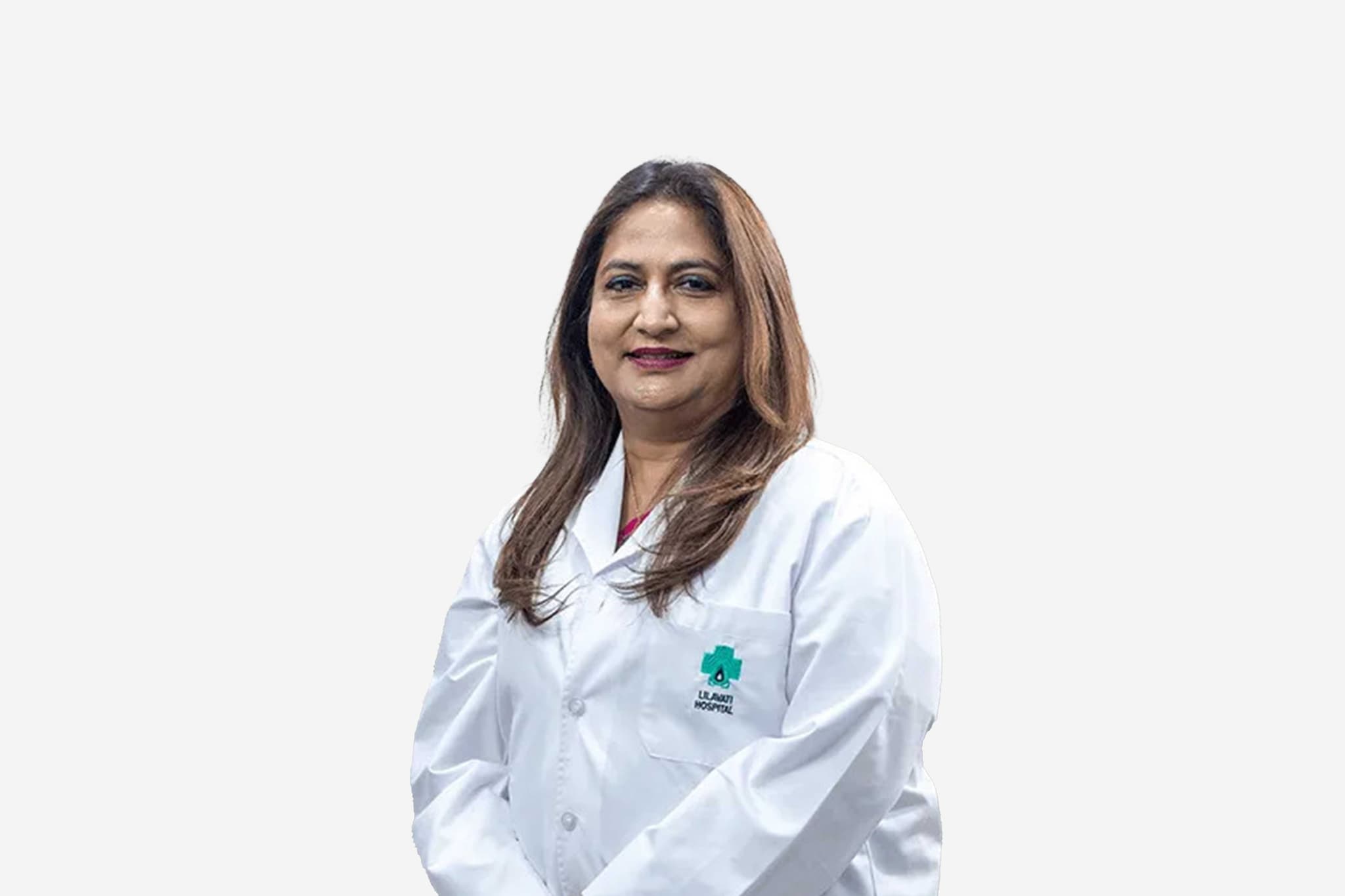
Dr Padmapriya Vivek

Director - Obstetrics & Gynecology

Gleneagles Hospital, Perumbakkam, Chennai

Fertility & IVF SpecialistGynecologistObstetrician

18+ years experience

Dr Padmapriya Vivek, Director - Obstetrics & Gynecology at Gleneagles Hospital, Perumbakkam, Chennai - Fortis Healthcare
  1. Home
  2. Doctors
  3. Dr Padmapriya Vivek

About Dr Padmapriya Vivek

Dr. Padmapriya Vivek is a highly distinguished specialist in obstetrics, gynecology, and fertility medicine, recognized for her technical mastery in complex reproductive healthcare and minimally invasive interventions. She specializes in the comprehensive management of high-risk pregnancies, advanced fertility protocols, and intricate reproductive disorders, utilizing state-of-the-art technological platforms to optimize maternal, fetal, and reproductive outcomes.

Mastery in Advanced Fertility Medicine and Assisted Reproduction

She specializes in running and institutionalizing advanced programs for comprehensive fertility treatment and reproductive preservation. Her clinical expertise encompasses sophisticated assisted reproductive technologies (ART), including Intracytoplasmic Sperm Injection (ICSI), Intracytoplasmic Morphologically Selected Sperm Injection (IMSI), Preimplantation Genetic Diagnosis (PGD), Preimplantation Genetic Screening (PGS), and structured gamete donor programs to support patients through complex infertility challenges.

Complex Reproductive Reconstructions and Uterine Transplantation

Dr. Padmapriya Vivek possesses profound surgical skill in executing highly complex gynaecological interventions, including pioneering procedures such as uterine transplantation. Her operative capabilities extend to advanced cosmetic gynaecology and intricate structural reconstructions, combining meticulous surgical precision with state-of-the-art modalities to restore anatomical functionality and enhance patient quality of life.

High-Risk Obstetrics and Critical Maternal-Fetal Care

An expert in managing critical obstetric scenarios, she is proficient in establishing clinical protocols for high-risk obstetrics and complex maternal-fetal tracking. Her clinical practice encompasses the successful delivery of over 18,000 babies, demonstrating exceptional proficiency in navigating severe pregnancy complications and ensuring maternal safety during demanding clinical situations.

Innovation in Natural Birth Pathways and Laparoscopic Surgeries

Her practice focuses heavily on promoting safe, natural birthing pathways, including the implementation of precise methodologies for painless normal deliveries and Vaginal Birth After Cesarean (VBAC). She integrates these birthing approaches with super-abundant experience in advanced vaginal and laparoscopic surgeries, utilizing minimal access techniques to minimize tissue trauma and accelerate post-operative recovery.

Clinical Governance and Institutional Leadership

Throughout her extensive career, Dr. Padmapriya Vivek has integrated modern reproductive innovations with rigorous clinical protocols to lead dedicated departments in obstetrics, gynecology, and fertility medicine. Her evidence-based framework and compassionate care are structured to deliver predictable, high-quality outcomes, establishing her as a trusted authority in transformative female healthcare.

Dr Padmapriya Vivek at a Glance

  • Specialist in Complex Gynecology, High-Risk Obstetrics, and Advanced Fertility Medicine.

  • Expert in advanced reproductive technologies including ICSI, IMSI, PGD, PGS, and fertility preservation.

  • Proficient in highly complex surgical interventions, including uterine transplantation and cosmetic gynecology.

  • Specialized in natural childbirth pathways, painless deliveries, and Vaginal Birth After Cesarean (VBAC).

  • Track record of managing over 18,000 successful deliveries and leading advanced institutional fertility programs.

  • Focused on combining cutting-edge laparoscopy and vaginal surgery with patient-centric clinical governance.

MBBS
MS
DRM (Germany)
Board Certified in Fertility & IVF Specialist
Dr Padmapriya Vivek has carried out 18,000 plus deliveries till date with an exceptional
unparalleled track record of 90% normal delivery rate.
Dr Padmapriya’s samaritan nature has been brought forefront to this World, when she toiled for more than 90 hours in delivering 17 babies safely in 2015 chennai appalled floods.
Back then, she had garnered significant accolades from people from all the walks of life including the state government.
She had validated her charitable nature in 2016 Vardah cyclone time too, which has ravaged the length
breadth of Chennai city in a dismayed manner.
Times health excellence award 2022 – Service excellence in obstetrics, Gynaecology
fertility medicine.
Excellence in Obstetrics – India’s most prominent women empowerment award 2019 in Delhi.
ET – Excellence award 2020 for the service in the field of obstetrics
gynaecology
fertility medicine.
State award in 2015 for working during floods – Award of appreciation.

Affiliated Hospitals

Gleneagles Hospital, Perumbakkam, Chennai

Multi Specialty

Chennai, Tamil Nadu

2008

Estd.

200+

Beds

250+

Doctors

May we help you?

Enter digits only (6-15 digits). We'll add your country code.

Related Treatments

Frozen Embryo Transfer (FET)

Frozen Embryo Transfer (FET) is a cornerstone of modern fertility treatment, consistently providing high success rates by utilizing cryopreserved embryos from previous cycles. This procedure allows for the transfer of a thawed embryo into a perfectly prepared uterine environment, bypassing the hormonal stress of immediate egg retrieval. Modern vitrification techniques and advanced endometrial monitoring ensure that embryos are preserved with nearly 100% viability, offering patients a safe and highly flexible path to pregnancy.

When You Should Consider FET

  • Presence of surplus high-quality embryos following a previous IVF or ICSI cycle.

  • Intention to undergo Preimplantation Genetic Testing (PGT-A) for chromosomal screening.

  • Risk of Ovarian Hyperstimulation Syndrome (OHSS) during a fresh stimulation cycle.

  • Desire to space out pregnancies and build a family over several years.

  • Recommended "freeze-all" strategy to allow the body to recover from hormonal medications.

  • Need for a donor embryo cycle to overcome age-related or genetic fertility challenges.

Conditions That Require Specialized Care

  • Recurrent Implantation Failure (RIF) where precise cycle timing is critical.

  • Thin endometrial lining requiring specialized medicated preparation protocols.

  • Hormone imbalances that make a fresh transfer medically inadvisable.

  • Polycystic Ovary Syndrome (PCOS), which increases the risk of complications during fresh cycles.

  • Advanced maternal age requiring the transfer of genetically screened euploid embryos.

How Frozen Embryo Transfer Is Performed

  • The uterine lining is prepared through either a natural monitored cycle or a medicated hormonal protocol.

  • Regular ultrasound scans and blood tests track endometrial thickness to ensure it reaches the ideal 7mm–12mm range.

  • On the day of transfer, the selected embryo is thawed using high-precision vitrification technology.

  • The embryo’s viability and cell integrity are assessed by an embryologist post-thaw.

  • A thin, flexible catheter is used to gently place the embryo into the uterus under ultrasound guidance.

  • Progesterone support is administered following the procedure to maintain a receptive environment for implantation.

Innovations in Embryo Transfer

  • High-Survival VitrificationAn ultra-rapid freezing method that prevents ice crystal formation, ensuring maximum embryo integrity.

  • AI-Enhanced Implantation TimingSoftware that analyzes uterine data to identify the precise "window of implantation" for each patient.

  • Non-Invasive Genetic ScreeningAnalyzing chromosomal health without a biopsy, reducing the physical stress on the frozen embryo.

  • Endometrial Receptivity Analysis (ERA)A genetic test of the lining to customize the exact hour of the transfer for patients with prior failures.

  • Time-Lapse Thaw MonitoringConstant observation of the embryo during the thawing phase to ensure optimal metabolic recovery.

  • Platelet-Rich Plasma (PRP) TherapyAn advanced technique used to "prime" and thicken the uterine lining in patients with a history of thin endometrium.

Pre-Procedure Preparation

  • Baseline pelvic scans to evaluate the current status of the uterus and ovaries.

  • Consultation to choose between a "Natural Cycle" or a "Medicated Cycle" based on your history.

  • Nutritional optimization, including a Mediterranean-style diet, to support uterine blood flow.

  • Screening for infectious diseases to ensure all safety and regulatory protocols are met.

  • Review of the "embryo freezing certificate" and signing of consent forms for thawing.

Pre-Procedure Tests

  • Transvaginal Ultrasound (TVS) to monitor the growth and texture of the endometrial lining.

  • Blood work to measure Estrogen, Progesterone, and TSH (Thyroid) levels.

  • Saline Infusion Sonogram (SIS) or Hysteroscopy to confirm the uterine cavity is clear.

  • Mock Transfer ("Trial Run") to map the cervical path and ensure a smooth procedure.

  • Repeat infectious disease screening as required by modern ART safety standards.

Why This Treatment Is Highly Effective

  • Boasts success rates that often surpass fresh transfers due to a more receptive, natural uterine state.

  • Virtually eliminates the risk of OHSS, making it the safest option for high-responder patients.

  • Allows for the use of PGT-A, ensuring only chromosomally normal (euploid) embryos are transferred.

  • Reduces the physiological and emotional stress of repeating egg retrieval for subsequent pregnancies.

  • Children born from FET cycles often show healthier birth weights and lower rates of preterm birth.

Recovery and Monitoring

  • The transfer is a 10–15 minute outpatient procedure that requires no anesthesia or downtime.

  • Patients can usually return to light daily activities within 24 hours of the transfer.

  • Hormonal support (Progesterone and Estrogen) is continued until the first pregnancy milestone.

  • A blood pregnancy test (Beta-hCG) is performed 10–14 days after the transfer.

  • If successful, an early "heartbeat ultrasound" is scheduled around the 6-week mark.

Life After Frozen Embryo Transfer

  • Seamless transition to routine prenatal care for a healthy, full-term pregnancy.

  • Long-term storage options remain available for any additional cryopreserved embryos.

  • Significant reduction in the cost and physical burden of future family-building steps.

  • Ongoing gynecological monitoring to ensure continued reproductive and uterine health.

  • Confidence in having utilized the most advanced, personalized technology in modern fertility care.

₹From From 300

Gamete Intrafallopian Transfer (GIFT)

Gamete Intrafallopian Transfer (GIFT) is a specialized assisted reproductive technology (ART) where fertilization occurs naturally inside the woman's body rather than in a laboratory. While modern IVF is more common, GIFT remains a unique option for patients who prefer in-vivo fertilization. This procedure mimics the natural conception process by placing a mixture of eggs and sperm directly into the fallopian tubes, allowing the body’s natural environment to facilitate the earliest stages of life.
When You Should Consider GIFT

  • Ethical or religious preferences that favor fertilization occurring inside the human body.

  • Presence of at least one healthy, functional, and unobstructed fallopian tube.

  • History of unexplained infertility where multiple cycles of IUI have been unsuccessful.

  • Desire to avoid the laboratory culture of embryos or the freezing of surplus embryos.

  • Cases where previous IVF attempts failed specifically during the laboratory fertilization stage.

  • Couples seeking a treatment that aligns with specific personal or philosophical beliefs regarding conception.

Conditions That Require Specialized Care

  • Cervical factor infertility where sperm cannot naturally pass into the uterus.

  • Minimal or mild endometriosis that has not compromised the fallopian tube structure.

  • Specific cases of mild male factor infertility where sperm quality is adequate for natural fertilization.

  • Healthy ovarian reserve requiring carefully monitored stimulation protocols.

  • Patients who are comfortable with a laparoscopic surgical approach for the transfer phase.

How Gamete Intrafallopian Transfer Is Performed

  • Ovarian stimulation is used to encourage the development of multiple mature follicles.

  • Egg maturity is monitored closely via ultrasound and hormonal blood panels.

  • Mature eggs are retrieved through a standard ultrasound-guided aspiration procedure.

  • A sperm sample is simultaneously prepared to isolate the most healthy and motile cells.

  • The eggs and sperm (gametes) are combined within a specialized transfer catheter.

  • Under general anesthesia, a laparoscopic procedure is performed to inject the gametes directly into the fallopian tube.

  • Fertilization occurs naturally within the tube, and the resulting embryo travels to the uterus for implantation.
Innovations in GIFT Procedures

  • Advanced Laparoscopic PrecisionThe use of high-definition 4K imaging during laparoscopy ensures the most accurate placement of gametes within the fallopian tube.

  • Micro-Volume Transfer CathetersSpecially designed soft-tip catheters that minimize tubal irritation and maximize the stability of the gamete mixture.

  • AI-Optimized StimulationAdvanced algorithms that predict the ideal egg retrieval window to ensure gametes are at peak viability for natural fertilization.

  • High-Gradient Sperm ProcessingRefined laboratory techniques that ensure only the highest-quality sperm are mixed with the eggs for transfer.

  • Minimally Invasive EntryModern surgical ports and techniques that reduce recovery time and post-operative discomfort following the laparoscopic phase.

  • Real-Time Hormonal MonitoringContinuous tracking of the luteal environment to ensure the uterus is perfectly primed for the naturally descending embryo.

Pre-Procedure Preparation

  • Diagnostic Laparoscopy or HSG to confirm that the fallopian tubes are healthy and functional.

  • Coordination with an anesthesiology team to prepare for the surgical component of the procedure.

  • Lifestyle and nutritional optimization to support a healthy fallopian tube environment.

  • Discussion of the ethical and clinical differences between GIFT and standard IVF.

  • Scheduling of the procedure to ensure the surgical theater and embryology lab are perfectly synchronized.

Pre-Procedure Tests

  • Hysterosalpingogram (HSG) to ensure there are no blockages or scarring in the fallopian tubes.

  • Comprehensive ovarian reserve testing, including AMH and follicle counts.

  • Standard infectious disease screening for both partners as required by ART regulations.

  • Semen analysis to ensure the sperm sample is compatible with a natural fertilization process.

  • Cardiac and general health clearance for laparoscopic surgery under general anesthesia.

Why This Treatment Is Highly Effective

  • Maintains the biological integrity of the fertilization process by keeping it within the body.

  • Provides a successful alternative for couples who have failed traditional laboratory-based ART.

  • Utilizes the fallopian tube’s natural secretions, which may support embryo development better in certain patients.

  • Offers high success rates for patients with unexplained infertility and healthy tubal anatomy.

  • Bridges the gap between natural conception and advanced science for those with specific ethical requirements.

Recovery and Monitoring

  • Patients typically remain in a recovery suite for several hours following the laparoscopic surgery.

  • Minor surgical discomfort or bloating may occur but usually subsides within 24 to 48 hours.

  • Progesterone support is provided to ensure the uterine lining remains receptive.

  • Pregnancy status is confirmed via a blood-based Beta-hCG test 14 days after the transfer.

  • If pregnancy occurs, early ultrasounds track the embryo's arrival and development in the uterus.

Life After GIFT

  • Transition to routine prenatal care following the confirmation of a healthy intrauterine pregnancy.

  • The psychological satisfaction of having conceived through a process that aligns with personal beliefs.

  • Ongoing monitoring of tubal health to maintain future reproductive potential.

  • Access to specialized fertility counseling to discuss the success and journey of the GIFT process.

  • Continued gynecological wellness checks to support long-term maternal health.

₹From From 800

IVF with Donor Eggs
IVF with Donor Eggs

IVF with Donor Eggs is a premier fertility treatment for individuals facing age-related fertility decline, premature ovarian failure, or poor egg quality. This procedure utilizes mature eggs from a healthy, thoroughly screened donor, which are fertilized in a state-of-the-art laboratory before the resulting embryo is transferred to the recipient. By integrating advanced donor matching and non-invasive genetic screening, modern protocols ensure the highest possible success rates for intended parents.

When You Should Consider Donor Egg IVF

  • Advanced maternal age where natural egg quality and quantity have declined.

  • Diagnosis of Premature Ovarian Insufficiency (POI) or early-onset menopause.

  • Repeated IVF failures using your own eggs due to poor embryo development.

  • History of multiple miscarriages linked to chromosomal abnormalities in the eggs.

  • Known genetic conditions or hereditary disorders you wish to avoid passing to offspring.

  • Diminished ovarian reserve where hormonal stimulation fails to produce viable eggs.

Conditions That Require Specialized Care

  • Severe Endometriosis affecting the quality of the primary ovarian environment.

  • Turner Syndrome or other chromosomal variations requiring donor gametes.

  • Previous cancer treatments (chemotherapy or radiation) that have impacted ovarian function.

  • Recurrent Implantation Failure (RIF) requiring specialized uterine lining synchronization.

  • Surgical removal of ovaries due to medical necessity or emergency.

[Image Placeholder: Healthy blastocyst-stage embryo developed from a donor egg]

How Donor Egg IVF Is Performed

  • Advanced digital platforms are used to match recipients with anonymous, altruistic donors.

  • Menstrual cycles are synchronized using hormonal monitoring to ensure optimal uterine receptivity.

  • Ovarian stimulation of the donor is monitored via high-precision automated follicle measurement.

  • Mature eggs are retrieved from the donor using a minimally invasive, ultrasound-guided procedure.

  • Fertilization is achieved using ICSI, where a single selected sperm is injected into each donor egg.

  • Resulting embryos are cultured in time-lapse incubators to monitor growth 24/7.

  • One healthy embryo is transferred into the recipient’s uterus using a high-precision catheter.

Modern Donor Egg Innovations

  • Advanced Donor MatchingAlgorithms that analyze medical, genetic, and phenotypic data to find the most compatible donor.

  • Vitrified Egg BankingAdvanced "flash-freezing" that allows for flexible timelines and immediate access to high-quality eggs.

  • Non-Invasive PGT-A (niPGT)Screening embryos for chromosomal health by analyzing culture fluid instead of performing a biopsy.

  • Real-Time SynchronizationPrecision hormone sensors that track the recipient's cycle for perfectly timed transfers.

  • Mitochondrial SupportEmerging techniques aimed at supporting recipient eggs using donor cellular components.

  • AI-Enhanced Embryo RankingDeep-learning models that predict the "live birth potential" of embryos with high clinical accuracy.

[Image Placeholder: High-tech IVF laboratory featuring integrated workstations and incubators]

Pre-Procedure Preparation

  • Review of the legal framework under current ART Acts to ensure all altruistic guidelines are met.

  • Comprehensive psychological counseling to prepare for the emotional journey of donor conception.

  • Uterine preparation including a healthy diet and vitamins to support the endometrium.

  • Synchronization of hormonal protocols using estradiol and progesterone medications.

  • Financial planning and coordination for all donor-related medical steps.

Pre-Procedure Tests

  • Saline Infusion Sonogram (SIS) or Hysteroscopy to confirm a clear uterine cavity.

  • Endometrial Receptivity Analysis (ERA) to identify the optimal "window of implantation."

  • Standardized viral screening (HIV, Hepatitis, Syphilis) for both the donor and the recipient.

  • Detailed semen analysis for the male partner, including a DNA Fragmentation Index.

  • Mock embryo transfer to ensure a smooth and painless procedure on the actual day.

Why This Treatment Is Highly Effective

  • Delivers exceptional success rates, often reaching 60% to 80% per embryo transfer.

  • Utilizes younger, high-quality eggs which significantly reduces the risk of miscarriage.

  • Modern screening tools reduce "trial and error" by selecting the healthiest embryos.

  • Provides a high cumulative success rate within three cycles for most recipients.

  • Allows women in their late 40s and early 50s to experience a healthy pregnancy.

Recovery and Monitoring

  • Recipients generally require only 24 hours of light activity following the embryo transfer.

  • Continued hormonal support (progesterone) for several weeks to maintain the early pregnancy.

  • A "Two-Week Wait" (2WW) period monitored via clinical health portals to track progress.

  • Blood-based pregnancy test (Beta-hCG) performed 10–14 days after the transfer.

  • Early ultrasound at 6–7 weeks to confirm the presence of a healthy fetal heartbeat.

Life After Donor Egg IVF

  • Transition to routine obstetric care with a specialized focus on healthy prenatal development.

  • Options for cryopreservation of remaining donor-egg embryos for future family building.

  • Long-term follow-up to ensure both the mother and child are meeting health milestones.

  • Access to support groups for families who have successfully conceived via egg donation.

  • Empowerment through the successful resolution of long-term infertility struggles.

₹From From 2000

IVF with Donor Sperm

IVF with Donor Sperm is a highly effective fertility solution for couples facing severe male factor infertility, single women, and same-sex couples. This procedure involves fertilizing a recipient's eggs with high-quality sperm from a registered ART bank, ensuring the highest standards of safety and genetic screening. Modern protocols emphasize the use of high-magnification selection and AI-assisted embryo grading to provide the most reliable path to a successful pregnancy.

When You Should Consider Donor Sperm IVF

  • Diagnosis of Azoospermia (complete absence of sperm) where surgical retrieval is not an option.

  • Severe male factor infertility, including low count, poor motility, or abnormal morphology.

  • History of repeated IVF failures attributed specifically to sperm-related issues.

  • Known hereditary genetic conditions in the male partner that could be passed to offspring.

  • Single women or same-sex couples wishing to start a family.

  • Cases where prior miscarriages were linked to high sperm DNA fragmentation.

Conditions That Require Specialized Care

  • Non-obstructive Azoospermia where the testes are not producing viable sperm.

  • Severe Rh-isoimmunization where a donor with a compatible blood type is required.

  • Recurrent Implantation Failure (RIF) necessitating a change in the genetic component of the embryo.

  • High-risk genetic disorders requiring specific donor screening and selection.

  • Advanced maternal age requiring the most potent sperm to maximize egg fertilization potential.

How Donor Sperm IVF Is Performed

  • Donor samples are sourced from registered ART banks, having undergone rigorous infectious disease and genetic screening.

  • The female recipient undergoes ovarian stimulation to produce multiple mature eggs.

  • Mature eggs are retrieved via a minor, ultrasound-guided surgical procedure under light sedation.

  • Selected donor sperm is prepared and "washed" to isolate the most motile and healthy cells.

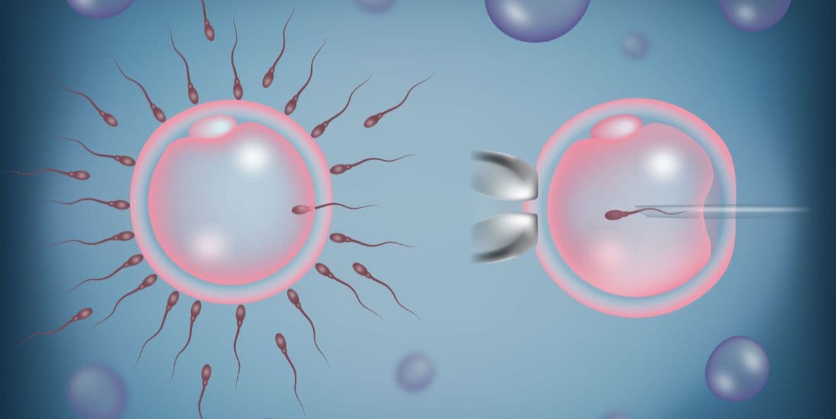
  • Fertilization is typically achieved via ICSI, where a single sperm is injected directly into each egg.

  • Resulting embryos are cultured in high-tech incubators with constant environmental monitoring.

  • The highest-quality embryo is selected and transferred into the recipient's uterus via a fine catheter.

Modern Innovations in Donor Sperm IVF

  • Microfluidic Sperm SelectionA gentle filtering technique that mimics the natural cervical path to select sperm with the lowest DNA damage.

  • AI-Based Embryo GradingSophisticated algorithms that analyze developmental patterns to identify embryos with the highest implantation potential.

  • Non-Invasive PGT-AGenetic screening performed by analyzing the culture media rather than biopsying the embryo itself.

  • High-Magnification Selection (IMSI)Using ultra-high-power microscopes (up to 6000x) to detect subtle defects in the donor sperm head.

  • Time-Lapse IncubationContinuous video monitoring (EmbryoScope) allows embryologists to observe growth without disturbing the embryos.

  • Advanced VitrificationAn ultra-rapid freezing method that ensures nearly 100% survival rates for frozen-thawed sperm and embryos.

Pre-Procedure Preparation

  • Completion of a comprehensive recipient health panel, including uterine and hormonal assessments.

  • Formal selection of a donor profile from a registered ART bank based on physical and genetic traits.

  • Legal counseling and signing of consent forms under current ART (Regulation) Acts.

  • Ovarian "priming" with supplements or medications to optimize the response to stimulation.

  • Discussion of the ethical and emotional aspects of donor conception with a fertility counselor.

Pre-Procedure Tests

  • Transvaginal Ultrasound (TVS) to assess ovarian reserve and uterine health.

  • Blood panels for Antral Follicle Count (AFC) and Anti-Müllerian Hormone (AMH).

  • Screening for infectious diseases (HIV, Hepatitis B/C, Syphilis) as per mandatory clinical guidelines.

  • Uterine cavity check (Hysteroscopy or saline ultrasound) to ensure a healthy environment for transfer.

  • Compatibility testing to ensure the donor’s blood type and genetic markers align with the recipient's needs.

Why This Treatment Is Highly Effective

  • Boasts high success rates (typically 60%–80%) due to the use of pre-screened, high-potency sperm.

  • Eliminates the uncertainty of male factor infertility by using proven-fertile donor samples.

  • Modern AI-selection tools significantly reduce the "time to pregnancy" by choosing the best embryo first.

  • Rigorous screening processes virtually eliminate the risk of transmitting infectious or common genetic diseases.

  • Provides a predictable and scientifically optimized environment for fertilization and early growth.

Recovery and Monitoring

  • Recovery from egg retrieval is rapid, usually requiring only a few hours of clinical observation.

  • The embryo transfer is a painless, 15-minute procedure that does not require anesthesia.

  • Follow-up blood tests (Beta-hCG) are conducted 10–14 days post-transfer to confirm pregnancy.

  • Progesterone support is administered for several weeks to ensure a receptive uterine lining.

  • Initial pregnancy ultrasounds are scheduled around 6 weeks to monitor the fetal heartbeat.

Life After Donor Sperm IVF

  • Transition to standard prenatal care following a confirmed and stable pregnancy.

  • Legal peace of mind, as the donor has no parental rights or claims under modern ART laws.

  • Option to cryopreserve additional embryos for future siblings from the same donor.

  • Access to specialized support networks for parents of donor-conceived children.

  • Ongoing gynecological follow-ups to maintain long-term reproductive and uterine health.

₹From From 1000

IVF with Intracytoplasmic Sperm Injection (ICSI)

In Vitro Fertilization (IVF) with Intracytoplasmic Sperm Injection (ICSI) is an advanced laboratory procedure designed to maximize the chances of fertilization by directly assisting the sperm's entry into the egg. In 2026, ICSI has become the gold standard for treating severe male factor infertility and overcoming barriers like hardened egg shells. By utilizing AI-driven sperm selection and microfluidic technology, this procedure ensures that a single, high-quality sperm is precisely injected into a mature egg to facilitate successful embryo development.

When You Should Consider ICSI

  • Diagnosis of low sperm count (oligospermia) or poor sperm motility (asthenozoospermia).

  • Presence of high numbers of abnormally shaped sperm (teratozoospermia).

  • History of failed or poor fertilization in previous traditional IVF cycles.

  • Use of eggs that have been previously frozen and thawed for a "delayed" cycle.

  • Requirement for sperm collected through surgical retrieval (TESE/TESA).

  • Couples undergoing Preimplantation Genetic Testing (PGT) who require precise fertilization data.

Conditions That Require Specialized Care

  • Severe Male Factor Infertility involving blockages or non-obstructive azoospermia.

  • Advanced Maternal Age where the egg's outer shell (zona pellucida) may be thicker or harder.

  • Unexplained Infertility where traditional fertilization methods have been unsuccessful.

  • Genetic conditions that require the selection of sperm with the highest DNA integrity.

  • Cryopreserved egg cycles where the freezing process has altered the egg's membrane.

How IVF with ICSI Is Performed

  • Ovarian stimulation is used to produce multiple mature eggs, which are then surgically retrieved.

  • Microfluidic selection devices (Zymot) are utilized to isolate sperm with the highest DNA integrity.

  • AI-driven imaging analyzes sperm morphology and movement to select the optimal candidate.

  • The egg is stabilized under a high-powered microscope using a specialized holding pipette.

  • A single sperm is carefully injected into the center of the egg using a microscopic glass needle.

  • Fertilized eggs are placed in time-lapse incubators (EmbryoScope) for undisturbed monitoring.

Types of ICSI Innovations in 2026

  • AI-Driven Sperm Selection Advanced software that identifies the sperm most likely to result in a healthy, viable embryo.

  • Microfluidic Sperm Sorting A gentle selection method that mimics the natural cervical tract to filter out sperm with DNA damage.

  • Time-Lapse Embryo Monitoring Continuous 24/7 video observation of the embryo, allowing for grading without removing it from the incubator.

  • AI Embryo Grading Algorithms that predict "live birth potential" by analyzing thousands of developmental data points.

  • Non-Invasive Genetic Testing (niPGT) Analyzing the liquid environment around the embryo for chromosomal health, avoiding the need for a biopsy.

  • Surgical Sperm Retrieval (TESA/TESE) Minor procedures to collect sperm directly from the reproductive tract when it is absent in the ejaculate.

Pre-Procedure Preparation

  • Complete a comprehensive semen analysis, including DNA fragmentation testing.

  • Undergo a "mock transfer" to map the uterine path for the eventual embryo implantation.

  • Adhere to a specialized vitamin and antioxidant protocol to optimize egg and sperm quality.

  • Manage lifestyle factors such as smoking and alcohol use to improve cellular health.

  • Participate in a consultation with an embryologist to discuss the specific ICSI plan.

Pre-Procedure Tests

  • Anti-Müllerian Hormone (AMH) and antral follicle count to assess ovarian reserve.

  • Sperm DNA Fragmentation Index (DFI) to determine the need for microfluidic selection.

  • Infectious disease screening for both partners as required by 2026 regulatory standards.

  • Uterine cavity evaluation (Hysteroscopy or saline ultrasound) to ensure a receptive environment.

  • Genetic carrier screening to identify any potential risks of inherited conditions.

Why IVF with ICSI Is Highly Effective

  • Achieves successful fertilization in 70% to 80% of mature eggs injected.

  • Overcomes severe male infertility barriers that previously made biological parenthood impossible.

  • Minimizes the risk of "total fertilization failure," providing more embryos for selection.

  • Utilizes 2026 AI technology to select sperm and embryos with the highest genetic potential.

  • Offers a standardized solution for patients using frozen-thawed eggs or donor tissue.

Recovery and Monitoring

  • Minor recovery time following egg retrieval, usually requiring one day of rest.

  • Monitoring of embryo development via a digital portal provided by the embryology lab.

  • Administration of progesterone and other hormones to prepare the uterine lining for transfer.

  • Follow-up consultations to discuss the results of genetic testing or embryo grading.

  • Emotional support and counseling integrated throughout the waiting period before the pregnancy test.

Life After IVF with ICSI

  • Transition to prenatal care once a successful pregnancy is confirmed via blood test and ultrasound.

  • Options for cryopreservation of remaining high-quality embryos for future family building.

  • Long-term follow-up to monitor the health and development of children born via assisted technology.

  • Continued management of underlying fertility conditions for future reproductive health.

  • Engagement with support communities for families who have utilized assisted reproduction

₹From From 1000

Standard In Vitro Fertilization (IVF)

Standard In Vitro Fertilization (IVF) is the global gold standard for treating diverse fertility challenges, including tubal blockages, endometriosis, and unexplained infertility. This advanced procedure involves fertilizing a woman's eggs with sperm in a specialized laboratory environment to create viable embryos. By utilizing modern hormonal protocols and high-tech incubation, IVF offers a highly controlled and effective pathway to biological parenthood.

When You Should Consider Standard IVF

  • Blocked or damaged fallopian tubes that prevent natural egg and sperm interaction.

  • Moderate to advanced endometriosis affecting ovarian or tubal function.

  • Ovulation disorders where simpler treatments like IUI have been unsuccessful.

  • Unexplained infertility where no clear cause has been identified after extensive testing.

  • History of repeated miscarriages or genetic concerns requiring embryo screening.

  • Diminished ovarian reserve where a more controlled stimulation environment is needed.

Conditions That Require Specialized Care

  • Polycystic Ovary Syndrome (PCOS) requiring careful management of stimulation drugs.

  • Male factor infertility involving low sperm count or poor motility.

  • Advanced maternal age where egg quality and quantity are significantly reduced.

  • Previous tubal ligation (sterilization) where the patient wishes to conceive again.

  • Presence of uterine fibroids or polyps that may affect embryo implantation.

How Standard IVF Is Performed

  • Ovarian stimulation is initiated using daily hormonal medications to encourage multiple egg growth.

  • Continuous monitoring via ultrasound and blood tests ensures follicles reach optimal maturity.

  • A "trigger injection" is administered to finalize the maturation of the eggs.

  • Egg retrieval is performed under light sedation using a fine, ultrasound-guided needle.

  • The retrieved eggs are combined with prepared sperm in a controlled culture dish.

  • Embryos are cultured for 3 to 5 days until they reach the robust blastocyst stage.

  • The highest-quality embryo is gently transferred into the uterus via a thin, flexible catheter.

Modern Innovations in Standard IVF

  • Time-Lapse Embryo ImagingAdvanced incubators that capture continuous photos, allowing embryologists to monitor growth without disturbing the embryos.

  • AI-Assisted Embryo SelectionDeep-learning algorithms that analyze cell division patterns to identify the most viable embryo for transfer.

  • Blastocyst Culture OptimizationRefined nutrient media that supports embryo growth to day 5, increasing the likelihood of successful implantation.

  • Microfluidic Sperm SortingA gentle selection process that mimics natural biological barriers to isolate the healthiest sperm cells.

  • Vitrification TechnologyAn ultra-rapid freezing method that ensures nearly 100% survival rates for surplus embryos.

  • Laser-Assisted HatchingA precision technique that creates a small opening in the embryo’s outer shell to help it attach to the uterine wall.

Pre-Procedure Preparation

  • Comprehensive fertility assessment for both partners to customize the stimulation protocol.

  • Lifestyle optimization, including specialized prenatal vitamins and a balanced diet.

  • Mandatory pre-treatment counseling and informed consent as per modern clinical regulations.

  • Financial and logistical planning to ensure a stress-free experience during the 4-to-6-week cycle.

  • Coordination with the nursing team to master the administration of hormonal medications.

Pre-Procedure Tests

  • Anti-Müllerian Hormone (AMH) and Antral Follicle Count (AFC) to predict ovarian response.

  • Transvaginal Ultrasound (TVS) to evaluate uterine health and ovarian accessibility.

  • Comprehensive semen analysis to determine if additional techniques like ICSI are needed.

  • Viral markers and infectious disease screening (HIV, Hepatitis, Syphilis) for both partners.

  • Hysterosalpingogram (HSG) or saline ultrasound to ensure a healthy path for embryo transfer.

Why This Treatment Is Highly Effective

  • Boasts significant success rates per cycle, particularly for patients under the age of 37.

  • Provides a controlled environment that bypasses damaged or blocked fallopian tubes entirely.

  • Allows for the selection of the strongest embryos, reducing the "time to pregnancy."

  • Offers the opportunity to freeze surplus embryos for future family-building steps.

  • Operates under strict regulatory frameworks to ensure patient safety and ethical standards.

Recovery and Monitoring

  • Recovery from egg retrieval is typically swift, with most patients returning to light activity within 24 hours.

  • Luteal phase support (Progesterone) is prescribed immediately following retrieval to assist the uterine lining.

  • Frequent contact with the medical team to monitor any signs of Ovarian Hyperstimulation Syndrome (OHSS).

  • A blood pregnancy test (Beta-hCG) is performed approximately 10 to 14 days after the transfer.

  • Post-transfer instructions focus on maintaining a low-stress environment and light daily movement.

Life After Standard IVF

  • Transition to high-quality prenatal care once a healthy pregnancy is confirmed via ultrasound.

  • Significant improvement in long-term family-building options through embryo cryopreservation.

  • Resolution of chronic infertility struggles, providing a clear path to parenthood.

  • Access to supportive communities and professional guidance for life after successful conception.

  • Continued monitoring of reproductive health to plan for potential future cycles.

₹From From 800

Vaginal Hysterectomy (Open/Laparoscopic/Robotic)

A vaginal hysterectomy is a surgical procedure to remove the uterus through the vaginal canal. Unlike an abdominal hysterectomy, this approach requires no external incisions on the abdomen, which typically results in a shorter hospital stay, lower costs, and a significantly faster recovery. It is a preferred method for treating various non-cancerous gynecological conditions.

When You Should Consider a Vaginal Hysterectomy

  • Uterine Fibroids: Benign growths in the uterine wall that cause heavy menstrual bleeding, pelvic pressure, or persistent pain.

  • Uterine Prolapse: When the pelvic floor muscles and ligaments weaken, causing the uterus to slip down into or even out of the vaginal canal.

  • Adenomyosis: A condition where the uterine lining grows into the muscular wall of the uterus, causing severe cramping and heavy periods.

  • Endometriosis: When tissue similar to the uterine lining grows outside the uterus, leading to chronic pain and scarring.

  • Abnormal Uterine Bleeding: When heavy or irregular bleeding cannot be managed with medication or less invasive procedures.

Surgical Approaches

  • Traditional Vaginal Hysterectomy: The entire procedure is performed through an incision inside the vagina. This is the least invasive method and leaves no visible scars.

  • Laparoscopically Assisted Vaginal Hysterectomy (LAVH): A thin camera (laparoscope) and micro-instruments are inserted through 3–4 tiny "keyhole" incisions in the belly to detach the upper uterus. The uterus is then removed through the vagina.

  • Robotic-Assisted Hysterectomy: Similar to the laparoscopic approach, but the surgeon uses a robotic console for superior 3D visualization and enhanced precision, which is ideal for complex cases like extensive endometriosis.

  • Vaginal Vault Creation: After the uterus is removed, the surgeon sews the top of the vagina closed, creating what is known as a "vaginal cuff."

How Is Performed

  • Anesthesia: The surgery is performed under general anesthesia or regional anesthesia (spinal/epidural) and typically takes 1 to 2 hours.

  • Access: The surgeon makes an incision inside the vagina to reach the uterus.

  • Detachment: Using specialized tools, the surgeon clamps and cuts the blood vessels and ligaments that support the uterus.

  • Removal: The uterus (and sometimes the fallopian tubes or ovaries) is withdrawn through the vaginal opening.

  • Closure: The internal incisions are closed with dissolvable stitches. Because there are no abdominal cuts in the traditional approach, there is no external scarring.

Pre-Procedure Preparation

  • Pelvic Examination: To assess the size of the uterus and ensure it can be safely removed through the vaginal canal.

  • Fasting: Adhering to strict "nothing by mouth" instructions for at least 8 hours prior to your general anesthesia.

  • Medication Audit: You may need to stop taking blood thinners, aspirin, or certain herbal supplements 7–10 days before the procedure.

  • Bowel Prep: In some cases, your surgeon may recommend a mild laxative or a specific diet the day before surgery.

  • Smoking Cessation: Stopping smoking at least 4 weeks before surgery is vital to promote healthy tissue healing and reduce the risk of infection.

Tests Before Vaginal Hysterectomy

  • Pelvic Ultrasound: To map the size and location of fibroids and check the health of the ovaries.

  • Endometrial Biopsy: To rule out any cancerous or precancerous cells within the uterine lining.

  • Pap Smear: To ensure there are no cervical abnormalities before the uterus and cervix are removed.

  • Blood Panels: Checking hemoglobin levels and blood type to prepare for the rare possibility of a transfusion.

  • ECG: A standard heart check to ensure you are healthy enough for the administration of anesthesia.

Life After Vaginal Hysterectomy

  • Hospital Stay: Most patients are discharged the same day or after one night of observation.

  • Physical Activity: Walking is encouraged immediately to prevent blood clots, but you must avoid heavy lifting (over 5 kg) and strenuous exercise for 4 to 6 weeks.

  • Vaginal Health: You must not use tampons or have sexual intercourse for 6 to 8 weeks to allow the vaginal cuff to heal completely.

  • Hormonal Changes: If the ovaries are left intact, you will not enter menopause prematurely, though you will no longer have periods. If ovaries are removed, menopause begins immediately.

  • Follow-up Care: A post-operative checkup is typically scheduled for 2 to 6 weeks after surgery to ensure the internal stitches are dissolving correctly.

Why Specialized Treatment Is Highly Effective

  • Faster Recovery: Patients generally return to normal activities much sooner than those undergoing traditional abdominal surgery.

  • No Visible Scarring: Because the entry point is internal, there are no cosmetic changes to the abdomen.

  • Reduced Complication Rates: Vaginal approaches are associated with lower risks of wound infection and less post-operative pain.

  • Cost-Efficient: Shorter hospital stays and quicker operating times often result in lower overall medical costs.

  • High Patient Satisfaction: Most women report a significant improvement in quality of life once the symptoms of fibroids or prolapse are resolved.

₹From From 500

Myomectomy (Open/Laparoscopic/Robotic)

A myomectomy is a surgical procedure to remove uterine fibroids (benign growths) while keeping the uterus intact. Unlike a hysterectomy, it preserves fertility and is the preferred option for women who wish to become pregnant or maintain their uterine health. This procedure specifically targets the fibroids while surgically repairing the uterine wall.

When You Should Consider a Myomectomy

  • Fertility Preservation: For women who plan to have children in the future and need to keep the uterus functional.

  • Heavy Menstrual Bleeding: When fibroids cause anemia, flooding, or prolonged periods that disrupt daily life.

  • Pelvic Pain or Pressure: Large fibroids pressing on the bladder (causing frequent urination) or the rectum.

  • Infertility or Miscarriage: When fibroids distort the uterine cavity and interfere with embryo implantation or maintenance of pregnancy.

  • Rapid Growth: If ultrasound monitoring shows fibroids are growing quickly, even if they are currently asymptomatic.

Surgical Approaches

  • Abdominal Myomectomy (Open): A traditional "bikini cut" horizontal incision is made in the lower abdomen. This is used for very large fibroids (typically over 10 cm), numerous growths, or those buried deep in the uterine wall.

  • Laparoscopic Myomectomy: Performed through 3–4 tiny "keyhole" incisions. The surgeon uses a camera and specialized tools to cut the fibroids into smaller pieces (morcellation) for removal through the small ports.

  • Robotic-Assisted Myomectomy: Similar to the laparoscopic approach, but the surgeon operates via a robotic console. This provides superior 3D visualization and greater precision, which is helpful for complex or hard-to-reach fibroids.

  • Hysteroscopic Myomectomy: No abdominal incisions are made. A specialized scope is inserted through the cervix to remove fibroids that are bulging into the uterine cavity (submucosal fibroids).

How Is Performed

  • Anesthesia: The surgery is performed under general anesthesia and typically takes between 1 to 3 hours depending on the number of fibroids.

  • Incision and Access: Depending on the approach, the surgeon accesses the uterus either through the abdomen or the vaginal canal/cervix.

  • Fibroid Removal: Each fibroid is carefully "shelled out" from the surrounding healthy uterine muscle.

  • Uterine Reconstruction: The surgeon meticulously sutures the layers of the uterine muscle back together to ensure the wall is strong enough to support a future pregnancy.

  • Closure: Abdominal incisions are closed with stitches or surgical glue. For hysteroscopic cases, there are no external wounds to close.

Pre-Procedure Preparation

  • Pelvic Imaging: An MRI or detailed ultrasound is mandatory to map the exact size, number, and depth of all fibroids.

  • Hormonal Therapy (Optional): In some cases, medication (like GnRH agonists) may be prescribed for 2–3 months before surgery to shrink the fibroids and reduce blood loss.

  • Iron Supplementation: If you are anemic due to heavy bleeding, you may need to take iron to build up your blood count before surgery.

  • Fasting: Adhering to strict "nothing by mouth" instructions for at least 8 hours prior to your general anesthesia.

  • Medication Audit: Stopping blood thinners, aspirin, or anti-inflammatory drugs 7–10 days before the procedure.

Tests Before Myomectomy

  • Saline Infusion Sonogram (SIS): Injecting sterile water into the uterus during an ultrasound to get a clear view of the uterine lining.

  • Hysteroscopy (Diagnostic): A quick office procedure to look inside the uterus and confirm if fibroids are affecting the cavity.

  • Blood Panels: Checking hemoglobin and hematocrit levels to assess for anemia.

  • ECG: A standard heart check to ensure you are healthy enough for the administration of anesthesia.

Life After Myomectomy

  • Hospital Stay: Usually 1–2 nights for open surgery; often same-day discharge or one night for laparoscopic and robotic approaches.

  • Physical Activity: Walking is encouraged immediately to prevent blood clots. Avoid heavy lifting and strenuous exercise for 4 weeks (laparoscopic) to 6 weeks (open).

  • Pregnancy Timing: Surgeons usually recommend waiting 3 to 6 months before trying to conceive to allow the uterine wall to heal completely.

  • Delivery Method: If the surgery involved deep incisions into the uterine wall, a C-section is often required for future deliveries to prevent uterine rupture during labor.

  • Follow-up Imaging: A repeat ultrasound is often done 3–6 months post-op to ensure the uterus has healed well and no small fibroids were left behind.

Why Specialized Treatment Is Highly Effective

  • Preserves Reproductive Potential: Unlike a hysterectomy, this procedure keeps the option of biological motherhood open.

  • Targeted Symptom Relief: Successfully removing fibroids eliminates the source of heavy bleeding and pelvic pressure while keeping the organ intact.

  • High Success Rates: Modern robotic and laparoscopic techniques allow for the removal of complex fibroids with minimal blood loss and faster recovery.

  • Improved Fertility Outcomes: For many women, removing cavity-distorting fibroids significantly increases the chances of successful natural conception or IVF.

  • Maintains Pelvic Integrity: Keeping the uterus helps maintain the natural support of the pelvic floor and avoids the early onset of surgical menopause.

₹From From 800

Endometrial Ablation

Endometrial ablation is a minimally invasive medical procedure used to treat heavy menstrual bleeding (menorrhagia). It works by destroying or removing the endometrium (the lining of the uterus) using various energy-based methods. Unlike a hysterectomy, this procedure leaves the uterus intact, providing a less invasive alternative for women seeking relief from debilitating cycles.

When You Should Consider Endometrial Ablation

  • Heavy Menstrual Flow: If you find yourself soaking through a pad or tampon every hour for several consecutive hours.

  • Prolonged Bleeding: When your menstrual period consistently lasts longer than eight days.

  • Anemia: If chronic blood loss has led to iron deficiency or low hemoglobin levels, causing persistent fatigue.

  • Failed Medical Therapy: For premenopausal women who have not found relief from hormonal contraceptives or other medications.

  • Lifestyle Disruption: When your menstrual cycle significantly interferes with your ability to work, exercise, or maintain daily activities.

Methods of Endometrial Ablation

  • Radiofrequency Ablation: A flexible, specialized mesh device is used to deliver controlled electrical energy to the lining.

  • Cryoablation: A thin probe uses extreme cold to freeze and destroy the endometrial tissue.

  • Thermal Balloon: A small balloon is inserted into the uterus and filled with heated fluid to treat the lining.

  • Hydrothermal Ablation: Heated saline fluid is circulated directly within the uterine cavity to reach all areas of the lining.

  • Microwave Ablation: Precise microwave energy is applied through a specialized wand to treat the uterine wall.

  • Electrosurgery: A wire loop or rollerball uses an electric current to remove the lining, typically performed under general anesthesia.

How Is Performed

  • Access: The procedure is performed entirely through the vagina and cervix, meaning there are no surgical incisions or external scars.

  • Anesthesia: Depending on the method, it can be performed under local, regional, or general anesthesia and typically takes 15 to 45 minutes.

  • Dilation: The cervix is gently and precisely opened (dilated) to allow the insertion of the ablation instruments.

  • Ablation: The chosen energy-based method is applied to the uterine lining to carefully remove or destroy the tissue.

  • Completion: Once the lining is treated, the instruments are removed, and the patient is moved to a recovery area for short-term observation.

Pre-Procedure Preparation

  • Endometrial Biopsy: A mandatory sample of the lining is taken before the procedure to rule out any cancerous or precancerous cells.

  • Hysteroscopy: A quick visualization of the inside of the uterus to ensure there are no large fibroids or polyps that might interfere with the ablation.

  • Pregnancy Test: To confirm you are not pregnant, as the procedure is strictly contraindicated during pregnancy.

  • Hormonal Thinning (Optional): You may be prescribed medication for a few weeks prior to the procedure to thin the uterine lining, which can improve results.

  • Fasting: Following standard "nothing by mouth" instructions if your procedure involves sedation or general anesthesia.

Tests Before Endometrial Ablation

  • Pelvic Ultrasound: To measure the thickness of the uterine lining and check the overall size and shape of the uterus.

  • Hemoglobin/Ferritin Levels: To assess the severity of anemia caused by heavy bleeding.

  • Cervical Screening (Pap Smear): To ensure there are no cervical abnormalities prior to instrument insertion.

  • ECG: A routine heart check to ensure you are healthy enough for the chosen level of anesthesia.

Life After Endometrial Ablation

  • Hospital Stay: Most patients are discharged the same day and can return to their normal daily activities within a few days.

  • Short-term Side Effects: You may experience minor cramping (1–3 days), mild nausea, or frequent urination during the first 24 hours.

  • Vaginal Discharge: A watery discharge mixed with a small amount of blood is normal and may last for 2 to 4 weeks as the uterus heals.

  • Menstrual Changes: Periods usually become significantly lighter, and in many cases, stop entirely (amenorrhea) within a few months.

  • Birth Control Necessity: While the procedure makes pregnancy unlikely and dangerous, it is not a sterilization method; birth control is required until you reach menopause.

Why Specialized Treatment Is Highly Effective

  • Incision-Free Recovery: Because no cuts are made to the body, the recovery time is exceptionally fast compared to traditional surgery.

  • Targeted Relief: Successfully destroys only the tissue responsible for heavy bleeding while preserving the rest of the reproductive anatomy.

  • High Success Rates: The majority of women report a dramatic reduction in menstrual flow and a significant improvement in their quality of life.

  • Versatile Options: With multiple energy-based methods available, the treatment can be tailored to the specific size and shape of your uterus.

  • Avoids Hysterectomy: Provides a effective, low-risk alternative for women who wish to avoid major organ removal and longer hospital stays.

₹From From 400

Ovarian Cyst Removal (Laparoscopic Cystectomy)

Laparoscopic ovarian cystectomy is a minimally invasive surgery to remove an ovarian cyst while preserving the healthy part of the ovary and the fallopian tube. It is the preferred method for treating benign (non-cancerous) cysts that are causing pain, growing rapidly, or at risk of twisting (torsion). This technique allows for precise removal of the cyst wall with minimal impact on future fertility.

When You Should Consider an Ovarian Cystectomy

  • Persistent Cysts: Functional cysts that do not disappear naturally after several menstrual cycles.

  • Endometriomas: Also known as "chocolate cysts," these are caused by endometriosis and can significantly affect fertility if left untreated.

  • Dermoid Cysts: Complex cysts containing different types of tissue (such as skin or hair) that will not resolve on their own and carry a risk of rupture.

  • Chronic Pelvic Pain: Large cysts that create constant pressure on the bladder or rectum, or cause sharp pain during physical activity.

  • Rapid Growth: If ultrasound monitoring shows a cyst is increasing in size, even if it is currently asymptomatic.

How Is Performed

  • Anesthesia: The surgery is performed under general anesthesia and typically takes between 45 to 90 minutes.

  • Incision: The surgeon makes 3–4 tiny "keyhole" incisions (5–10 mm) in the abdomen, including one inside the belly button for the camera (laparoscope).

  • Visualization: Carbon dioxide gas is used to gently inflate the abdomen, providing the surgical team with a clear, magnified view of the pelvic organs.

  • Cyst Removal: Using specialized micro-instruments, the surgeon carefully peels the cyst wall away from the healthy ovarian tissue, ensuring the ovary remains functional.

  • Extraction: The cyst is placed in a small surgical bag and removed through one of the incisions. Large cysts may be drained first to fit through the small opening.

  • Closure: The small incisions are closed with dissolvable stitches or surgical glue, resulting in minimal scarring.

Pre-Procedure Preparation

  • Pelvic Ultrasound or MRI: To determine the exact size, location, and internal structure of the cyst before surgery.

  • Blood Testing: Checking "tumor markers" (such as CA-125) to confirm the cyst appears benign and to assess overall health.

  • Fasting: Adhering to strict "nothing by mouth" instructions for at least 8 hours prior to your general anesthesia.

  • Medication Audit: Stopping blood thinners, aspirin, or certain supplements 7–10 days before the procedure as directed.

  • Bowel Prep (Optional): In some cases, a mild laxative may be recommended the day before to clear the pelvic area for better visualization.

Tests Before Ovarian Cystectomy

  • Transvaginal Ultrasound: The primary tool used to distinguish between fluid-filled simple cysts and complex solid masses.

  • Pregnancy Test: To rule out ectopic pregnancy or ensure the safety of the developing fetus if pregnancy is known.

  • Coagulation Profile: To ensure your blood's ability to clot is optimal for a safe, minimally invasive recovery.

  • ECG: A standard heart check to confirm you are healthy enough for the administration of anesthesia.

Life After Ovarian Cystectomy

  • Hospital Stay: Most patients are discharged the same day or stay for one night of observation.

  • Shoulder Pain: It is common to feel temporary pain in the shoulders for 24–48 hours; this is caused by the gas used during surgery irritating the diaphragm.

  • Activity Restrictions: You can usually return to light daily activities within 3 to 5 days. However, avoid heavy lifting and strenuous exercise for 2 to 3 weeks.

  • Wound Care: Keep the small incisions clean and dry. Most stitches are internal and will dissolve on their own within 10–14 days.

  • Sexual Activity: Most doctors recommend waiting at least 2 weeks before resuming sexual intercourse to allow internal healing.

Why Specialized Treatment Is Highly Effective

  • Preserves Fertility: By removing only the cyst wall and saving the healthy ovarian tissue, the "ovarian reserve" and hormone production are protected.

  • Minimal Scarring: The use of tiny incisions results in superior cosmetic outcomes compared to traditional open surgery.

  • Rapid Recovery: The minimally invasive approach allows patients to return to their jobs and families much faster than with a laparotomy.

  • High Precision: The magnified view provided by the laparoscope allows surgeons to identify and protect the delicate fallopian tubes and surrounding nerves.

  • Lower Complication Rates: Laparoscopy is associated with less post-operative pain, lower risk of infection, and a reduced chance of developing internal scar tissue (adhesions).

₹From From 500

Oophorectomy (Ovary Removal)

An oophorectomy is the surgical removal of one or both ovaries. It is often performed in conjunction with other procedures, such as a hysterectomy or salpingectomy (removal of the fallopian tubes). This procedure is a critical intervention for managing ovarian diseases, reducing cancer risk, or addressing severe pelvic conditions.

When You Should Consider an Oophorectomy

  • Ovarian Cancer: As a primary treatment for a confirmed cancerous mass or a highly suspicious complex cyst.

  • Prophylactic (Risk-Reducing): For women with high-risk genetic mutations, such as BRCA1 or BRCA2, to prevent future cancer development.

  • Severe Endometriosis: When the ovaries are extensively damaged or causing chronic, debilitating pelvic pain that does not respond to medication.

  • Ovarian Torsion: An emergency situation where the ovary twists on its supporting ligaments, cutting off its own blood supply.

  • Chronic Pelvic Inflammatory Disease (PID): Persistent and severe infection that has caused irreversible damage to the ovarian tissue.

Types of Oophorectomy

  • Unilateral Oophorectomy: Removal of only one ovary. In most cases, menstruation continues and natural pregnancy remains possible.

  • Bilateral Oophorectomy: Removal of both ovaries. This results in immediate "surgical menopause" if the patient has not yet reached natural menopause.

  • Salpingo-Oophorectomy: The surgical removal of an ovary along with its attached fallopian tube, often performed to reduce the risk of future tubal issues.

  • Prophylactic Bilateral Salpingo-Oophorectomy (RRBSO): A specific preventive surgery for high-risk patients to remove both sets of ovaries and tubes simultaneously.

How Is Performed

  • Anesthesia: The procedure is performed under general anesthesia and typically takes between 45 to 90 minutes.

  • Laparoscopic/Robotic Approach: The most common method, involving 3–4 tiny "keyhole" incisions. The surgeon uses a camera to detach and remove the ovaries through these small openings.

  • Laparotomy (Open Surgery): A single, larger abdominal incision is used if the ovaries are very large, if there is extensive scar tissue, or if cancer is suspected.

  • Vaginal Approach: If performed alongside a vaginal hysterectomy, the ovaries may be detached and removed through the vaginal canal.

  • Closure: Small laparoscopic incisions are closed with dissolvable stitches or surgical glue, while open incisions may require staples or standard sutures.

Pre-Procedure Preparation

  • Genetic Counseling: For patients undergoing prophylactic surgery, a review of genetic markers (BRCA) is essential.

  • Pelvic Imaging: A detailed ultrasound or MRI to map the size, location, and blood supply of the ovaries.

  • Fasting: Adhering to strict "nothing by mouth" instructions for at least 8 hours prior to your general anesthesia.

  • Hormone Consultation: Discussing a plan for Hormone Replacement Therapy (HRT) if a bilateral procedure is planned for a premenopausal patient.

  • Medication Audit: Stopping blood thinners or aspirin 7–10 days before the procedure to minimize the risk of internal bleeding.

Tests Before Oophorectomy

  • Tumor Marker Tests (CA-125): Blood tests used to help evaluate the likelihood of malignancy in ovarian masses.

  • Transvaginal Ultrasound: To provide high-resolution images of the ovarian follicles and any abnormal growths.

  • CT Scan of the Abdomen/Pelvis: Often used if cancer is suspected to check for involvement of nearby lymph nodes or organs.

  • ECG: A standard heart check to ensure you are healthy enough for the duration of the surgical procedure.

Life After Oophorectomy

  • Hospital Stay: Usually a same-day discharge for laparoscopic procedures; 1–2 nights of observation are typical for open surgery.

  • Surgical Menopause: If both ovaries are removed, the sudden drop in estrogen can lead to immediate hot flashes, night sweats, and vaginal dryness.

  • Long-term Health: Without estrogen, there is an increased risk of osteoporosis (bone thinning) and heart disease; HRT is often started immediately to mitigate these risks.

  • Activity Restrictions: Light walking is encouraged within 24 hours. Avoid heavy lifting and strenuous exercise for 2–4 weeks (laparoscopic) or 6 weeks (open).

  • Fertility Impact: If both ovaries are removed, natural conception is no longer possible. Patients wishing to preserve fertility should discuss egg freezing prior to surgery.

Why Specialized Treatment Is Highly Effective

  • Prevents Life-Threatening Cancer: For high-risk genetic carriers, this surgery reduces the risk of ovarian cancer by up to 90%.

  • Immediate Pain Resolution: Provides definitive relief for patients suffering from chronic pain due to torsion or severe endometriosis.

  • Minimally Invasive Options: Modern laparoscopic and robotic techniques allow for precise removal with very short recovery times and minimal scarring.

  • Comprehensive Hormonal Care: Integrated treatment plans ensure that surgical menopause symptoms are managed effectively with modern HRT.

  • High Technical Success: When performed by specialists, the risks of injury to the bladder or ureters are exceptionally low, ensuring a safe return to health.

₹From From 500

Salpingectomy (Fallopian Tube Removal)

Salpingectomy is the surgical removal of one or both fallopian tubes. Unlike a tubal ligation (where tubes are tied), a salpingectomy involves the complete physical removal of the tube. It is a highly effective permanent procedure often performed to treat medical emergencies or as a proactive measure against cancer while maintaining hormonal balance.

When You Should Consider a Salpingectomy

  • Ectopic Pregnancy: An emergency situation where a fertilized egg implants in the tube; removal is necessary if the tube has ruptured or is severely damaged.

  • Hydrosalpinx: When a tube becomes blocked and fills with fluid, causing chronic pain or interfering with the success of IVF treatments.

  • Permanent Birth Control: A preferred alternative to "tying tubes" for women seeking the most effective form of permanent sterilization.

  • Cancer Prevention: For women with high-risk genetic mutations (like BRCA1/2), as research suggests many "ovarian" cancers actually originate in the fallopian tubes.

  • Pelvic Inflammatory Disease (PID): When severe, chronic infection has caused irreversible damage or abscesses within the tubal structures.

Types of Salpingectomy

  • Unilateral Salpingectomy: Removal of only one fallopian tube. If the other tube and both ovaries remain, natural pregnancy is still possible.

  • Bilateral Salpingectomy: Removal of both tubes. This results in permanent sterilization; future pregnancy would require In Vitro Fertilization (IVF).

  • Salpingo-oophorectomy: The surgical removal of the fallopian tube along with its attached ovary, often performed for localized disease or cancer risk reduction.

How Is Performed

  • Anesthesia: The procedure is typically performed under general anesthesia and takes approximately 30 to 60 minutes.

  • Laparoscopic (Keyhole) Approach: The most common method, involving 2–3 tiny incisions (5–10 mm) in the abdomen for a camera and specialized micro-instruments.

  • Laparotomy (Open) Approach: A larger abdominal incision used in emergencies, such as a ruptured ectopic pregnancy with significant internal bleeding.

  • Detachment: The surgeon carefully seals the blood vessels and detaches the tube from the uterus and the ovary.

  • Extraction: The tube is removed through one of the small incisions, and the surgical sites are inspected for any signs of bleeding.

  • Closure: Small incisions are closed with dissolvable stitches or surgical glue, leaving minimal scarring.

Pre-Procedure Preparation

  • Pelvic Ultrasound: To confirm the location of an ectopic pregnancy or to visualize the extent of tubal disease like hydrosalpinx.

  • Pregnancy Test: A mandatory check to confirm pregnancy status and determine the appropriate surgical urgency.

  • Fasting: Adhering to strict "nothing by mouth" instructions for at least 8 hours prior to your general anesthesia.

  • Medication Audit: You may be asked to stop taking aspirin or blood thinners 7–10 days before the procedure to minimize surgical risks.

  • Emergency Stabilization: In cases of rupture, intravenous fluids and blood type cross-matching are performed immediately.

Tests Before Salpingectomy

  • Transvaginal Ultrasound: The primary imaging tool used to evaluate the health of the tubes and ovaries.

  • Beta-hCG Blood Test: Specifically used to monitor hormone levels in suspected ectopic pregnancies.

  • Complete Blood Count (CBC): To check for signs of infection or anemia caused by internal bleeding.

  • ECG: A standard heart check to ensure you are healthy enough for the administration of anesthesia.

Life After Salpingectomy

  • Hospital Stay: Most patients are discharged the same day as the surgery following a laparoscopic procedure.

  • Shoulder Pain: It is common to feel referred pain in the shoulders for 24–48 hours; this is caused by the gas used to inflate the abdomen during surgery.

  • Activity Restrictions: You can usually return to work and light daily activities within 3 to 7 days. Avoid heavy lifting (over 5 kg) for 2 to 3 weeks.

  • Sexual Activity: Most doctors recommend waiting at least 2 weeks before resuming sexual intercourse to allow for proper internal healing.

  • Hormonal Balance: Because the ovaries are typically left intact, you will not enter menopause prematurely and your hormonal cycle will remain unchanged.

Why Specialized Treatment Is Highly Effective

  • Definitive Cancer Protection: Removing the tubes significantly lowers the long-term risk of developing high-grade serous ovarian cancer.

  • Life-Saving in Emergencies: Provides immediate resolution for ruptured ectopic pregnancies, preventing life-threatening internal hemorrhage.

  • Improved IVF Success: Removing a fluid-filled tube (hydrosalpinx) increases the chances of a successful embryo implantation during fertility treatments.

  • Minimal Recovery Time: The laparoscopic approach ensures that patients can return to their normal lives within a week with very little post-operative pain.

  • Superior Birth Control: Offers a higher degree of protection against unintended pregnancy than traditional tubal ligation methods.

₹From From 500

Ectopic Pregnancy Surgery

Surgery for an ectopic pregnancy is an emergency procedure performed when a fertilized egg implants outside the uterus, most commonly in a fallopian tube. Because an ectopic pregnancy cannot survive and poses a life-threatening risk of internal hemorrhage, surgical intervention is often necessary to protect the mother’s health and future fertility.

When You Should Consider Surgery for Ectopic Pregnancy

  • Confirmed Ectopic Pregnancy: When ultrasound and blood tests confirm the embryo has implanted outside the uterine cavity.

  • Severe Abdominal Pain: Sharp, stabbing pelvic pain, often on one side, that may come and go or vary in intensity.

  • Signs of Rupture: If you experience extreme lightheadedness, fainting, or shoulder tip pain, which indicates internal bleeding.

  • Failed Medical Management: When medication (such as methotrexate) has not successfully dissolved the pregnancy tissue.

  • High hCG Levels: If pregnancy hormone levels are too high for medical treatment to be effective or safe.

  • Unstable Vital Signs: An emergency situation where low blood pressure or a rapid heart rate suggests an active tubal rupture.

Surgical Approaches

  • Laparoscopic Surgery (Keyhole): The most common and preferred approach. The surgeon makes 2–3 tiny incisions in the abdomen to insert a high-definition camera and specialized micro-instruments.

  • Salpingectomy: The complete removal of the fallopian tube containing the pregnancy. This is often the safest choice if the tube is severely damaged or if the other tube is healthy.

  • Salpingostomy: A small, precise slit is made in the fallopian tube to remove the pregnancy tissue while leaving the tube intact. This is typically considered if the other tube is already damaged or missing.

  • Laparotomy (Open Surgery): A larger abdominal incision used in critical emergencies where a tube has ruptured and severe internal bleeding requires immediate, direct control.

How Is Performed

  • Anesthesia: The procedure is performed under general anesthesia and typically takes between 30 to 60 minutes, though emergency cases may require more time.

  • Access: Depending on the stability of the patient, the surgeon accesses the pelvic area through small laparoscopic ports or a traditional abdominal incision.

  • Tissue Removal: All pregnancy tissue is meticulously removed to prevent it from continuing to grow or causing further internal damage.

  • Hemostasis: The surgeon carefully seals all blood vessels to stop internal bleeding and ensure the pelvic cavity is clear of blood and debris.

  • Closure: Small laparoscopic incisions are closed with dissolvable stitches or surgical glue, while open incisions are secured with standard sutures or staples.

Pre-Procedure Preparation

  • Emergency Ultrasound: A final scan to locate the pregnancy and assess whether the fallopian tube has already ruptured.

  • Serial hCG Testing: Monitoring the levels of the pregnancy hormone to determine the urgency and type of surgical approach needed.

  • Blood Type and Cross-match: Immediate testing to ensure compatible blood is available in the event a transfusion is necessary due to blood loss.

  • Intravenous Access: Starting IV fluids and potentially medications to stabilize blood pressure before entering the operating room.

  • Fasting: In non-emergency cases, following "nothing by mouth" instructions; however, in emergencies, the surgical team proceeds immediately for patient safety.

Tests Before Surgery for Ectopic Pregnancy

  • Transvaginal Ultrasound: The primary diagnostic tool used to visualize the empty uterus and the mass in the fallopian tube.

  • Quantitative Beta-hCG: A precise blood test to measure the exact amount of pregnancy hormone in the system.

  • Complete Blood Count (CBC): To check for signs of anemia or internal blood loss through hemoglobin and hematocrit levels.

  • Coagulation Profile: Ensuring the blood’s ability to clot is normal prior to making surgical incisions.

Life After Surgery for Ectopic Pregnancy

  • Hospital Stay: Most laparoscopic patients are discharged the same day or after one night. Laparotomy patients typically stay for 2–3 days for observation.

  • Immediate Recovery: It is normal to experience temporary shoulder pain (from the gas used in laparoscopy), abdominal soreness, and light vaginal bleeding for 1–2 weeks.

  • Activity Restrictions: Avoid heavy lifting and strenuous exercise for 2 to 4 weeks (laparoscopic) or 6 weeks (open surgery) to allow internal healing.

  • Hormone Monitoring: Weekly blood tests to monitor hCG levels are often required until they reach zero to ensure no pregnancy tissue remains.

  • Future Pregnancy: If one healthy tube remains, the chances of a successful future pregnancy remain high. Most doctors recommend waiting at least two menstrual cycles before trying to conceive again.

Why Specialized Treatment Is Highly Effective

  • Life-Saving Intervention: Rapidly stops internal bleeding and removes the risk of a life-threatening tubal rupture.

  • Preserves Future Fertility: Specialized techniques like salpingostomy or careful laparoscopic salpingectomy protect the remaining reproductive anatomy.

  • Minimally Invasive Success: Laparoscopic methods result in less post-operative pain, smaller scars, and a much faster return to normal life.

  • Definitive Resolution: Unlike medical management, surgery provides an immediate solution for cases where the pregnancy tissue is large or the tube is compromised.

  • Comprehensive Emergency Care: Operating in a specialized surgical environment ensures that advanced monitoring and blood replacement are available if complications arise.

₹From From 400

Tubal Recanalization

Tubal recanalization is a medical procedure used to reopen or reconnect the fallopian tubes to restore fertility. It is most commonly performed to reverse a previous tubal ligation (sterilization) or to clear blockages caused by infection, scarring, or debris. This procedure offers a path to natural conception by restoring the continuous pathway required for an egg to meet sperm.

When You Should Consider Tubal Recanalization

  • Reversal of Tubal Ligation: If you previously had your "tubes tied" and now wish to restore natural fertility.

  • Proximal Tubal Occlusion: When the portion of the tube closest to the uterus is blocked by mucus plugs or minor scarring.

  • Secondary Infertility: If you have successfully conceived in the past but now face blockages due to pelvic inflammatory disease (PID) or endometriosis.

  • Desire for Natural Conception: For patients who prefer natural pregnancy over assisted reproductive technologies like IVF.

  • Post-Infection Blockage: When a previous pelvic infection has left debris or light adhesions within the tubal canal.

Methods of Tubal Recanalization

  • Non-Surgical (Fluoroscopic) Recanalization: An interventional radiologist uses X-ray guidance to pass a fine catheter through the cervix into the tubes to clear "plugs" without incisions.

  • Laparoscopic Tubal Anastomosis: A minimally invasive "keyhole" surgery where a surgeon uses micro-instruments to reconnect the severed ends of the tubes.

  • Hysteroscopic Cannulation: A small camera is inserted through the vagina and cervix to clear blockages specifically located at the opening of the tubes.

  • Open Microsurgical Repair: Performed through a traditional "bikini cut" incision, typically reserved for complex cases where laparoscopy is not feasible.

How Is Performed

  • Anesthesia: Depending on the method, the procedure can be performed under local anesthesia (non-surgical), regional, or general anesthesia (surgical).

  • Duration: Non-surgical methods typically take about 30 minutes, while surgical reconnection can take between 1 to 3 hours.

  • Access: The surgeon or radiologist accesses the tubes either through the natural opening of the cervix or via small abdominal incisions.

  • Clearing or Joining: The blockage is either physically cleared with a catheter or the scarred ends of the tube are trimmed and precisely sutured together.

  • Dye Test (Chromotubation): A colored dye is often injected through the tubes during the procedure to confirm they are completely open and functional.

  • Closure: For surgical cases, small incisions are closed with dissolvable stitches or surgical glue.

Pre-Procedure Preparation

  • Hysterosalpingogram (HSG): A specialized X-ray used to map the exact location of the blockage and assess the health of the remaining tubal segments.

  • Semen Analysis: It is often mandatory for the partner to undergo testing to ensure there are no male fertility factors before proceeding with surgery.

  • Laparoscopic Review: In some cases, a diagnostic laparoscopy is done first to ensure there is enough healthy tube (at least 4–5 cm) for a successful repair.

  • Fasting: Following "nothing by mouth" instructions for 8 hours if the procedure involves sedation or general anesthesia.

  • Medication Audit: Stopping blood thinners or aspirin 7–10 days prior to the procedure to minimize surgical risks.

Tests Before Tubal Recanalization

  • Pelvic Ultrasound: To check for other fertility-related issues such as fibroids or ovarian cysts.

  • Ovarian Reserve Testing: Blood tests (such as AMH) to ensure the patient has a healthy egg supply, particularly for those over age 35.

  • Infection Screening: Testing for Chlamydia or Gonorrhea to ensure no active infection is present that could cause immediate re-scarring.

  • ECG: A standard heart check to confirm safety for the chosen level of anesthesia.

Life After Tubal Recanalization

  • Hospital Stay: Most procedures are performed on an outpatient basis, allowing you to return home the same day.

  • Immediate Recovery: Most women can resume light daily activities within a few days to two weeks, depending on the surgical approach.

  • Conception Timeline: It may take several months to a year to conceive naturally; doctors often recommend waiting one to two menstrual cycles before trying.

  • Monitoring for Ectopic Pregnancy: Because there is a higher risk (5% to 15%) of a tubal pregnancy after repair, any positive pregnancy test must be followed by an early ultrasound.

  • Follow-up Imaging: A repeat HSG may be performed 3–6 months later to confirm the tubes remain open (patent).

Why Specialized Treatment Is Highly Effective

  • Restores Natural Fertility: Allows for multiple future pregnancies without the need for repeated medical interventions or hormonal stimulation.

  • Technically Advanced Microsurgery: The use of high-magnification laparoscopy or robotic platforms ensures the most precise reconnection of delicate tissues.

  • Non-Invasive Options: Fluoroscopic techniques offer a highly effective way to clear blockages without the need for surgery or general anesthesia.

  • High Success Rates: For ideal candidates (under 35 with sufficient tubal length), the chances of successful pregnancy are significantly improved.

  • Cost-Effective Long Term: For many couples, a one-time successful recanalization is more affordable than multiple rounds of IVF.

₹From From 800

Diagnostic Hysteroscopy

Diagnostic hysteroscopy is a minimally invasive procedure used to look inside the uterus to diagnose the cause of abnormal bleeding, infertility, or repeated miscarriages. It involves inserting a thin, lighted tube with a camera (hysteroscope) through the vagina and cervix, requiring no surgical incisions. This procedure allows for direct visualization of the uterine cavity, providing more accuracy than an ultrasound alone for certain conditions.

When You Should Consider a Diagnostic Hysteroscopy

  • Abnormal Uterine Bleeding: To investigate heavy, prolonged, or unexpected post-menopausal bleeding.

  • Infertility Evaluation: To check for structural abnormalities, such as polyps or a uterine septum, that might prevent an embryo from implanting.

  • Recurrent Miscarriage: Looking for internal scarring (Asherman’s Syndrome) or congenital uterine defects that could affect a pregnancy.

  • Displaced IUD: To locate and potentially retrieve an intrauterine device that has moved out of its proper position.

  • Abnormal Imaging Results: If a prior ultrasound or saline sonogram showed a suspicious shadow or growth within the uterine lining.

How It Is Performed

  • Anesthesia: Depending on your comfort level and the doctor's preference, it can be done with no anesthesia, local anesthesia (numbing the cervix), or light sedation.

  • Duration: The procedure is typically performed in an outpatient setting or doctor’s office and takes only 5 to 15 minutes.

  • Preparation: You are positioned similarly to a routine pelvic exam. No abdominal cuts are required as the hysteroscope uses the natural opening of the cervix.

  • Expansion: A saline liquid or carbon dioxide gas is gently introduced to expand the uterus, providing the doctor with a clear, panoramic view of the uterine walls and the openings of the fallopian tubes.

  • Inspection: The doctor moves the camera to examine the lining for fibroids, polyps, or structural defects.

  • Biopsy/Sampling: If a suspicious area is identified, a small tissue sample (biopsy) can be taken at the same time for laboratory testing.

Pre-Procedure Preparation

  • Timing Your Cycle: The procedure is best performed during the first week after your period ends, when the uterine lining is at its thinnest.

  • Pregnancy Test: A mandatory check to ensure you are not pregnant, as the procedure cannot be performed during pregnancy.

  • Medication Audit: You may be advised to take an over-the-counter pain reliever, like ibuprofen, about an hour before the procedure to minimize cramping.

  • Cervical Ripening (Optional): In some cases, medication may be inserted vaginally a few hours prior to help soften and slightly open the cervix.

  • Fasting: If you are receiving light sedation, you must follow "nothing by mouth" instructions for 6–8 hours before the appointment.

Tests Before Diagnostic Hysteroscopy

  • Pelvic Ultrasound: To provide a baseline view of the uterus and ovaries and to identify the location of any known fibroids.

  • Infection Screening: Testing for active pelvic infections or STIs to ensure it is safe to pass instruments into the uterus.

  • Blood Panels: A routine check of your blood count, especially if you have been experiencing heavy bleeding and anemia.

  • Hysterogram (HSG): Sometimes performed prior to hysteroscopy to check the patency of the fallopian tubes.

Life After Diagnostic Hysteroscopy

  • Recovery Time: Most patients can return to work and normal daily activities the same day or the following morning.

  • Short-term Symptoms: It is normal to experience mild cramping and light vaginal spotting for 1 to 2 days.

  • Gas/Shoulder Pain: If carbon dioxide was used for expansion, you may feel temporary referred pain in your shoulder for 24 hours.

  • Activity Restrictions: Most doctors recommend avoiding sexual intercourse and the use of tampons for a few days to allow the cervix to close and reduce the risk of infection.

  • Follow-up: Your doctor will discuss the visual findings immediately, though biopsy results typically take 5–7 days.

Why Specialized Treatment Is Highly Effective

  • Direct Visualization: Provides a "real-time" view that is often more definitive than X-rays or ultrasounds for identifying small polyps or adhesions.

  • Incision-Free: The lack of surgical cuts means there is no external scarring and a near-instantaneous recovery period.

  • Immediate Transition: If a problem is found, it can often be treated during the same session by switching to an operative hysteroscopy to remove the growth.

  • High Safety Profile: Complications like uterine perforation or infection are extremely rare when performed by experienced specialists.

  • Fertility Friendly: It is the "gold standard" for evaluating the uterine environment before proceeding with expensive fertility treatments like IVF.

₹From From 200

Polypectomy (Uterine Polyp Removal)

A uterine polypectomy is a minimally invasive surgical procedure to remove polyps—small, usually benign (non-cancerous) growths—from the lining of the uterus (endometrium). It is the most effective way to stop abnormal bleeding and improve fertility related to polyps. This procedure targets the growths directly, providing immediate diagnostic and therapeutic results.

When You Should Consider a Uterine Polypectomy

  • Abnormal Uterine Bleeding: If you experience heavy periods, bleeding between your cycles, or any bleeding after menopause.

  • Infertility: Polyps can interfere with the uterine environment, sometimes acting like a natural "IUD" and preventing an embryo from implanting.

  • Recurrent Miscarriage: When polyps distort the uterine cavity or interfere with the blood supply to a developing fetus.

  • Irregular Cycles: Persistent spotting or unpredictable cycles that are not resolved by hormonal medications.

  • Post-Menopausal Spotting: Any amount of bleeding after menopause should be investigated, as polyps are a common cause.

How Is Performed

  • Access: This is a "scarless" surgery. The surgeon inserts a hysteroscope (a thin telescope with a camera) through the vagina and cervix into the uterus. No abdominal incisions are made.

  • Anesthesia: The procedure is typically performed in an outpatient setting under local anesthesia, light sedation, or general anesthesia, taking 15 to 45 minutes.

  • Expansion: The uterus is filled with a saline solution or gas to expand the cavity, providing the surgeon with a high-definition, panoramic view of the polyps.

  • Removal: The surgeon uses specialized micro-instruments—such as a small wire loop (resectoscope), tiny scissors, or a mechanical morcellator—to snip or shave the polyp off at its base.

  • Biopsy: The removed polyps are almost always sent to a laboratory for pathology testing to confirm they are benign and rule out any cellular abnormalities.

Pre-Procedure Preparation

  • Timing Your Procedure: Ideally performed during the first week after your period ends, when the uterine lining is at its thinnest and polyps are easiest to see.

  • Pregnancy Test: A mandatory check to ensure you are not pregnant before any instruments are introduced into the uterus.

  • Pelvic Imaging: A recent ultrasound or saline sonogram is required to map the location and size of the polyps.

  • Fasting: Following "nothing by mouth" instructions for 6–8 hours if your procedure involves sedation or general anesthesia.

  • Medication Audit: You may be advised to take an anti-inflammatory pain reliever an hour before the procedure to minimize post-operative cramping.

Tests Before Uterine Polypectomy

  • Saline Infusion Sonogram (SIS): A specialized ultrasound that uses fluid to outline the uterine cavity and highlight the polyps.

  • Diagnostic Hysteroscopy: Often performed just prior to the removal to confirm the surgical plan.

  • Cervical Screening: To ensure there are no active infections or cervical issues that could complicate the entry of the hysteroscope.

  • ECG: A routine heart check for patients undergoing sedation or general anesthesia.

Life After Uterine Polypectomy

  • Recovery Time: Most patients are able to return to their normal daily routine within 1 to 2 days.

  • Immediate Symptoms: It is normal to experience mild cramping and light vaginal spotting or a "watery" discharge for 3 to 7 days.

  • Pain Management: Over-the-counter pain relief (like ibuprofen) is usually sufficient to manage any temporary discomfort.

  • Activity Restrictions: You should avoid sexual intercourse, tampons, and douching for 1 to 2 weeks to prevent infection and allow the uterine lining to heal.

  • Cycle Regulation: Most women see an immediate improvement in their menstrual regularity and a significant reduction in heavy bleeding.

Why Specialized Treatment Is Highly Effective

  • Incision-Free Procedure: The lack of external cuts means no visible scarring and a near-instantaneous recovery compared to traditional surgery.

  • Fertility Boost: Removing polyps significantly increases the success rates of both natural conception and assisted reproductive treatments like IVF.

  • Definitive Results: Provides both a cure for the bleeding and a tissue sample for biopsy in a single, short session.

  • High Precision: Specialized mechanical morcellators allow for the rapid removal of multiple polyps with minimal trauma to the surrounding healthy endometrium.

  • Immediate Symptom Relief: Most patients notice a dramatic difference in their very next menstrual cycle following the procedure.

₹From From 300

Dilation and Curettage (D&C)

Dilation and Curettage (D&C) is a minor surgical procedure used to diagnose or treat various uterine conditions by removing tissue from the lining of the uterus. This procedure remains a fundamental intervention for managing endometrial health and addressing reproductive complications.

When You Should Consider D&C

  • Abnormal Uterine Bleeding: Investigation of heavy periods, bleeding between cycles, or post-menopausal bleeding.

  • Retained Tissue Management: Clearing the uterus of tissue following a miscarriage or abortion to prevent infection.

  • Postpartum Complications: Removing fragments of the placenta that remain in the uterus after childbirth.

  • Detection of Anomalies: Identifying uterine polyps, endometrial hyperplasia, or potential uterine cancer.

  • Pregnancy Complications: Treatment of molar pregnancies where abnormal tissue growth occurs.

  • Diagnostic Investigation: Collecting tissue samples to determine the cause of unexplained pelvic symptoms.

Methods of Uterine Treatment

  • Dilation: The process of widening the cervix using specialized rods called dilators or medication to soften the cervical canal.

  • Curettage: Use of a spoon-shaped instrument known as a curette to gently scrape tissue from the uterine lining.

  • Suction Curettage: A minimally invasive vacuum method used to remove uterine contents efficiently.

  • Diagnostic Sampling: Extraction of endometrial tissue for laboratory analysis and histopathology.

How D&C Is Performed

  • Specialized Access: The healthcare provider widens the cervix to allow instruments to enter the uterine cavity safely.

  • Tissue Removal: A curette or suction device is inserted to clear or sample the endometrium.

  • Anesthesia Integration: Patients receive general, regional, or local anesthesia with sedation to ensure complete comfort.

  • Real-Time Precision: The procedure typically takes 10 to 15 minutes and is conducted in a controlled clinical environment.

  • Outpatient Protocol: Performed as a day-care surgery, allowing for same-day discharge.

Technological Innovations

  • Softening Agents: Advanced pharmacological options to dilate the cervix more naturally and with less trauma.

  • Precision Curettes: Modern, low-profile instruments designed for high-accuracy tissue collection.

  • Suction Technology: Specialized vacuum devices that provide a gentler alternative to traditional scraping.

  • Enhanced Imaging: Integration of real-time monitoring to ensure the total removal of targeted tissue.

Tests Before D&C

  • Pelvic Ultrasound: To visualize the uterine lining and identify areas of thickening or retained tissue.

  • Blood Tests: Assessment of overall health and screening for pregnancy or infection markers.

  • Physical Examination: A comprehensive pelvic exam to evaluate the cervix and uterine position.

  • ECG and Chest X-ray: Standard pre-operative checks to ensure safety under anesthesia.

Benefits of D&C

  • Accurate Diagnosis: Provides clear evidence for treating conditions like polyps or precancerous thickening.

  • Infection Prevention: Eliminates retained tissue that could lead to severe pelvic inflammatory issues.

  • Hemorrhage Control: Effectively stops excessive bleeding caused by incomplete miscarriages or thick linings.

  • Rapid Results: Offers immediate therapeutic relief for many patients suffering from acute uterine issues.

  • High Success Rates: A well-established procedure with high technical success and low complication rates.

Life After D&C

  • Rapid Recovery: Most individuals can return to their home within a few hours of the procedure.

  • Short Downtime: Regular activities can typically be resumed within one to two days.

  • Post-Operative Monitoring: Mild cramping and light spotting are monitored for up to two weeks as part of normal healing.

  • Restored Health: Immediate improvement in symptoms such as heavy bleeding or pelvic discomfort.

₹From From 200

Hormone Replacement Therapy (HRT)

Hormone Replacement Therapy (HRT) is a medical treatment used to augment or replace the body’s natural hormones when levels decline or become imbalanced. In 2026, HRT remains a cornerstone of care for managing menopausal symptoms in women, age-related androgen decline in men, and gender-affirming transitions.

When You Should Consider Hormone Replacement Therapy

  • To relieve menopausal symptoms such as hot flashes, night sweats, and vaginal dryness.

  • If you are experiencing bone density loss (osteoporosis) related to hormonal decline.

  • For men with clinical hypogonadism seeking to improve energy, libido, muscle mass, and mood.

  • When seeking gender-affirming care to align physical characteristics with gender identity.

  • If started within the "window of opportunity"—typically within 10 years of menopause onset or before age 60.

Methods of Hormone Replacement Therapy

  • Systemic Therapy: Hormones that circulate through the entire bloodstream via oral tablets, transdermal patches, or topical gels.

  • Localized Therapy: Targeted relief for vaginal symptoms using creams or rings with minimal systemic absorption.

  • Injections: Long-acting delivery where hormones are injected directly into the muscle.

  • Pellet Implantation: Small pellets implanted under the skin that release a steady dose over several months.

  • Bioidentical Hormones: Use of substances chemically identical to those naturally produced by the human body.

How Hormone Replacement Therapy Is Performed

  • Individualized Dosing: Protocols are tailored based on the specific hormone deficiencies and patient goals.

  • Application: Depending on the method, patients may take a daily pill, apply a gel, or change a patch every few days.

  • Combination Therapy: For women with a uterus, progesterone is administered alongside estrogen to protect the uterine lining.

  • Estrogen-Only Therapy: Reserved typically for patients who have undergone a hysterectomy.

  • Surgical Implantation: For pellets, a small incision is made (usually in the hip area) to place the hormone pellets under the skin.

Pre-Procedure Preparation

  • Undergo a comprehensive "risk-benefit" analysis based on age and health history.

  • Discuss family history of blood clots, stroke, or hormone-sensitive cancers.

  • Identify the most suitable delivery method (oral vs. transdermal) to minimize specific risks like thrombosis.

  • Establish baseline symptom tracking to measure the effectiveness of the treatment.

  • Ensure no undiagnosed vaginal bleeding or active liver disease is present.

Tests Before Hormone Replacement Therapy

  • Blood Hormone Panels: To measure current levels of estrogen, progesterone, or testosterone.

  • Bone Density Scan (DEXA): To assess the baseline health of the skeletal system.

  • Mammogram/Pelvic Exam: To rule out pre-existing hormone-sensitive conditions.

  • Liver Function Tests: To ensure the body can safely process systemic hormones.

  • Blood Pressure and Lipid Profile: To evaluate cardiovascular baseline before starting therapy.

Life After Hormone Replacement Therapy

  • Experience a significant reduction in vasomotor symptoms and improved sleep quality.

  • Attend regular clinical follow-ups and blood tests to ensure dosage remains effective and safe.

  • Monitor for common adjustment effects such as breast tenderness, bloating, or headaches.

  • Maintain regular screenings for breast and endometrial health as recommended by your provider.

  • Benefit from long-term protective effects against bone fractures.

Benefits of Hormone Replacement Therapy

  • Effectively manages the physical and emotional transitions associated with hormonal shifts.

  • Reduces the long-term risk of osteoporosis and associated bone fractures.

  • Enhances quality of life by restoring libido, energy levels, and mood stability.

  • Offers protective cardiovascular and cognitive benefits for specific patient populations.

  • Provides highly customizable delivery options to fit different lifestyles and risk profiles.

₹From From 300

Normal Delivery
Normal Delivery

Revisional bariatric surgery is a specialized procedure performed on patients who have already undergone a primary weight-loss surgery but require a second operation. This may be due to medical complications, insufficient weight loss, or significant weight regain. Because of existing scar tissue and altered anatomy, these procedures are more complex and carry higher risks than initial weight-loss surgeries.

When You Should Consider Revisional Surgery

  • Inadequate Weight Loss or Regain: If the initial stomach pouch has stretched over time or the body has adapted to the metabolic changes of the first surgery.

  • Severe Acid Reflux (GERD): Persistent or worsening reflux that does not respond to medication, frequently occurring after a gastric sleeve procedure.

  • Mechanical Complications: Issues such as a "slipped" gastric band, a staple line leak, or a narrowing (stricture) of a previous surgical connection.

  • Chronic Ulcers or "Dumping Syndrome": Persistent digestive issues following a bypass that cannot be managed through dietary changes alone.

  • Nutritional Malabsorption: Severe deficiencies in vitamins or protein that remain uncontrolled despite high-dose supplementation.

Common Revisional Procedures

  • Sleeve to Bypass Conversion: Converting a gastric sleeve into a Roux-en-Y gastric bypass. This is highly effective for treating severe reflux and triggering further weight loss.

  • Gastric Band Conversion: Removing a "Lap-Band" and converting the anatomy into either a sleeve or a bypass, often performed in a single or two-stage process.

  • Bypass Revision (Stoma Tightening): For a stretched gastric bypass, a surgeon may "re-tighten" the connection (stoma) to restore the feeling of fullness.

  • Intestinal Lengthening: Adjusting the "Roux" limb to increase the bypassed section of the intestine, thereby increasing calorie malabsorption.

  • Sleeve Re-Sleeving: Trimming a previously stretched gastric sleeve to restore its original narrow, restrictive shape.

How Is Performed

  • Anesthesia: The surgery is performed under general anesthesia and typically takes between 2 to 4 hours due to the precision required.

  • Surgical Approach: Almost always performed laparoscopically or robotically to navigate through previous scar tissue with high-definition visualization and accuracy.

  • Managing Adhesions: The surgeon must carefully separate internal scarring (adhesions) from previous operations before the new revision can begin.

  • Stapling and Joins: New surgical joins are created using specialized stapling instruments designed for thicker or scarred tissue.

  • Leak Testing: Extensive intra-operative testing is performed to ensure all new connections are watertight and airtight before finishing.

Pre-Procedure Preparation

  • Anatomy Mapping: Extensive imaging is required to visualize the current state of the stomach and intestines before entering the operating room.

  • Nutritional Optimization: Correcting any existing vitamin deficiencies through intensive supplementation prior to surgery.

  • Psychological Assessment: A thorough review to ensure the patient is prepared for the "reset" in dietary habits and lifestyle expectations.

  • Smoking Cessation: Patients must stop smoking at least 6 to 8 weeks before surgery, as nicotine significantly increases the risk of staple line leaks in scarred tissue.

  • Weight Management: Following a specific pre-operative diet to reduce liver size and improve surgical access.

Tests Before Revisional Surgery

  • Upper Endoscopy (EGD): Inserting a camera into the stomach to inspect the pouch and check for ulcers, inflammation, or stretching.

  • Barium Swallow (UGI Series): A specialized X-ray where the patient swallows contrast liquid to show the exact size and flow of the current anatomy.

  • Abdominal CT Scan: To provide a 3D view of the internal organs and identify any hidden hernias or mechanical issues.

  • Comprehensive Blood Panels: Detailed analysis of iron, B12, Vitamin D, and protein levels to establish a baseline.

Life After Revisional Surgery

  • Hospital Stay: Usually involves 2 to 3 nights of close monitoring to ensure the new surgical joins are healing correctly.

  • Dietary Reset: Patients must return to a "Phase 1" clear liquid diet and progress slowly through pureed and soft foods over a 4–6 week period.

  • Slower Weight Loss: It is important to note that weight loss after a revision is typically slower and less dramatic than after the first surgery.

  • Lifelong Monitoring: Regular follow-up appointments and blood tests are essential to monitor for malabsorption and nutritional health.

  • Activity Restrictions: Light walking is encouraged immediately, but heavy lifting and core-straining exercises are restricted for 6 weeks.

Why Specialized Treatment Is Highly Effective

  • Resolves Chronic Pain and Reflux: Provides immediate relief for patients suffering from the mechanical failures or side effects of a primary surgery.

  • Second Chance for Health: Allows patients who did not reach their goals to restart their weight-loss journey with a more powerful metabolic tool.

  • Precision Technology: The use of robotic platforms allows surgeons to operate safely in "hostile" abdomens filled with previous scar tissue.

  • Customized Anatomy: Every revision is tailored specifically to the patient’s existing anatomy, ensuring the most effective solution for their unique situation.

  • Prevents Long-Term Damage: Addressing issues like chronic ulcers or severe reflux prevents permanent damage to the esophagus and digestive tract.

₹From From 200

Surgical Delivery
Surgical Delivery

A surgical delivery, commonly known as a C-section (Caesarean Section), is the delivery of a baby through surgical incisions made in the mother's abdomen and uterus. This procedure is performed when a vaginal delivery is considered unsafe or impossible for the mother or the baby, ensuring a controlled and secure birth environment.

When You Should Consider a Surgical Delivery

  • Labor Dystocia: When labor fails to progress because the cervix stops dilating or the baby stops moving down the birth canal.

  • Fetal Distress: If the baby’s heart rate indicates they are not tolerating the stress of labor contractions well.

  • Abnormal Positioning: When the baby is in a breech (feet or bottom first) or transverse (sideways) position.

  • Placental Issues: Conditions such as Placenta Previa (where the placenta covers the cervix) or placental abruption.

  • Multiple Gestation: In the case of twins or triplets, particularly if the first baby is not in a head-down position.

  • Previous C-section: While many are candidates for a VBAC (Vaginal Birth After Caesarean), a repeat surgical delivery is often planned for safety.

How Is Performed

  • Anesthesia: The procedure is typically performed under regional anesthesia (spinal or epidural), which numbs the mother from the waist down while allowing her to remain awake.

  • Duration: The total surgery usually takes 45 to 60 minutes, though the baby is typically delivered within the first 10–15 minutes.

  • Abdominal Incision: The surgeon makes a horizontal incision (often called a "bikini cut") just above the pubic hairline to minimize visible scarring.

  • Uterine Opening: A second incision is made in the lower segment of the uterus to reach the baby.

  • Delivery and Placenta: The surgeon manually eases the baby out, suctions the nose and mouth, and cuts the umbilical cord. The placenta is then removed and the uterus is inspected.

  • Closure: The uterus is closed with dissolvable stitches, while the abdominal layers and skin are secured with stitches, staples, or surgical glue.

Pre-Procedure Preparation

  • Blood Testing: To determine blood type and hemoglobin levels in case a transfusion is required during the procedure.

  • Fasting: If the C-section is planned, you must follow strict "nothing by mouth" instructions for 8 hours prior to surgery.

  • Fetal Monitoring: Continuous monitoring of the baby's heart rate leading up to the start of the procedure.

  • Abdominal Prep: The surgical site is cleaned and sometimes clipped of hair to reduce the risk of infection.

  • IV Fluids and Catheterization: An intravenous line is started for fluids and medication, and a bladder catheter is inserted to keep the bladder empty and protected during surgery.

Tests Before Surgical Delivery

  • Ultrasound: To confirm the baby's position, placental location, and estimated fetal weight.

  • Biophysical Profile (BPP): A test that combines fetal heart rate monitoring and ultrasound to check the baby's well-being.

  • Coagulation Profile: To ensure the mother’s blood clots normally before making surgical incisions.

  • Non-Stress Test (NST): To measure the baby's heart rate in response to its own movements.

Life After Surgical Delivery

  • Hospital Stay: The typical stay is 2 to 4 days for monitoring and initial recovery.

  • Pain Management: Incision site pain is managed with prescribed oral medications. Early walking (within 24 hours) is highly encouraged to prevent blood clots.

  • Incision Care: The wound must be kept clean and dry. Any staples or non-dissolvable stitches are usually removed by a healthcare provider after one week.

  • Physical Restrictions: You must avoid heavy lifting (nothing heavier than the baby) and strenuous exercise for at least 6 weeks.

  • Vaginal Bleeding: It is normal to experience vaginal discharge (lochia) for 4–6 weeks as the uterus heals and returns to its original size.

Why Specialized Treatment Is Highly Effective

  • Life-Saving Intervention: Provides a rapid and safe alternative when vaginal delivery poses a risk to the life of the mother or child.

  • Controlled Environment: Allows for a scheduled, calm delivery in cases where medical complications are known in advance.

  • Advanced Surgical Techniques: Modern horizontal incisions and suturing methods result in stronger uterine healing and more discreet cosmetic outcomes.

  • Immediate Pediatric Access: Being in an operating room ensures that specialized pediatric teams are immediately available to care for the newborn if needed.

  • Predictable Outcomes: For high-risk pregnancies involving placental issues or multiples, a surgical delivery offers the most predictable path to a healthy birth.

₹From From 300

Related Specialists

Dr Rishma Dhillon Pai
Fertility & IVF Specialist, Gynecologist, Obstetrician
Lilavati Hospital & Research Centre
32+years experience
View Profile
Dr S N Basu
Dr S N Basu
Fertility & IVF Specialist, Gynecologist, Obstetrician
Max Super Specialty Hospital, Dwarka
46+years experience
View Profile
Dr Sowjanya Aggarwal
Fertility & IVF Specialist, Gynecologist, Obstetrician
Max Super Specialty Hospital, Vaishali
21+years experience
View Profile
Dr Tripat Choudhary
Dr Tripat Choudhary
Gynecologist, Obstetrician
Fortis La Femme, Greater Kailash II, New Delhi
43+years experience
View Profile
Dr Vimal Grover
Dr Vimal Grover
Gynecologist, Obstetrician
Fortis La Femme, Greater Kailash II, New Delhi
45+years experience
View Profile
Dr Dinesh Kansal
Dr Dinesh Kansal
Gynecologist, Obstetrician
BLK-Max Super Specialty Hospital, Delhi
36+years experience
View Profile
Dr Gayatri Deshpande
Gynecologist, Obstetrician
Nanavati-Max Super Specialty Hospital, Mumbai
32+years experience
View Profile
Dr Harmeet Malhotra
Dr Harmeet Malhotra
Gynecologist, Obstetrician
Apollo Hospital, Noida
44+years experience
View Profile
Dr Hemangi Negi
Dr Hemangi Negi
Gynecologist, Obstetrician
Max Multi Specialty Centre, Sector 19, Noida
27+years experience
View Profile
Dr Kiran Coelho
Dr Kiran Coelho
Gynecologist, Obstetrician
Lilavati Hospital & Research Centre
32+years experience
View Profile
Dr Krishnaveni Nayini
Gynecologist, Obstetrician
Yashoda Hospital, Hitec City
26+years experience
View Profile
Dr Manju Khemani
Dr Manju Khemani
Gynecologist, Obstetrician
Max Smart Super Specialty Hospital, Saket
45+years experience
View Profile
Dr Molshri Misra
Dr Molshri Misra
Gynecologist, Obstetrician
Aditya Birla Memorial Hospital, Pune
11+years experience
View Profile
Dr Nandita Palshetkar
Fertility & IVF Specialist, Gynecologist
Lilavati Hospital & Research Centre
25+years experience
View Profile
Dr Neera Aggarwal
Dr Neera Aggarwal
Gynecologist, Obstetrician
Max Super Specialty Hospital, Patparganj
58+years experience
View Profile
Dr Nozer Sheriar
Dr Nozer Sheriar
Gynecologist, Obstetrician
Breach Candy Hospital
43+years experience
View Profile
Dr Vedant Kabra
Dr Vedant Kabra
Surgical Oncologist
Fortis Memorial Research Institute, Gurugram
25+years experience
View Profile
Dr Vinod Raina
Dr Vinod Raina
Medical Oncologist
Fortis Memorial Research Institute, Gurugram
40+years experience
View Profile
Dr Vivek Jawali
Dr Vivek Jawali
Cardiothoracic & Vascular Surgeon, Heart Transplant Surgeon, Cardiac Surgeon
Fortis Hospital, BG Road, Bengaluru
39+years experience
View Profile
Dr Z S Meharwal
Dr Z S Meharwal
Heart Transplant Surgeon, Cardiac Surgeon
Fortis Escorts Heart Institute, New Delhi
35+years experience
View Profile

May we help you?

Enter digits only (6-15 digits). We'll add your country code.