Endovascular Aneurysm Repair
Endovascular Aneurysm Repair (EVAR)
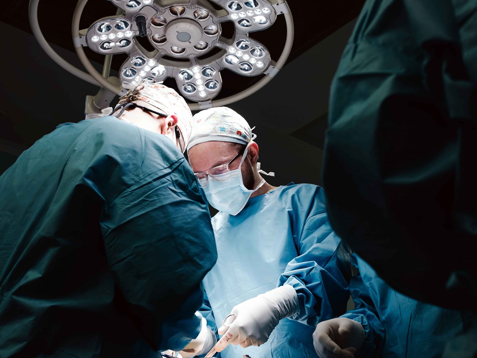
Endovascular Aneurysm Repair (EVAR) is a minimally invasive surgical procedure used to treat aortic aneurysms, most commonly Abdominal Aortic Aneurysms (AAA). By placing a stent graft inside the weakened portion of the aorta, the procedure creates a new pathway for blood flow, effectively "re-lining" the vessel to prevent a life-threatening rupture. EVAR is the preferred treatment for patients with suitable anatomy, offering a safer alternative to traditional open surgery.
When You Should Consider EVAR
Aneurysm Size: When the diameter exceeds 5.5 cm in men or 5.0 cm in women, where rupture risk increases significantly.
Rapid Expansion: Growth of more than 0.5 cm within a 6-month period.
Symptomatic Presentation: Any aneurysm causing persistent abdominal, flank, or back pain.
High Surgical Risk: For patients whose age, heart disease, or lung complications make open surgery dangerous.
Suitable Anatomy: Presence of an infrarenal aneurysm with a healthy "landing zone" of non-dilated aorta for secure anchoring.
Methods of EVAR
Standard EVAR: Use of a bifurcated (Y-shaped) stent graft for typical abdominal aneurysms located below the kidney arteries.
TEVAR (Thoracic EVAR): A specialized version used for aneurysms located in the thoracic (chest) section of the aorta.
FEVAR (Fenestrated EVAR): Custom-made grafts with "windows" (fenestrations) to maintain blood flow to vital branching arteries, such as those leading to the kidneys.
Stent Grafting: Deployment of a fabric-covered metal frame (Nitinol or stainless steel) to seal the aneurysm sac.
Real-time Fluoroscopy: High-definition X-ray guidance used to ensure precise placement of the device through the femoral arteries.
How Is Performed
Access: Small incisions or needle punctures are made in both groins to reach the femoral arteries.
Navigation: A delivery catheter carrying the collapsed stent graft is guided to the aneurysm site under X-ray imaging.
Deployment: The graft is released and expands to seal against the healthy artery walls above and below the weakened bulge.
Verification: An intraoperative angiogram (contrast dye injection) confirms there are no leaks and blood is flowing correctly through the graft.
Finalization: The delivery tools are removed, and the small access sites in the groin are closed with sutures or collagen plugs.
Pre-Procedure Preparation
Fasting: Required for 8–12 hours before the procedure, as it may require general or regional anesthesia.
Lab Work: Blood tests to evaluate kidney function (crucial for processing contrast dye) and clotting status.
Medication Review: Adjusting current medications, particularly antiplatelet drugs or diabetic treatments.
Anatomical Mapping: Detailed measurement using high-resolution CT scans to select the correct graft size and shape.
Allergy Check: Discussing any sensitivities to iodine, contrast dye, or metals like Nitinol (nickel-titanium).
Tests Before EVAR
CT Angiography (CTA): The primary tool for measuring aneurysm size and planning the precise graft path.
Duplex Ultrasound: To assess blood flow velocity and provide initial sizing of the aneurysm.
Cardiac Clearance: ECG and stress tests to ensure the heart can handle the procedure.
Blood Panel: Comprehensive checks including Creatinine (kidney function) and Hemoglobin levels.
Ankle-Brachial Index (ABI): To check for peripheral artery disease that might complicate access through the leg arteries.
Life After EVAR
Hospital Stay: Typically 1–2 days, with most patients encouraged to walk within 24 hours.
Lifelong Monitoring: Regular imaging (CT or Ultrasound) is mandatory to ensure the graft hasn't moved or developed leaks (endoleaks).
Follow-up Schedule: Imaging typically occurs at 1 month, 6 months, 12 months, and annually thereafter.
Activity Restrictions: Avoid heavy lifting and strenuous physical activity for approximately 2–4 weeks post-surgery.
Rupture Prevention: While the graft provides immediate protection, strict blood pressure control remains vital for long-term health.
Benefits of EVAR
Lower Mortality: Significantly lower initial mortality rates compared to open surgical repair.
Less Invasive: Avoids large abdominal or chest incisions, which reduces blood loss and the risk of infection.
Rapid Recovery: Faster healing time, allowing a quicker return to work and daily activities.
Accessible for High-Risk Patients: Provides an option for those who would not survive traditional open vascular surgery.
Durable Solution: Offers a long-term mechanical barrier to prevent the aorta from bursting.