Medivisor India Treatment Logo
  • Home
  • About Us
    • About Us
    • Our Services
    • Our Team
    • Our Medical Advisors
    • Our Safety Measures
  • Treatment
    • Loading treatments...
  • Hospitals
    • Loading hospitals...
  • Patient Guide
    • Treatment Cost
    • Treatment Process
    • Visa Process
    • Travel Guide
    • FAQs
    • Why Medivisor
  • Gallery
    • Success Stories
    • Patient Activities
    • Social Activities
    • News Coverage
    • Blog
  • Become a Partner

+91 83407 80250

Medivisor Logo

Medivisor India Treatment is a trusted New Delhi–based medical travel partner connecting global patients to advanced, affordable, high-quality healthcare in India.

  • Medivisor House 359, Sector 1, Vaishali, Ghaziabad, (Delhi/NCR) India
  • +91 8340 780 250
  • info@medivisorhealth.com

About Us

  • About Us
  • Our Services
  • Our Team
  • Our Medical Advisors
  • Our Safety Measures
  • Join Our Network
  • Contact Us

India Treatment

  • Hospitals
  • Treatments
  • Treatment Cost
  • Treatment Process
  • Visa Process
  • Travel Guide
  • FAQs
  • Why Medivisor

Gallery

  • Patient Testimonials
  • Patient Activities
  • News Coverage
  • Blog

Get in Touch

© 2026 Medivisor India Treatment. All rights reserved.

Cancer Care/Oncology hospital

Cancer Care/Oncology

  1. Home
  2. Departments
  3. Cancer Care/Oncology

Oncology Department (Cancer Care)

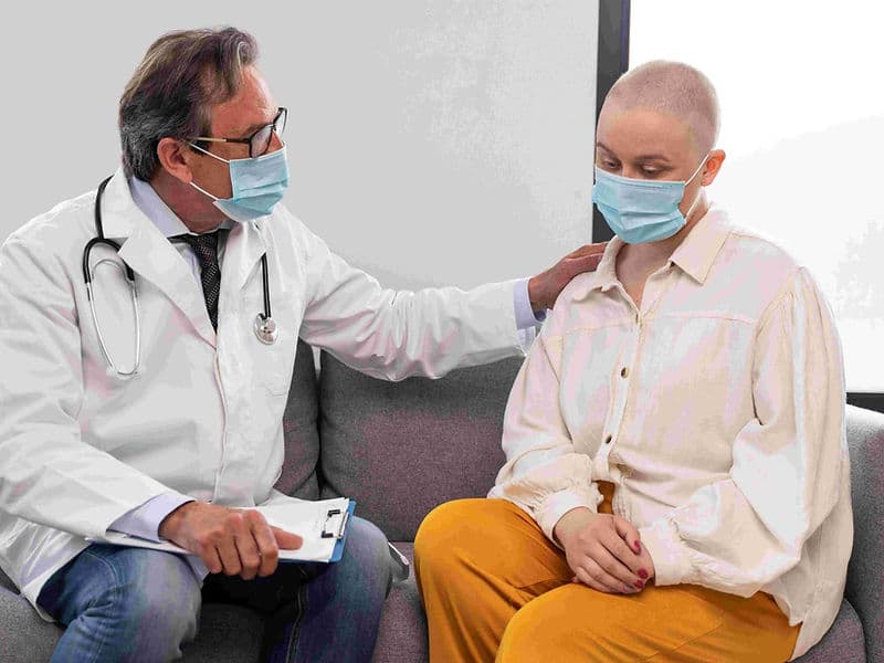
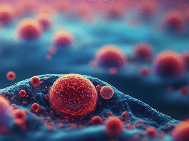
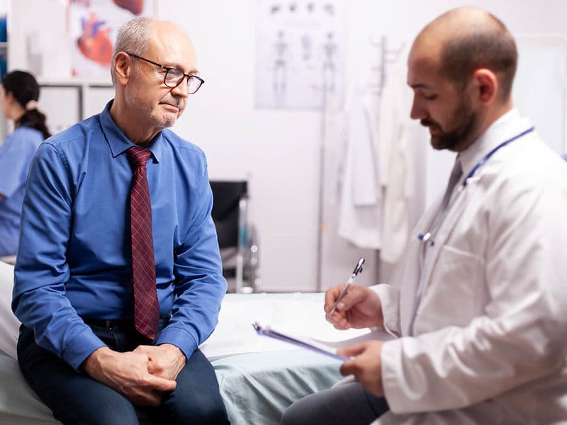
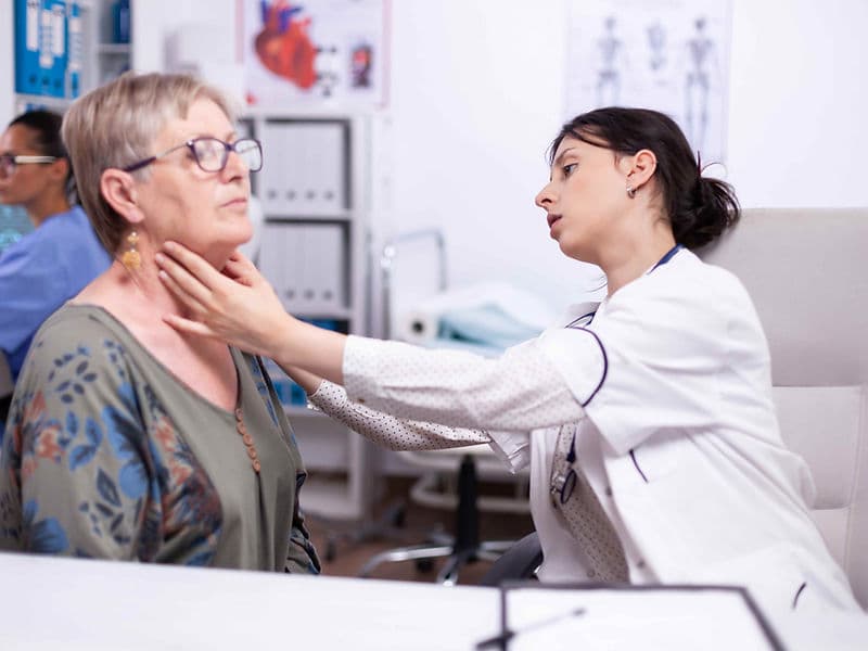
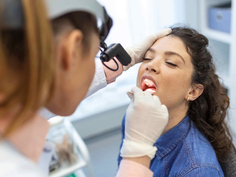
The Oncology Department is a premier center for comprehensive cancer care, designed as a sanctuary of hope and advanced healing. Modern oncology at this level is built upon a Multidisciplinary Team (MDT) framework, where every patient’s journey is overseen by a collective of experts. For international patients, this department represents the pinnacle of precision medicine, combining high-energy radiation, molecular systemic therapies, and surgical mastery to provide a holistic, 360-degree approach to recovery.

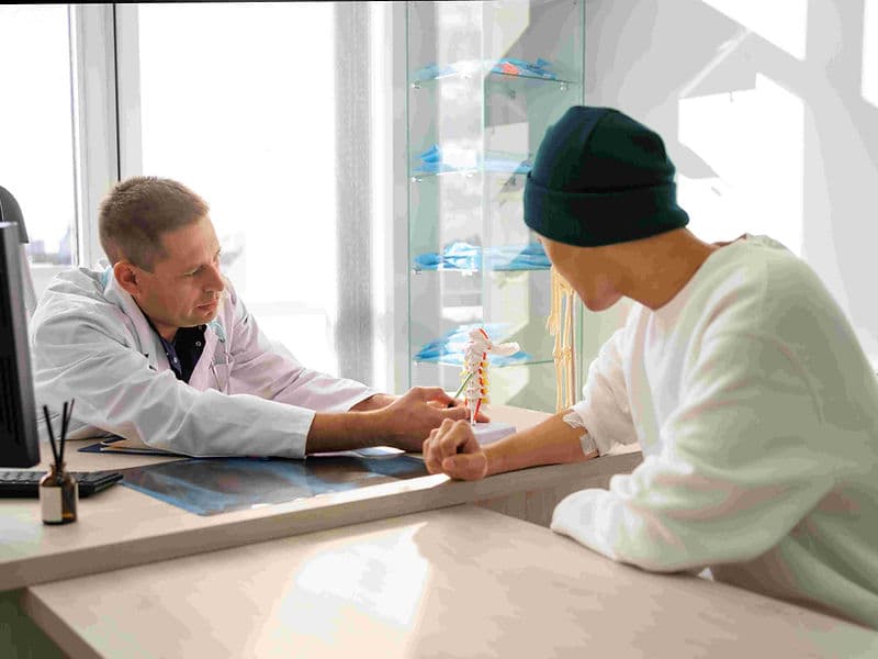
A Multidisciplinary Circle of Expertise

Effective cancer care requires a "Tumor Board" approach, where specialists collaborate to design a personalized treatment roadmap:

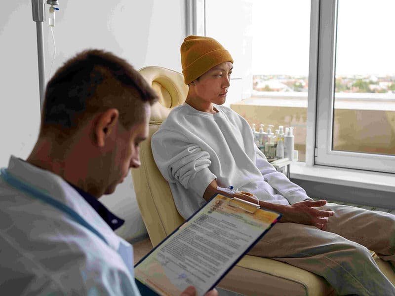
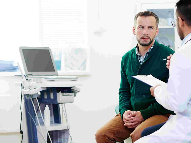
  • Medical Oncologists: The lead strategists who manage systemic treatments, including advanced Chemotherapy, Immunotherapy, and Targeted Therapy.

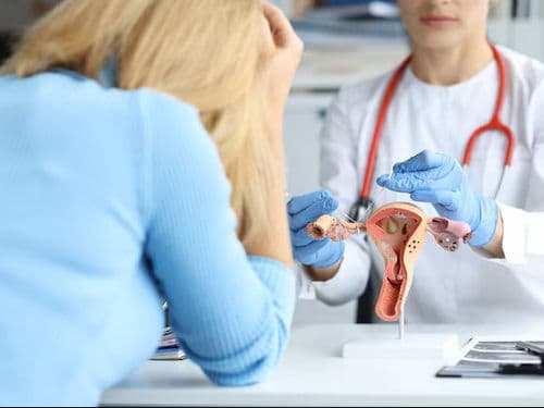
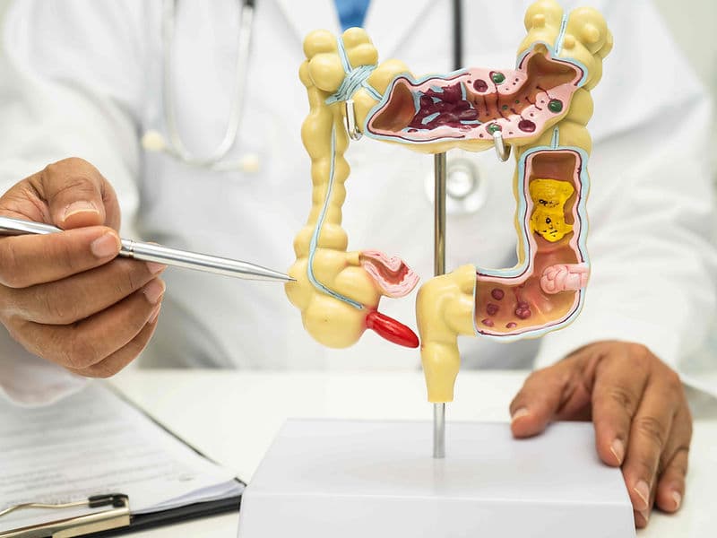
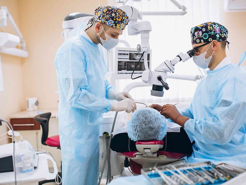
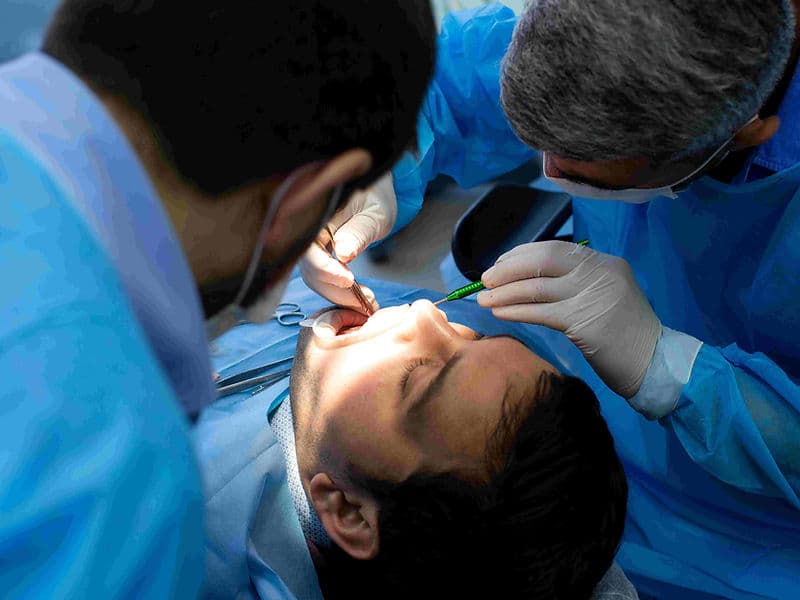
  • Surgical Oncologists: Specialists focused on the precision removal of tumors while preserving healthy surrounding tissue (margins).

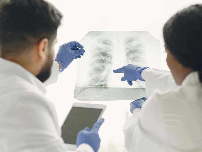
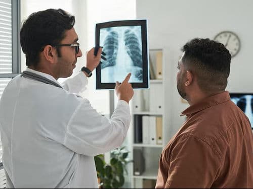
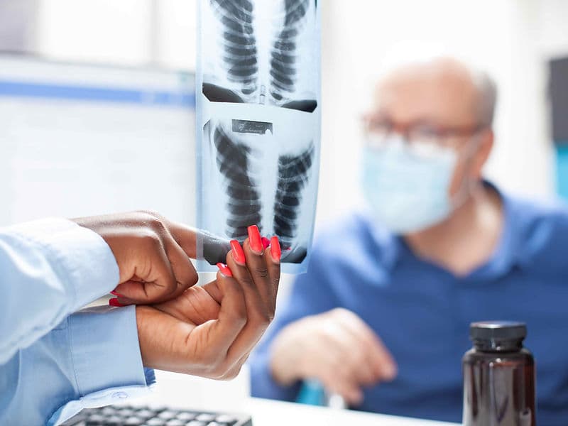
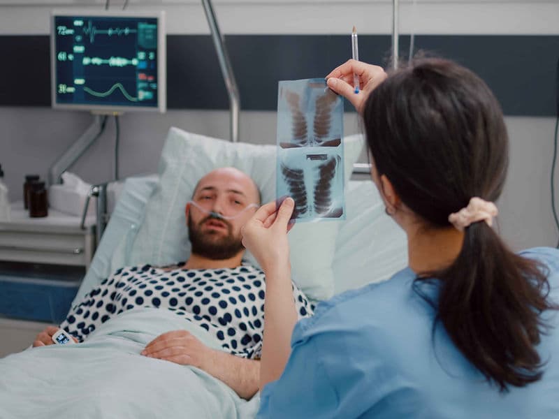
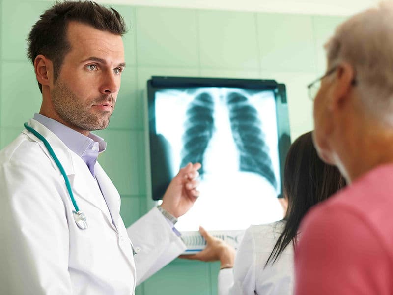
  • Radiation Oncologists: Experts who use high-energy subatomic particles to target and destroy cancer cells without the need for a scalpel.

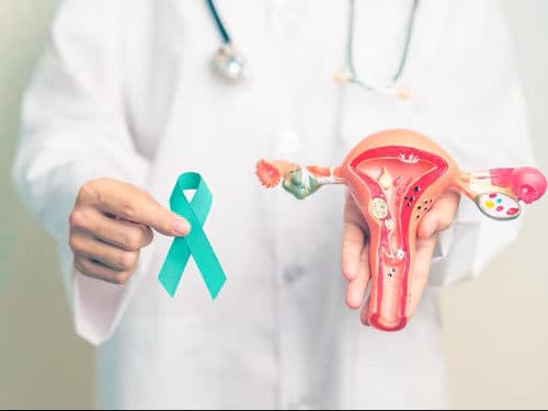
  • Haemato-Oncologists: Dedicated specialists for blood-based cancers, such as Leukemia and Lymphoma, often overseeing Bone Marrow Transplants.

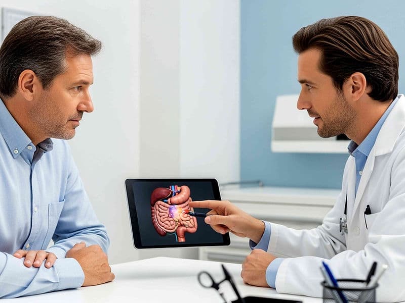
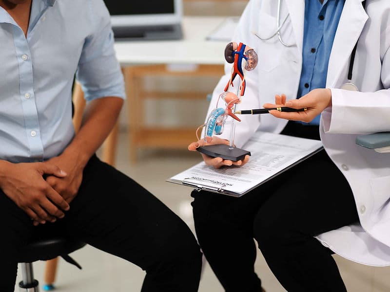
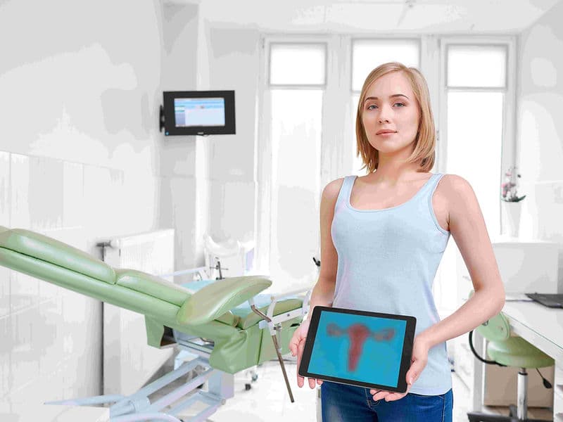
  • Onco-Pathologists: Vital researchers who decode the genetic signature of a tumor to identify the most effective drug matches.

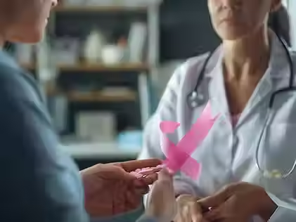
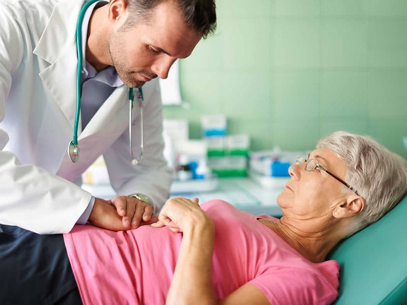
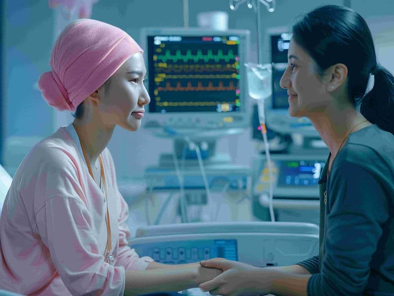
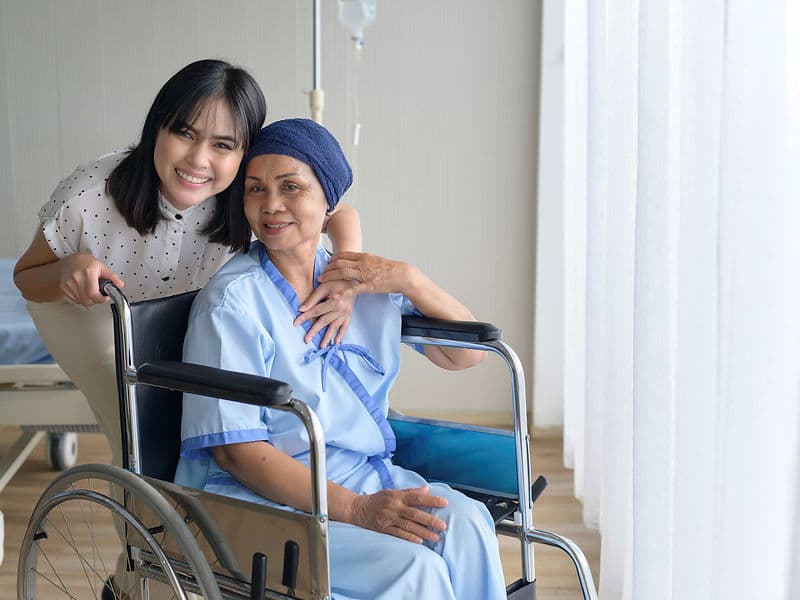
  • Palliative Care Specialists: A compassionate team dedicated to symptom management and maintaining the highest possible quality of life.

Advanced Technology and Precision Tools

Our department utilizes the latest global innovations to ensure treatment is as effective as it is safe:

  • LINAC (TrueBeam) Technology: High-energy linear accelerators that synchronize with your breathing to target tumors with pinpoint accuracy, sparing healthy organs.

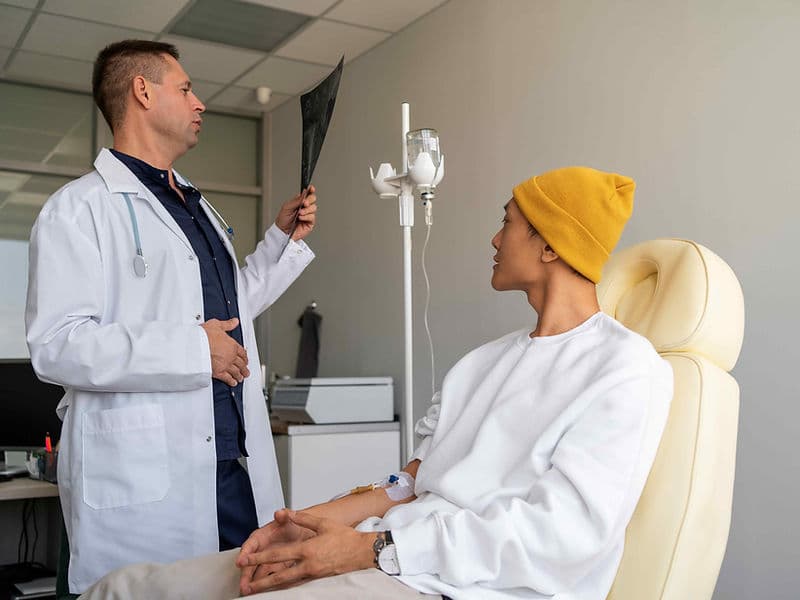
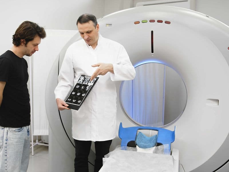
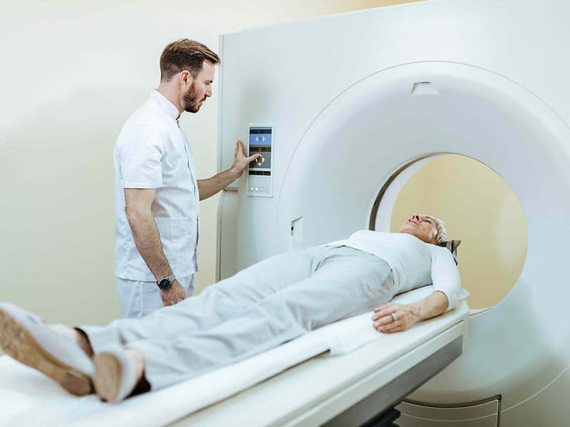
  • PET-CT Whole-Body Imaging: Advanced scanners that use radioactive markers to visualize active disease, helping to track treatment progress in real-time.

  • Molecular Targeted Therapy: Drugs designed to attack specific genetic mutations within cancer cells, sparing healthy cells and reducing side effects.

  • Scalpel-Free Radiosurgery: Non-invasive systems like Brachytherapy and localized radiation "seeds" that treat the tumor from the inside out.

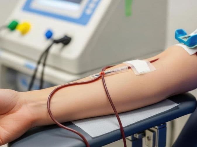
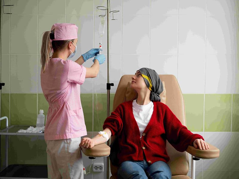
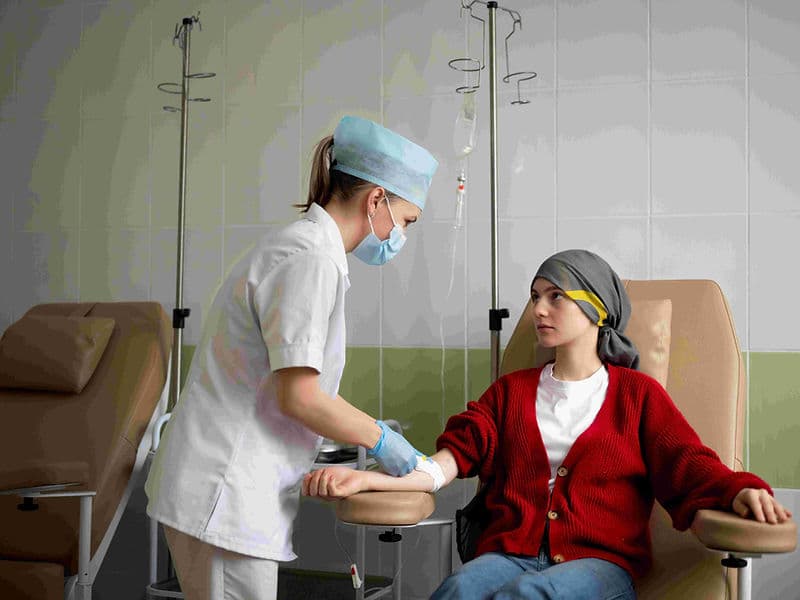
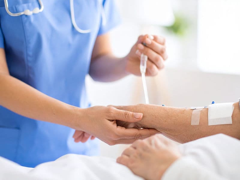
  • Digital Infusion Suites: High-tech "Daycare" centers equipped with precise infusion pumps and Cold Caps to minimize hair loss during therapy.

Specialized Care Units and Patient Facilities

To ensure safety and comfort, the department is organized into dedicated functional zones:

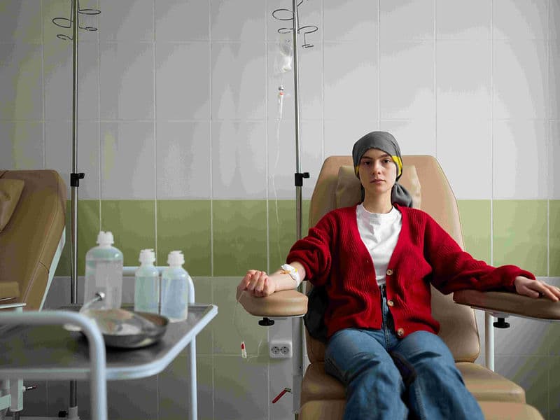
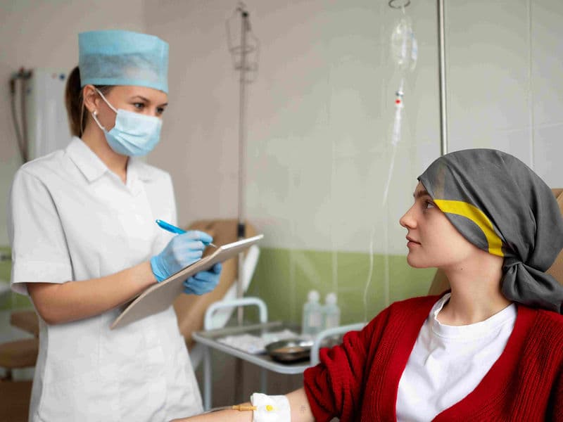
  • The Oncology Daycare Center: A premium, comfortable environment for outpatient chemotherapy infusions.

  • Bone Marrow Transplant (BMT) Unit: Ultra-sterile "Clean Rooms" with HEPA filtration systems designed for patients with sensitive immune systems.

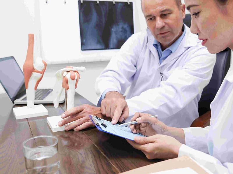
  • The Tumor Board Room: A collaborative hub where surgeons, radiologists, and pathologists meet to review and refine individual patient cases.

  • Nuclear Medicine Wing: A lead-lined facility for specialized diagnostic scans and targeted isotope treatments like Radio-Iodine therapy.

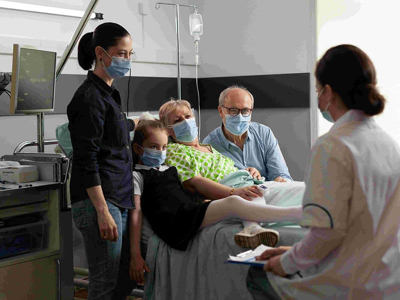
  • International Patient Sanctuary: A dedicated lounge for global families to coordinate medical records, travel logistics, and long-term survivorship plans.

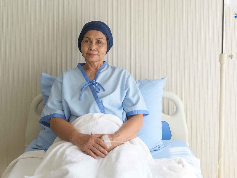
A Commitment to Long-Term Wellness

The care journey extends well beyond the final treatment session to ensure lasting health:

  • Survivorship Roadmaps: Comprehensive follow-up plans covering nutrition, surveillance scans, and exercise.

  • Maintenance Therapy: Ongoing access to the latest targeted pills to prevent recurrence and ensure long-term remission.

  • Psychosocial Support Groups: Integrated networks and counseling to help patients and families navigate the emotional transition to life after cancer.

May we help you?

Enter digits only (6-15 digits). We'll add your country code.

Cancer Care/Oncology Treatments

Acute Lymphoid Leukemia

Acute Lymphoid Leukemia

Acute Lymphoblastic Leukemia (ALL), also known as acute lymphoid or lymphocytic leukemia, is a fast-growing cancer of the blood and bone marrow. It occurs when the body overproduces immature white blood cells, called lymphoblasts, which crowd out healthy red blood cells, platelets, and normal white blood cells.

When You Should Consider ALL Evaluation

  • Persistent fatigue or weakness due to low red blood cell counts.

  • Frequent or unexplained fevers and infections.

  • Easy bruising, frequent nosebleeds, or tiny red spots under the skin (petechiae).

  • Bone or joint pain caused by the buildup of lymphoblasts.

  • Swelling in the neck, armpits, or groin (lymph nodes) or a mass in the chest.

Methods of ALL Classification

  • B-cell ALL: The most common form, accounting for about 85% of childhood cases and 75–80% of adult cases.

  • T-cell ALL: More common in adults (25%) and often associated with a mediastinal (chest) mass.

  • Philadelphia Chromosome-Positive (Ph+ ALL): A high-risk subtype involving a specific genetic translocation that requires specialized targeted therapy.

How ALL Treatment Is Performed

  • Induction Therapy: Intensive chemotherapy lasting 4–6 weeks aimed at killing most cancer cells to achieve remission.

  • CNS-Directed Therapy: Intrathecal chemotherapy injected into spinal fluid to prevent cancer from hiding in the brain or spinal cord.

  • Consolidation Therapy: High-dose therapy lasting 6–8 months to destroy any remaining "hidden" cells after remission.

  • Maintenance Therapy: Lower-dose oral and IV drugs administered over 2–3 years to prevent the cancer from returning.

  • Advanced Options: Includes Targeted Therapy (blocking specific enzymes), Immunotherapy (monoclonal antibodies), and CAR T-cell Therapy (genetically modified T-cells).

Pre-Procedure Preparation

  • Detailed genetic testing and chromosomal analysis to identify specific ALL subtypes like Ph+.

  • Placement of a central venous catheter (port) to facilitate long-term chemotherapy and blood draws.

  • Discussion of fertility preservation options before starting intensive chemotherapy or radiation.

  • Baseline heart and lung function tests to ensure the body can tolerate intensive induction therapy.

Tests Before ALL Treatment

  • Bone Marrow Aspiration and Biopsy: To confirm the percentage of lymphoblasts in the marrow.

  • Lumbar Puncture (Spinal Tap): To check if leukemia cells have spread to the central nervous system.

  • Complete Blood Count (CBC): To evaluate the levels of red cells, white cells, and platelets.

  • Flow Cytometry: To determine the exact immunophenotype (B-cell vs. T-cell) of the leukemia cells.

Life After ALL Treatment

  • Children (Ages 1–10) see the best prognosis, with 5-year survival rates exceeding 90%.

  • Adolescents and young adults have an estimated 5-year survival rate of roughly 65–75%.

  • Regular follow-up for 2 to 3 years is required during the maintenance phase to monitor for relapse.

  • Long-term monitoring for "late effects" of treatment, such as cardiac issues or secondary cancers.

Benefits of ALL Treatment

  • Achieves high rates of complete remission through structured therapy phases.

  • Prevents central nervous system involvement through proactive CNS-directed treatments.

  • Offers curative potential for relapsed cases using modern advances like CAR T-cell therapy.

  • Restores normal bone marrow function and healthy blood cell production.

Acute Myeloid Leukemia

Acute Myeloid Leukemia

Acute Myeloid Leukemia (AML) is a fast-growing cancer where the bone marrow makes abnormal myeloblasts, red blood cells, or platelets. These "leukemia cells" quickly crowd out healthy cells, leading to a high risk of infection, anemia, and easy bleeding.

When You Should Consider AML Evaluation

  • Sudden bruising or tiny red spots on the skin called petechiae.

  • Shortness of breath and extreme pale skin indicating anemia.

  • Persistent fevers that do not respond to standard antibiotics.

  • Evidence of high risk for infection or unexplained, easy bleeding.

Methods of AML Classification

  • FLT3 Mutation: Found in about 30% of cases and usually requires specific targeted drugs.

  • IDH1/IDH2 Mutations: Subtypes targeted by newer oral therapies.

  • TP53 Mutation: Often indicates a more resistant form of the disease.

  • APL (Acute Promyelocytic Leukemia): A unique, highly curable subtype treated with non-chemo drugs like arsenic trioxide.

  • Secondary AML: Often found in older adults (60+) arising from previous blood disorders.

How AML Treatment Is Performed

  • Induction Therapy: Typically a "7+3" regimen involving 7 days of one chemotherapy and 3 days of another to achieve complete remission.

  • Consolidation (Post-remission): Additional chemotherapy or a Stem Cell Transplant to kill remaining microscopic cells.

  • Targeted Therapy: Use of specific drugs for mutations like FLT3 or IDH1/IDH2.

  • Low-Intensity Options: Use of Venetoclax pills or Hypomethylating Agents (HMA) like Azacitidine for patients who cannot handle high-dose chemo.

  • Non-Chemo Regimens: Use of All-Trans Retinoic Acid (ATRA) specifically for the APL subtype.

Pre-Procedure Preparation

  • Cytogenetic profiling to determine the specific genetic mutations and treatment plan.

  • Assessment of age and physical tolerance for intensive chemotherapy.

  • Evaluation of heart or kidney function to determine if low-intensity options like Venetoclax are necessary.

  • Screening for previous blood disorders that may lead to secondary AML.

Tests Before AML Treatment

  • Bone Marrow Analysis: To identify abnormal myeloblasts and clear the marrow of visible blasts.

  • Genetic Testing: To check for FLT3, IDH1/IDH2, or TP53 mutations.

  • Blood Counts: To assess the severity of anemia and low platelet levels.

  • Cytogenetic Profiling: To map the "cytogenetic" profile which dictates the specific therapy.

Life After AML Treatment

  • For younger adults (<60), the 5-year survival rate is roughly 40% to 50%.

  • For older adults (60+), survival is lower, typically around 10% to 20%.

  • Patients with the APL subtype enjoy an excellent cure rate of over 90%.

  • Ongoing monitoring is required during the consolidation phase to prevent a relapse.

Benefits of AML Treatment

  • Clears the blood and bone marrow of visible leukemia blasts.

  • Provides "insurance" against relapse through consolidation or transplants.

  • Offers improved survival for older patients through modern low-intensity pill combinations.

  • Restores the production of healthy white blood cells, red cells, and platelets.

Adjuvant Chemotherapy (After Surgery)

Adjuvant Chemotherapy (After Surgery)

Adjuvant Chemotherapy is the administration of anti-cancer drugs after a primary treatment—usually surgery—to eliminate any microscopic cancer cells that may remain in the body but are too small to be detected by scans. Its goal is to reduce the risk of the cancer returning (recurrence) and to improve long-term survival. Modern protocols are increasingly personalized using "liquid biopsies" that detect circulating tumor DNA (ctDNA) to determine exactly who needs this "cleanup" treatment.

When You Should Consider Adjuvant Chemotherapy

  • Post-Surgical Security: When a surgeon has removed the visible tumor but the pathology report suggests a high risk of "micrometastases" (hidden cells).

  • Lymph Node Involvement: If cancer cells were found in the lymph nodes near the original tumor site, indicating the cancer may have begun to spread.

  • High-Grade Tumors: When the cancer cells look particularly aggressive under a microscope, making them more likely to multiply quickly.

  • Positive ctDNA Status: If a post-operative blood test detects microscopic fragments of tumor DNA in your bloodstream.

  • Genomic Risk: When genetic tests (such as Oncotype DX or MammaPrint) indicate your specific tumor has a high "recurrence score."

The Biological Rationale

  • The "Cleanup Crew": Even with "clear margins" after surgery, individual cells can detach and enter the blood or lymphatic systems. Adjuvant chemo acts as a safety net to destroy these cells.

  • Targeting Rapid Division: Chemo is most effective against cells that are actively dividing; by starting shortly after surgery, it catches any remaining cells before they can form a new mass.

  • The "Window of Opportunity": Adjuvant treatment is most effective when started within 4 to 8 weeks after surgery. Delaying beyond 12 weeks may significantly reduce the treatment's ability to prevent recurrence.

  • Combination Defense: By using 2 or 3 different drugs (like FOLFOX for colon cancer), doctors can attack hidden cells from multiple biological angles at once.

  • Systemic Protection: Unlike radiation, which is local, adjuvant chemo travels throughout the entire body to protect every organ.

How Adjuvant Chemotherapy Is Administered

  • Fixed Duration: Unlike treatment for advanced cancer, adjuvant therapy has a definite "finish line," typically lasting 3 to 6 months.

  • Cycle-Based Delivery: Treatment is given in cycles (e.g., 1 day of treatment followed by 2 or 3 weeks of rest) to allow your healthy bone marrow to recover.

  • Access Points: Depending on the regimen, it may be delivered via a temporary IV, a PICC line, or a surgically implanted chemo-port.

  • Pre-Medication: You will receive anti-nausea and anti-inflammatory medications before each session to minimize side effects.

  • Dose Calibration: Your oncology team will carefully adjust the dose based on how well you recovered from your recent surgery.

Pre-Procedure Preparation

  • Surgical Recovery: You must be sufficiently healed from your operation (usually 4+ weeks) before the body can safely handle chemotherapy.

  • Baseline Labs: A complete blood count (CBC) and metabolic panel are required to ensure your organs are ready for systemic treatment.

  • Dental Clearance: It is vital to address dental issues before starting, as chemo can make oral infections much harder to treat.

  • Nutritional Optimization: Focus on high-protein foods to help repair the tissues stressed by both the recent surgery and the upcoming chemo.

  • Support System: Since you are moving directly from surgical recovery into chemo, arrange for help with household tasks to combat cumulative fatigue.

Tests During Adjuvant Chemotherapy

  • CBC (Complete Blood Count): Frequent monitoring (often weekly) to check for a drop in white blood cells (neutropenia) or platelets.

  • ctDNA Monitoring: Regular blood draws to see if the "circulating tumor DNA" levels are dropping to zero.

  • Kidney & Liver Panels: To ensure these organs are successfully filtering the chemotherapy as they recover from the stress of anesthesia.

  • Heart Monitoring: If using certain drugs (like Herceptin or Anthracyclines), regular echocardiograms are performed to check heart strength.

  • Physical Assessment: Frequent checks of your surgical incision site to ensure the chemotherapy isn't interfering with the final stages of healing.

Life During Adjuvant Chemotherapy

  • Managing Fatigue: The "double hit" of surgery followed by chemo can cause significant exhaustion. Light, daily walks are the best proven way to maintain energy.

  • Infection Prevention: Your immunity will be lowest about 7–14 days after each dose (the "nadir"). Avoid crowds and people with active illnesses.

  • Wound Care: Keep your surgical scar protected from the sun, as chemo can make healing skin extra sensitive to UV rays.

  • Brain Health: Some patients experience "chemo-brain" (mild fogginess). Using planners and organizers can help manage daily tasks.

  • The Goal in Mind: Remember that adjuvant therapy is often the bridge between "remission" and a permanent cure.

Benefits of Adjuvant Chemotherapy

  • Reduced Recurrence Risk: Significantly lowers the chance of the cancer returning in the same spot or elsewhere in the body.

  • Improved Overall Survival: Clinical data shows that for many cancers (Stage II/III breast, colon, and lung), adjuvant chemo adds years to life expectancy.

  • Peace of Mind: Provides the reassurance that every possible measure has been taken to eliminate the disease.

  • Disease-Free Survival: Increases the amount of time you can live without any active signs of cancer.

  • Precision Medicine: Modern genomic tools ensure that adjuvant chemo is only given to those who will truly benefit, sparing others unnecessary treatment.

Allogeneic Bone Marrow Transplant

Allogeneic Bone Marrow Transplant

Allogeneic Bone Marrow Transplant—also known as an allogeneic stem cell transplant—is a life-saving procedure where a patient receives healthy stem cells from a donor to replace their own diseased or damaged marrow. Primarily used for aggressive blood cancers and non-malignant conditions like Thalassemia, this procedure introduces a new immune system into the patient’s body. Modern clinical protocols and advanced matching technologies have made this a highly successful intervention for patients with complex hematological disorders.

When You Should Consider Allogeneic BMT

  • Diagnosis of Acute Myeloid Leukemia (AML) or Acute Lymphoblastic Leukemia (ALL) with high-risk features.

  • Severe Aplastic Anemia where the bone marrow has stopped producing enough blood cells.

  • Presence of inherited blood disorders such as Thalassemia Major or Sickle Cell Anemia.

  • Myelodysplastic Syndromes (MDS) that show signs of progressing toward leukemia.

  • Certain types of aggressive Lymphoma that have relapsed after an autologous transplant.

  • Chronic Myeloid Leukemia (CML) that has become resistant to standard targeted therapies.

Conditions That Require Specialized Care

  • High-risk Leukemia requiring a powerful "Graft-versus-Tumor" effect to prevent relapse.

  • Pediatric Thalassemia requiring permanent correction of hemoglobin production.

  • Rare primary immunodeficiency disorders where the patient lacks a functional immune system.

  • Bone marrow failure syndromes requiring a complete replacement of the hematopoietic system.

  • Metabolic disorders that can be corrected by introducing healthy donor enzymes via stem cells.

How Allogeneic Bone Marrow Transplant Is Performed

  • A donor search is conducted to find a close Human Leukocyte Antigen (HLA) match.

  • The patient undergoes "Conditioning" with high-dose chemotherapy or radiation to suppress their immune system.

  • Healthy stem cells are collected from the donor's bone marrow or peripheral blood.

  • On the day of the transplant, donor cells are infused into the patient’s bloodstream through a central venous catheter.

  • The patient remains in a sterile, HEPA-filtered isolation room to prevent infection during the "neutral" phase.

  • Infused donor cells migrate to the marrow space and begin producing new, healthy blood cells (Engraftment).

Modern Innovations in Allogeneic BMT

  • Haploidentical (Half-Match) ProtocolsAdvanced techniques that allow parents or children to serve as donors with success rates comparable to full matches.

  • T-Cell Depletion & ModulationPrecision laboratory methods that remove specific donor cells responsible for GVHD while keeping those that fight cancer.

  • Next-Generation Sequencing (NGS) HLA MatchingUltra-high-resolution DNA matching that identifies the most compatible donor at the molecular level.

  • Reduced Intensity Conditioning (RIC)"Mini-transplants" that use lower doses of chemo, making the procedure safer for older or more fragile patients.

  • Microbiome-Preserving ProtocolsSpecialized nutritional and antibiotic strategies that protect the gut health to reduce the risk of post-transplant complications.

  • Post-Transplant Cyclophosphamide (PTCy)A breakthrough medication protocol that significantly lowers the incidence of Graft-versus-Host Disease in mismatched cases.

Donor Types and Selection

  • Matched Related Donor (MRD)The gold standard, typically a sibling who shares identical genetic markers (10/10 HLA match).

  • Matched Unrelated Donor (MUD)A compatible volunteer identified through international bone marrow registries.

  • Haploidentical DonorA biological parent or child who is a 50% genetic match, now widely used due to improved safety protocols.

  • Umbilical Cord BloodRich in stem cells, cord blood can be used for patients who cannot find a suitable adult donor.

Pre-Procedure Preparation

  • Rigorous HLA testing of the patient and potential family donors to find the best possible match.

  • Evaluation by a multidisciplinary team including hematologists, infectious disease specialists, and nutritionists.

  • Placement of a multi-lumen central venous catheter for chemotherapy, cell infusion, and blood sampling.

  • Extensive counseling on the long-term recovery process and the management of a new immune system.

  • Dental and sinus clearances to ensure there are no dormant infections prior to the conditioning phase.

Pre-Procedure Tests

  • High-resolution HLA typing (Class I and II) to confirm donor compatibility.

  • Bone marrow aspiration and biopsy to establish the baseline disease status.

  • Organ function tests including Echocardiogram, PFTs (Lungs), and Kidney function panels.

  • Comprehensive viral screening for CMV, EBV, HIV, and Hepatitis for both donor and recipient.

  • Cross-matching and donor-specific antibody (DSA) testing to prevent graft rejection.

Why This Treatment Is Highly Effective

  • Provides a "Graft-versus-Tumor" (GVT) effect, where the new immune system actively hunts and kills cancer cells.

  • Offers the only potential cure for many aggressive forms of leukemia and bone marrow failure.

  • Successfully cures pediatric Thalassemia in a high majority of cases, eliminating the need for lifelong transfusions.

  • Modern supportive care has significantly reduced the historical risks of infection and organ damage.

  • Technological advances allow for successful transplants even without a perfectly matched sibling.

Recovery and Monitoring

  • The "Engraftment" period (2–3 weeks) requires intensive monitoring for fever and blood count recovery.

  • Patients remain on immunosuppressant medications for several months to prevent Graft-versus-Host Disease (GVHD).

  • Full immune system reconstitution typically takes 6 to 12 months, during which special precautions are needed.

  • Frequent blood tests and chimeric studies are done to ensure the donor cells have successfully "taken over."

  • Gradual re-introduction to social environments occurs as white blood cell levels stabilize.

Life After Allogeneic BMT

  • Long-term remission and potential cure from previously fatal blood disorders.

  • A personalized re-vaccination schedule to rebuild immunity from the "donor's" perspective.

  • Regular monitoring for chronic GVHD, which can affect the skin, eyes, or liver.

  • Return to a full, active life, including school or work, once the immune system is mature.

  • Ongoing partnership with the transplant team to ensure long-term wellness and disease-free survival.

Amputation (Cancer)

Amputation (Cancer)

Amputation for cancer is a definitive surgical procedure to remove all or part of a limb when a malignant tumour, such as a bone or soft tissue sarcoma, cannot be safely or functionally treated with limb-salvage surgery. While limb salvage is now the gold standard (successful in up to 95% of cases), amputation remains a critical life-saving or palliative option for specific advanced scenarios. The focus has shifted toward "Pre-prosthetic Surgery," where nerves are rewired during the amputation to minimize phantom pain and improve control over high-tech bionic limbs.

When You Should Consider Amputation

  • Neurovascular Involvement: When the cancer has invaded the major nerves and blood vessels, meaning a functional limb cannot be preserved even if the tumour is removed.

  • Failed Limb Salvage: When previous attempts to save the limb have failed to achieve clear (negative) margins or the cancer has reoccurred multiple times.

  • Extensive Tissue Loss: If removing the tumour would require taking so much bone or muscle that the remaining limb would be non-functional or a source of chronic pain.

  • Palliative Management: To relieve intractable pain, manage severe bleeding, or treat an infected, "fungating" tumour in advanced metastatic disease.

  • Infection Risks: When a tumour is associated with a deep-seated infection that makes the use of internal metal implants (for limb salvage) too dangerous.

Common Types of Cancer Amputation

The level of amputation is determined by the need to achieve clear surgical margins while preserving the best possible site for a future prosthesis.

Lower Limb:

  • Transtibial (Below-Knee): Preserves the natural knee joint, offering significantly better functional outcomes and lower energy expenditure for walking.

  • Transfemoral (Above-Knee): Performed through the thigh bone; requires more energy for mobility due to the loss of the natural knee joint.

  • Hip Disarticulation: Removal of the entire leg at the hip joint.

  • Hemipelvectomy (Hindquarter): A radical procedure involving the removal of the entire leg and part of the pelvis, usually for tumours involving the pelvic bone itself.

Upper Limb:

  • Transradial / Transhumeral: Amputation below or above the elbow.

  • Forequarter Amputation: Removal of the entire arm, including the shoulder blade (scapula) and collarbone (clavicle), for advanced shoulder or chest wall tumours.

How an Amputation Is Performed

  • Anaesthesia: Performed under general anaesthesia, typically combined with a regional nerve block or epidural to "numb" the nerves before they are cut, which helps prevent future phantom pain.

  • Margin Verification: The surgeon identifies the highest point of the tumour and moves a safe distance (usually 5–10 cm) above it to ensure the bone is cut through a completely healthy area.

  • TMR (Targeted Muscle Reinnervation): A 2026 standard technique where severed nerves are "plugged" into nearby muscles. This prevents painful "neuromas" and allows for the future use of mind-controlled bionic prosthetics.

  • Myoplasty / Myodesis: The remaining muscles are carefully attached to the end of the bone or to each other to create a stable, well-padded "cushion" for the prosthesis.

  • Skin Flap Design: Surgeons create a "long flap" of healthy skin and fat to wrap around the end of the stump, ensuring the surgical scar is not located on a weight-bearing surface.

Pre-Surgery Preparation

  • Prosthetic Consultation: Meeting with a prosthetist before surgery to discuss the types of artificial limbs available and how the amputation level will affect your future mobility.

  • Psychological Support: Connecting with peer support groups or counselors who specialize in limb loss to prepare for the emotional transition.

  • Upper Body Strengthening: If a lower-limb amputation is planned, focusing on arm and core strength to prepare for using crutches or a wheelchair.

  • Phantom Pain Education: Learning about "Mirror Therapy" and other techniques to manage neurological sensations after surgery.

  • VTE Prophylaxis: Starting blood-thinning protocols to prevent clots, as amputation involves major blood vessel management.

Pre-Surgery Tests

  • Contrast-Enhanced MRI or CT: To determine the exact "proximal" extent of the tumour and ensure the planned amputation level is high enough to achieve clear margins.

  • Angiography: To assess the blood flow in the limb and ensure the remaining "stump" will have enough circulation to heal the surgical flaps.

  • Bone Scan or PET-CT: To rule out any other tumours in the same limb (skip lesions) that might require a higher level of amputation.

  • Baseline CBC and Nutrition: Checking for anaemia or low protein (albumin) levels, which are critical for healing a large surgical wound.

  • Cardiovascular Stress Test: To ensure the heart can handle the increased energy demands of walking with a prosthesis.

Life After Amputation (Recovery & Risks)

  • Wound Healing: Focuses on shaping the residual limb (stump) using elastic bandages or "shrinkers" to reduce swelling and prepare for a prosthesis.

  • Prosthetic Fitting: A temporary prosthesis may be used within weeks, followed by a permanent, custom-fitted device once swelling has subsided (usually 8–12 weeks).

  • Physical Therapy: Vital for gait training and strengthening remaining muscles. Walking with an above-knee prosthesis can require 60–100% more energy than natural walking.

  • Phantom Limb Pain: The feeling of pain or sensation in the missing part of the limb. 2026 treatments include nerve-mapping, specialized medications, and virtual reality therapy.

  • Activity Rules: You will work with occupational therapists to adapt your home and car to ensure you can return to independence as quickly as possible.

Why Specialized Treatment Is Highly Effective

  • Definitive Disease Control: Amputation provides the highest possible level of local cancer clearance for tumours that are too large for limb-sparing techniques.

  • Faster Return to Activity: For some complex sarcomas, a well-performed amputation and modern prosthesis allow a patient to return to walking and daily life faster than a multi-year limb-reconstruction process.

  • Advanced Bionics: 2026 prosthetic technology includes microprocessors and sensors that allow for a near-natural gait and even the restoration of some "touch" sensations.

  • TMR Integration: By performing Targeted Muscle Reinnervation during the initial surgery, 2026 surgeons have significantly reduced the incidence of chronic, debilitating phantom pain.

  • Multidisciplinary 2026 Care: The combination of oncology surgeons, prosthetists, and specialized physical therapists ensures a holistic recovery aimed at "total mobility."

Autologous Bone Marrow Transplant (ABMT)

Autologous Bone Marrow Transplant (ABMT)

Autologous Bone Marrow Transplant—also known as an autologous stem cell transplant—is a sophisticated procedure used to treat various blood cancers and severe autoimmune diseases. This treatment involves using the patient's own healthy stem cells to "rescue" the bone marrow after it has been cleared of disease by high-dose chemotherapy or radiation. By utilizing the patient's own biological material, this procedure eliminates the risk of donor-related complications and provides a powerful pathway to remission.

When You Should Consider ABMT

  • Diagnosis of Multiple Myeloma where transplant is recommended as a primary frontline therapy.

  • Relapsed or refractory Hodgkin’s or Non-Hodgkin’s Lymphoma that has not responded to standard chemotherapy.

  • Certain germ cell tumors that have returned after initial treatment.

  • Severe, treatment-resistant autoimmune diseases such as Multiple Sclerosis (MS) or Systemic Sclerosis.

  • Presence of high-risk neuroblastoma in pediatric cases where aggressive therapy is required.

  • Recommendation for high-dose "conditioning" therapy that would otherwise permanently damage bone marrow function.

Conditions That Require Specialized Care

  • Multiple Myeloma requiring long-term marrow stabilization and disease control.

  • Relapsed Lymphoma where the goal is to achieve deep, durable remission.

  • Severe Crohn's Disease or other autoimmune conditions that have failed all standard biologic therapies.

  • Amyloidosis, a rare protein disorder that can affect organ function.

  • Specific types of leukemia that are in remission but carry a high risk of recurrence.

How Autologous Bone Marrow Transplant Is Performed

  • Stem cells are mobilized from the bone marrow into the bloodstream using growth factor injections.

  • Healthy stem cells are harvested via Apheresis, where blood is filtered through a specialized machine.

  • The collected stem cells are cryopreserved (frozen) and safely stored in a laboratory.

  • The patient undergoes high-dose "Conditioning" (chemotherapy or radiation) to eliminate remaining cancer cells.

  • The frozen stem cells are thawed and reinfused into the bloodstream, much like a standard blood transfusion.

  • The infused cells migrate to the bone marrow (Engraftment) and begin producing new, healthy blood cells.

Innovations in Autologous Transplant

  • Advanced Apheresis TechnologyHigh-efficiency cell separators that maximize the yield of healthy stem cells while reducing the time spent on the machine.

  • Next-Generation Mobilization AgentsThe use of precision medications that more effectively push stem cells into the bloodstream, even for "poor mobilizers."

  • Real-Time CD34+ TrackingSophisticated laboratory monitoring that identifies the exact hour of peak stem cell concentration for optimal harvesting.

  • Targeted Conditioning RegimensRefined chemotherapy protocols designed to maximize cancer cell death while minimizing damage to healthy organs.

  • Rapid Engraftment MonitoringMolecular tools that detect the earliest signs of new blood cell production, allowing for faster discharge from the hospital.

  • Automated Thawing SystemsDigitally controlled warming devices that protect the delicate cell membranes during the transition from ice to infusion.

Pre-Procedure Preparation

  • Extensive physical evaluation to ensure the heart, lungs, and kidneys can tolerate high-dose therapy.

  • Dental clearance to eliminate any hidden sources of infection before the immune system is suppressed.

  • Placement of a central venous catheter (PICC or Hickman line) for easy blood access and infusion.

  • Coordination of a 3-to-6-week hospital stay in a specialized, HEPA-filtered isolation room.

  • Nutritional optimization and psychological counseling to prepare for the intensive recovery period.

Pre-Procedure Tests

  • Bone Marrow Aspiration and Biopsy to confirm the status of the underlying disease.

  • High-resolution PET/CT scans to map the location and extent of any remaining cancer cells.

  • Echocardiogram or MUGA scan to assess cardiac output and heart health.

  • Pulmonary Function Tests (PFTs) to ensure the lungs can handle systemic treatment.

  • Comprehensive blood panels, including viral markers and organ function profiles.

Why This Treatment Is Highly Effective

  • Eliminates the risk of Graft-versus-Host Disease (GVHD), as the body recognizes the cells as its own.

  • Allows for the use of "curative" doses of chemotherapy that would be impossible without a stem cell rescue.

  • Features high success rates, with durable remission seen in a significant majority of Multiple Myeloma patients.

  • Significantly improves survival outcomes and quality of life in relapsed lymphoma cases.

  • Offers a potential "reset" for the immune system in patients with aggressive autoimmune disorders.

Recovery and Monitoring

  • The "Engraftment" phase typically takes 10 to 14 days, during which the patient is closely monitored for infections.

  • Daily blood counts are performed to track the rise of white blood cells, red blood cells, and platelets.

  • Supportive care, including blood transfusions and IV antibiotics, is provided until the new marrow is functional.

  • Patients remain in a protective environment until their absolute neutrophil count reaches a safe level.

  • Long-term follow-up involves monitoring for "late effects" and ensuring the disease remains in remission.

Life After Autologous Transplant

  • Gradual return to daily activities as the immune system slowly recovers over several months.

  • Long-term disease management, which may include maintenance therapy to prevent recurrence.

  • Re-vaccination protocols, as the transplant often "wipes out" previous immunity to childhood diseases.

  • Regular oncology or hematology check-ups with advanced imaging and blood markers.

  • Empowerment through the successful completion of one of the most intensive and effective medical treatments available

Bladder Cancer Surgery

Bladder Cancer Surgery

Bladder cancer surgery ranges from minimally invasive procedures designed to preserve the organ to major reconstructive operations when the bladder must be removed. The specific surgical approach depends on the cancer's stage, its aggressiveness, and whether it has invaded the muscle layer of the bladder wall. These procedures are the primary treatment for eliminating tumors and preventing the spread of the disease to other organs.

When You Should Consider Bladder Cancer Surgery

  • Non-Muscle Invasive Bladder Cancer (NMIBC): For early-stage tumors that are located on the inner lining of the bladder and have not yet grown into the muscle.

  • Muscle-Invasive Bladder Cancer (MIBC): When the cancer has penetrated the deeper muscle layer of the bladder wall, requiring a more aggressive surgical approach.

  • Recurrent Tumors: If cancer returns after previous treatments or if the tumor grade is high (very aggressive).

  • Localized Muscle-Invasive Cancer: When the tumor is confined to a specific area that allows for partial removal while saving the rest of the bladder.

  • Diagnostic Necessity: A surgical biopsy is often the first step to accurately stage the cancer and determine the best long-term treatment plan.

How It Is Performed

  • Transurethral Resection of Bladder Tumor (TURBT): The most common procedure for early-stage cancer. A surgeon inserts a thin, lighted tool called a resectoscope through the urethra. An electric wire loop or laser is used to cut away or burn the tumor without any external incisions.

  • Partial Cystectomy: A specialized approach where only the cancerous portion of the bladder is removed. This is an option if the cancer is localized to one small area that can be removed without compromising the bladder's ability to hold urine.

  • Radical Cystectomy: The entire bladder is removed, along with nearby lymph nodes. In men, this often includes the prostate; in women, it may include the uterus and ovaries. This is the gold standard for treating muscle-invasive cancer.

  • Urinary Diversion (Reconstruction): If the entire bladder is removed, the surgeon creates a new way for the body to store and pass urine:
    Ileal Conduit (Urostomy): A piece of the small intestine creates a tube to carry urine to a stoma (opening) on the abdomen, draining into an external bag.
    Continent Urinary Reservoir: An internal pouch is made from the intestine. The patient empties the pouch several times a day using a thin catheter, avoiding an external bag.
    Neobladder Reconstruction: A new "bladder" is created from the intestine and connected to the urethra, allowing for more natural urination.

[Image showing the reconstruction of a neobladder using a segment of the intestine]

Pre-Procedure Preparation

  • Cystoscopy: A visual inspection of the bladder using a camera to map the tumor's location and size.

  • Imaging (CT or MRI): Detailed scans to determine if the cancer has spread to nearby lymph nodes or other organs.

  • Bowel Prep: For radical surgery involving intestinal reconstruction, you may be required to follow a liquid diet and take laxatives a day before.

  • Fasting: Following "nothing by mouth" instructions for 8 hours prior to your scheduled anesthesia.

  • Anesthesia Consultation: A meeting to discuss general anesthesia or spinal blocks used during the procedure.

Tests Before Bladder Cancer Surgery

  • Urinalysis and Cytology: Testing urine for blood, infection, and the presence of microscopic cancer cells.

  • Blood Panels: A routine check of your blood count, electrolytes, and kidney function (creatinine levels).

  • Chest X-ray: To ensure the lungs are clear and the cancer hasn't spread to the chest area.

  • ECG: A standard heart check to confirm cardiovascular stability for the duration of the surgery.

Life After Bladder Cancer Surgery

  • Hospital Stay: TURBT patients often go home the same day. Radical cystectomy requires a hospital stay of approximately one week for monitoring and recovery.

  • Immediate Symptoms: You may experience blood in the urine (hematuria), frequent urges to go, or a burning sensation during urination for a few days.

  • Recovery Timeline: Full recovery from major surgery can take several weeks to months. Most patients return to light activities within 4–6 weeks.

  • Follow-up Care: Regular check-ups with a urologist and oncologist are essential. This includes periodic cystoscopies and scans to ensure the cancer has not returned.

  • Adjusting to Diversion: Patients with a neobladder or stoma will receive specialized training from a wound and ostomy nurse to manage their new urinary system.

Why Specialized Treatment Is Highly Effective

  • Organ Preservation: Modern techniques like TURBT allow many patients to keep their natural bladder while effectively removing early-stage cancer.

  • Definitive Cancer Control: Radical cystectomy offers the highest chance of long-term survival for muscle-invasive bladder cancer by removing the primary source of the disease.

  • Reconstructive Excellence: Advanced neobladder surgery allows many patients to maintain a high quality of life with natural urinary function.

  • Minimally Invasive Options: Many bladder surgeries can now be performed robotically or laparoscopically, leading to less pain and faster healing.

  • Personalized Pathways: Surgeons can tailor the approach—from partial removal to complete reconstruction—based on the specific grade and location of your tumor.

Bone & Soft Tissue Tumor Surgery

Bone & Soft Tissue Tumor Surgery

Surgery for bone and soft tissue tumors (primarily sarcomas) is a highly specialized field where the goal is to remove the cancer completely while preserving as much physical function and appearance as possible. Because these tumors often grow near major nerves, blood vessels, and joints, the surgery requires meticulous planning. The standard of care involves using 3D-printed models and computer-assisted navigation to achieve precise "clear margins" while sparing the limb.

When You Should Consider Sarcoma Surgery

  • Primary Bone Cancer: For malignancies such as osteosarcoma, Ewing sarcoma, or chondrosarcoma.

  • Soft Tissue Sarcoma: When a cancerous mass is identified in the muscle, fat, nerves, or connective tissues (e.g., liposarcoma or synovial sarcoma).

  • Benign but Aggressive Tumors: For non-cancerous growths like Giant Cell Tumors (GCT) that can destroy local bone if not removed.

  • Metastatic Bone Disease: When cancer from another organ (like the lung or breast) has spread to a bone and threatens to cause a fracture.

  • Recurrent Tumors: When a previously treated tumor returns in the same anatomical compartment.

Types of Surgical Margins

The "margin" is the area of healthy tissue removed along with the tumor. Surgeons use specific classifications to define how much tissue to take:

  • Intralesional (Curettage): The tumor is scraped out from the inside. This is generally used only for benign (non-cancerous) bone tumors.

  • Marginal Excision: The tumor is removed exactly at its edge (pseudocapsule). This is often used for benign soft tissue tumors like lipomas.

  • Wide Excision: The tumor is removed with a continuous "cuff" of healthy tissue surrounding it. This is the standard of care for malignant tumors (sarcomas) to ensure no microscopic cells are left behind.

  • Radical Resection: Removal of the entire anatomical compartment (the whole bone or muscle group) containing the tumor.

Advanced Reconstruction Techniques

Once a tumor is removed, the resulting gap must be rebuilt to restore strength and mobility:

  • Biological Reconstruction: Uses the body's own ability to heal.
    Allograft: Uses donated bone from a bone bank to act as a scaffold.
    Autograft: Uses the patient's own bone, such as the fibula (calf bone), often moved with its blood vessels intact.

  • Mechanical Reconstruction: Uses artificial megaprostheses (large metal implants) to replace joints or long sections of bone. These allow for immediate weight-bearing.

  • Distraction Osteogenesis: Using devices like the Ilizarov fixator to slowly "grow" new bone to fill a gap.

Specialized Procedures for Children

Because children's bones are still growing, surgery requires unique solutions to prevent leg-length discrepancies:

  • Expandable Prostheses: Metal implants that can be lengthened non-invasively using magnets as the child grows to keep the legs equal in length.

  • Rotationplasty: A specialized procedure where the middle of the leg is removed, and the lower leg is rotated 180° and reattached to the thigh. The ankle then functions as a knee joint.

  • Growth Plate Sparing: Advanced 3D navigation allows surgeons to remove tumors while saving the natural growth plates whenever possible.

[Image showing an expandable "growing" prosthesis for a pediatric patient]

How Is Performed

  • Anesthesia: Performed under general anesthesia. Advanced nerve blocks are often used to provide long-term pain relief to the limb.

  • Computer-Assisted Navigation: Surgeons use "GPS for surgery" to follow a pre-planned 3D map, ensuring they cut exactly where the cancer ends and healthy bone begins.

  • Multidisciplinary Collaboration: If the tumor involves major blood vessels or leaves a large skin defect, vascular and plastic surgeons work simultaneously to perform bypasses or skin flaps.

  • 3D-Printed Cutting Guides: Custom-made templates are placed on the bone during surgery to guide the saw blade with sub-millimeter precision.

  • Intraoperative Imaging: Using O-arm or C-arm technology to verify the placement of implants and the completeness of the resection before the patient leaves the OR.

Pre-Procedure Preparation

  • Tumor Board Review: Your case is reviewed by a team of radiologists, pathologists, and oncologists to determine the best sequence of treatment.

  • 3D Virtual Planning: Surgeons use MRI/CT scans to create a virtual 3D model of your limb to practice the surgery before the actual procedure.

  • Physical Therapy Baseline: Establishing a baseline for your limb's strength and range of motion to guide your post-operative recovery.

  • Nutritional Optimization: Ensuring high protein intake to support the extensive bone and tissue healing required.

  • "Pre-hab" Exercise: Strengthening the healthy limbs to prepare for the period of restricted weight-bearing on the operated side.

Tests Before Bone and Soft Tissue Tumor Surgery

  • High-Resolution MRI: The most critical test for visualizing the tumor's relationship to muscles, nerves, and blood vessels.

  • Systemic CT Scan: To rule out "skip lesions" or spread to the lungs, which is common with certain sarcomas.

  • PET-CT Scan: To identify any other areas of metabolic activity that might indicate the cancer has moved elsewhere.

  • Core Needle Biopsy: To confirm the exact grade and type of the sarcoma, which dictates how wide the surgical margins must be.

  • Angiography: To map out the blood supply of the limb, especially if a vascularized bone graft (autograft) is planned.

Life After Sarcoma Surgery (Recovery & Risks)

  • Hospital Stay: Typically 5 to 10 days depending on the complexity of the reconstruction and the level of pain management needed.

  • Rehabilitation: This is the most critical phase. Physical therapy usually begins within 24–48 hours and can continue for 6 to 12 months.

  • Weight-Bearing Rules: Depending on the type of bone graft or prosthesis, you may need to use crutches or a walker for 3 to 6 months while the bone heals.

  • Mechanical Wear: Over many years, metal megaprostheses can wear out or loosen, potentially requiring a revision surgery.

  • Infection Monitoring: Large implants and pre-operative chemotherapy can increase the risk of infection, requiring long-term monitoring.

Why Specialized Treatment Is Highly Effective

  • Limb Salvage Success: Over 90% of sarcoma patients can have their limbs saved rather than amputated, with no loss in survival rates.

  • Precise Margin Control: Advanced 3D navigation has significantly lowered the risk of local recurrence by ensuring no microscopic cells are missed.

  • Functional Recovery: Modern megaprostheses and biological grafts allow many patients to return to walking, swimming, and an active lifestyle.

  • Growth Management: Expandable technology ensures that children can reach their full height without the need for multiple major open surgeries.

  • Integrated Care: When surgery is combined with modern immunotherapy and targeted radiation, the long-term cure rates for sarcomas are higher than ever before.

Bone Marrow Transplant

Bone Marrow Transplant

A bone marrow transplant (BMT), also called a Hematopoietic Stem Cell Transplant, is a procedure that replaces diseased or damaged bone marrow with healthy stem cells. These stem cells are the "factories" that produce your red blood cells, white blood cells, and platelets.

When You Should Consider Bone Marrow Transplant

  • To replace non-functioning marrow in conditions such as Aplastic Anemia.

  • To "rescue" the marrow after high-dose chemotherapy for Leukemia, Lymphoma, or Multiple Myeloma.

  • To replace "broken" or genetically abnormal cells in disorders like Sickle Cell Disease or Thalassemia.

  • When other primary treatments have failed and a transplant offers the only curative option.

  • Following the identification of a matched donor or the successful collection of own healthy stem cells.

Methods of Bone Marrow Transplant

  • Autologous Transplant: A procedure using the patient's own stem cells, which are collected and frozen before intensive treatment.

  • Allogeneic Transplant: A transplant using stem cells from a matched relative or an unrelated volunteer donor.

  • Matched Sibling Donor: Using a brother or sister who has the same human leukocyte antigen (HLA) type.

  • Haploidentical Transplant: A type of allogeneic transplant using a donor who is a "half-match," such as a parent or child.

  • Umbilical Cord Blood Transplant: Using stem cells harvested from the umbilical cord and placenta after a baby is born.

How Bone Marrow Transplant Is Performed

  • Conditioning: Administration of high-dose chemotherapy or radiation over 5–7 days to clear out old marrow.

  • Stem Cell Infusion: Healthy cells are infused through a central venous catheter (PICC or Hickman line), similar to a blood transfusion.

  • Engraftment: A 2–4 week period where the new cells travel to the bones and begin producing new blood cells.

  • Immune Reset: The process of the new immune system gradually maturing and learning to protect the body.

  • Continuous Monitoring: Intensive observation in the hospital to manage the high risk of infection during the recovery phase.

Pre-Procedure Preparation

  • Extensive work-up testing of the heart, lungs, and kidneys to ensure the body can handle the procedure.

  • Placement of a central venous catheter for the infusion of cells and administration of medications.

  • Coordination of stem cell collection (apheresis) for autologous patients or donor matching for allogeneic patients.

  • Understanding the "Point of No Return" during the conditioning phase where the old marrow is destroyed.

Tests Before Bone Marrow Transplant

  • HLA Typing: A specialized blood test used to match patients with the most compatible donors.

  • Bone Marrow Biopsy: To assess the current state of the marrow and the presence of any remaining cancer cells.

  • Organ Function Screens: Detailed evaluations including ECGs, lung function tests, and kidney filtration checks.

  • Infectious Disease Screening: Comprehensive testing for viruses or bacteria that could become dangerous during recovery.

Life After Bone Marrow Transplant

  • Most patients remain hospitalized for 3–5 weeks following the infusion.

  • Long-term recovery is a gradual process requiring close medical supervision for up to a year.

  • Patients must follow strict infection-prevention protocols while their immune system is "reset" to zero.

  • Ongoing management may include anti-rejection medications to prevent Graft-vs-Host Disease (GVHD).

Benefits of Bone Marrow Transplant

  • Provides a curative pathway for many blood cancers that are resistant to standard chemotherapy.

  • Restores the body's ability to produce healthy, functional red blood cells, white blood cells, and platelets.

  • Corrects the underlying genetic "blueprints" in patients with hereditary blood disorders.

  • Offers a chance for long-term remission and the restoration of a healthy immune system.

Breast Cancer Treatment

Breast Cancer Treatment

Breast Cancer Treatment involves a multidisciplinary approach to identify and eliminate abnormal cell growth within the breast tissue. Modern oncology focuses on precision medicine to target tumors in the milk ducts or lobules, aiming to restore physical health, maintain aesthetics, and prevent the spread of cancer cells to other parts of the body.

When You Should Consider Screening

  • Discovery of a new lump or thickened tissue in the breast or underarm area.

  • Visible changes in the size, shape, or symmetry of the breast.

  • Skin abnormalities such as dimpling, redness, or a texture resembling orange peel.

  • Nipple inversion or persistent pain in the nipple area.

  • Spontaneous discharge other than breast milk.

  • Persistent swelling or localized discomfort that does not correlate with the menstrual cycle.

Conditions That Require Specialized Care

  • HR-Positive breast cancer fueled by estrogen or progesterone hormones.

  • HER2-Positive or HER2-Low cancers with specific protein over-expression.

  • Triple-Negative Breast Cancer (TNBC) requiring aggressive systemic intervention.

  • Inflammatory Breast Cancer characterized by rapid skin changes and swelling.

  • Metastatic conditions where cells have migrated to the lymph nodes or distant organs.

How Breast Cancer Treatment Is Performed

  • Advanced imaging and biopsy are used to determine the tumor’s molecular profile.

  • Surgical intervention is performed to remove the tumor while preserving as much healthy tissue as possible.

  • Targeted drug therapies are administered to interrupt specific growth signals of cancer cells.

  • Systematic treatment like immunotherapy is used to enhance the body's natural defenses.

  • Precision radiation is applied to the affected area to eliminate any microscopic remnants.

  • Ongoing monitoring and hormonal therapy are utilized to prevent future recurrence.

Types of Breast Cancer Treatment

  • Lumpectomy (Breast-Conserving Surgery) The surgical removal of the tumor and a small margin of surrounding healthy tissue.

  • Mastectomy The removal of the entire breast tissue, often accompanied by immediate oncoplastic reconstruction.

  • Antibody-Drug Conjugates (ADCs) Advanced "smart" chemotherapy that delivers medication directly to the cancer cells to minimize side effects.

  • Targeted Therapy Medications designed to attack specific genetic mutations or proteins like HER2 or ESR1.

  • Immunotherapy Treatment that assists the immune system in identifying and destroying aggressive cancer cells.

  • Precision Radiation Therapy High-energy beams focused on the tumor site using shortened, highly effective schedules.

Pre-Surgery Preparation

  • Discuss surgical options and reconstruction preferences with the oncoplastic team.

  • Undergo a complete physical evaluation to ensure readiness for anesthesia.

  • Follow specific instructions regarding the cessation of certain medications or supplements.

  • Arrange for post-operative support and home care during the initial recovery phase.

  • Complete all pre-treatment mapping and diagnostic scans as scheduled.

Pre-Surgery Tests

  • Diagnostic Mammography to provide detailed views of the breast tissue.

  • Breast MRI for high-resolution imaging of the tumor's extent and location.

  • Ultrasound-guided biopsy to determine the specific molecular subtype of the cancer.

  • Blood chemistry panels including tumor markers and organ function tests.

  • Genetic testing to identify inherited mutations that may influence treatment choices.

Why Specialized Treatment Is Highly Effective

  • Utilizes real-time biomarkers to match patients with the most effective medications.

  • Minimizes damage to healthy cells through the use of targeted delivery systems.

  • Offers high survival rates through early detection and rapid intervention.

  • Integrates aesthetic considerations with life-saving surgical procedures.

  • Provides a personalized roadmap to recovery based on the unique biology of the tumor.

Recovery After Treatment

  • Specialized post-operative care to manage incision sites and physical comfort.

  • Early physical therapy to maintain range of motion in the arm and shoulder.

  • Transition from hospital care to home recovery within the recommended timeframe.

  • Gradual return to daily activities guided by the clinical oncology team.

  • Scheduled follow-up visits to monitor healing and long-term wellness.

Life After Breast Cancer Treatment

  • Regular surveillance through imaging to ensure continued remission.

  • Adherence to long-term hormonal or maintenance therapies as prescribed.

  • Participation in nutritional and lifestyle programs to support overall health.

  • Engagement with survivor support networks for emotional and psychological well-being.

  • Consistent physical activity to improve energy levels and metabolic health.

Cervical Cancer Treatment

Cervical Cancer Treatment

Cervical Cancer Treatment refers to a comprehensive range of medical and surgical protocols aimed at eliminating malignant cells within the tissues of the cervix. The focus is on highly precise interventions that target tumors while prioritizing the preservation of reproductive health. Advanced immunotherapies and Antibody-Drug Conjugates (ADCs) are now standard for managing advanced cases, ensuring long-term remission and the prevention of recurrence.

When You Should Consider Screening

  • Abnormal Bleeding: Unusual vaginal bleeding occurring between menstrual periods or specifically after intercourse.

  • Postmenopausal Bleeding: Any vaginal bleeding that begins after the onset of menopause.

  • Unusual Discharge: Persistent vaginal discharge that may have an unusual color, consistency, or odor.

  • Pelvic or Back Pain: Chronic pelvic discomfort or unexplained pain in the lower back region.

  • Dyspareunia: Pain or discomfort experienced during sexual activity.

  • Systemic Signs: General fatigue or persistent swelling in the lower extremities (legs).

Conditions That Require Specialized Care

  • Squamous Cell Carcinoma: Affecting the thin, flat cells lining the outer part of the cervix (most common type).

  • Adenocarcinoma: Originating in the glandular cells of the cervical canal that produce mucus.

  • Recurrent Cervical Cancer: Cancer that has returned after an initial period of successful remission.

  • Metastatic Disease: Advanced cancer that has spread to the pelvic lymph nodes or distant organs like the lungs or liver.

  • High-Risk Precancerous Lesions (CIN III): Lesions that require immediate removal to prevent them from turning into invasive malignancy.

Types of Cervical Cancer Treatment

  • Radical Trachelectomy: A specialized fertility-sparing surgery that removes the cervix and upper vagina while leaving the uterus intact, allowing for future pregnancy.

  • Hysterectomy: The surgical removal of the uterus and cervix; used for cases where fertility preservation is not a priority or the cancer is more advanced.

  • Antibody-Drug Conjugates (ADCs): Precision medications like Tisotumab Vedotin that deliver potent treatment directly to tumor proteins.

  • Immunotherapy: The use of checkpoint inhibitors (like Pembrolizumab) to help the immune system recognize and eliminate HPV-related cancer cells.

  • Brachytherapy (Internal Radiation): A procedure where radioactive material is placed directly into the cervix for high-dose, localized treatment.

  • Concurrent Chemoradiation: The strategic combination of chemotherapy and radiation given together to maximize the "kill rate" of cancer cells.

[Image showing the process of Brachytherapy with a radiation source placed near the cervix]

How Is Performed

  • Diagnostic Mapping: Primary HPV DNA testing and specialized pelvic imaging are completed to determine the tumor's exact boundaries.

  • Surgical Intervention: Depending on the stage, surgeons remove either the lesion (Cone Biopsy), the cervix (Trachelectomy), or the entire reproductive structure.

  • Targeted Delivery: If the cancer is advanced, ADCs or targeted drugs are administered via IV to seek out specific markers on the cancer cells.

  • Radiation Application: High-precision external beams or internal "seeds" are used to destroy cells in the pelvic area while sparing the bladder and bowel.

  • Immune Stimulation: Systematic immunotherapy is used to "unmask" HPV-driven cells so the body's natural defenses can attack them.

  • Surveillance: Rigorous follow-up including co-testing (Pap + HPV) is conducted to monitor cellular health.

Pre-Procedure Preparation

  • Fertility Consultation: Discuss long-term goals regarding reproductive health; options like egg freezing may be considered.

  • Physical Optimization: Follow recommended guidelines for nutrition and light activity to ensure the body is prepared for anesthesia.

  • Medication/Fasting Audit: Adhere to strict fasting (NPO) protocols and stop any blood-thinning supplements as directed.

  • Recovery Logistics: Arrange for a supportive home environment and a caregiver to assist during the initial 2-week healing phase.

  • Multidisciplinary Review: Ensure the case has been reviewed by a specialized team, including surgeons, radiation oncologists, and pathologists.

Tests Before Cervical Cancer Treatment

  • Primary HPV DNA Test: To identify the specific high-risk strain of the virus driving the cellular changes.

  • Colposcopy & Biopsy: A microscopic exam of the cervix with a targeted tissue sample to confirm the depth of the cancer.

  • Pelvic MRI: The standard for determining if the cancer has affected surrounding ligaments or moved toward the vaginal wall.

  • PET-CT Scan: To check for any increased metabolic activity in the lymph nodes or distant organs.

  • Renal & Immune Panels: Comprehensive blood work to ensure the kidneys can handle contrast dyes and the immune system is prepared for treatment.

Life After Cervical Cancer Treatment

  • Rigorous Follow-up: Adherence to a schedule involving regular HPV testing and physical exams every 3–6 months for the first few years.

  • Lifestyle Shifts: Commitment to a healthy lifestyle, including absolute smoking cessation, as smoking significantly increases the risk of recurrence.

  • Pelvic Health: Engagement with pelvic floor physical therapy to manage any changes in bladder, bowel, or sexual function.

  • Family Prevention: Continued education for family members regarding the benefits of the HPV vaccine.

  • Emotional Wellness: Participation in support programs to manage the psychological impact of a cancer diagnosis and potential fertility changes.

Why Specialized Treatment Is Highly Effective

  • Root Cause Targeting: Addresses the underlying HPV-driven changes rather than just treating the tumor surface.

  • Fertility Preservation: Modern surgical pathways prioritize keeping the uterus intact for young women whenever oncologically safe.

  • Reduced Toxicity: Targeted ADCs and precision radiation reduce the "collateral damage" to healthy pelvic organs like the bladder.

  • Prevention Standards: Benefit from standardized protocols designed to achieve high cure rates and prevent recurrence.

  • Individualized Care: Every treatment plan is biologically mapped to the specific genetic profile of the tumor.

Chronic Lymphoid Leukemia

Chronic Lymphoid Leukemia

Chronic Lymphocytic Leukemia (CLL) is the most common type of leukemia in adults. It is a slow-growing cancer of B-lymphocytes (a type of white blood cell) that originates in the bone marrow and spreads to the blood and lymph nodes. Unlike acute leukemias, many people with CLL live for years or even decades without needing immediate treatment.

When You Should Consider CLL Treatment

  • Extreme fatigue, drenching night sweats, or unexplained weight loss.

  • Massive or painful swelling of the spleen or lymph nodes.

  • Worsening anemia (low red cells) or thrombocytopenia (low platelets) indicating bone marrow failure.

  • Rapid lymphocyte doubling, where the white blood cell count doubles in less than 6 months.

Methods of CLL Management

  • Active Surveillance (Watch and Wait): The standard of care for early-stage, asymptomatic patients where starting chemotherapy early has not shown to increase lifespan.

  • BTK Inhibitors: Daily oral pills, such as Ibrutinib, Acalabrutinib, or Zanubrutinib, that block survival signals in B-cells.

  • BCL-2 Inhibitors: Targeted drugs like Venetoclax that trigger "cell death" in leukemia cells.

  • Monoclonal Antibodies: IV treatments like Obinutuzumab or Rituximab that "tag" cancer cells for the immune system to destroy.

  • IVIG Infusions: Regular immunoglobulin infusions to manage high infection risks for pneumonia or shingles.

How CLL Is Monitored

  • Regular Testing: Patients typically undergo blood tests and physical exams every 3 to 6 months.

  • Disease Progression Checks: Doctors monitor for signs of "active disease" that would necessitate a shift from surveillance to therapy.

  • Immune System Screening: Watching for autoimmune issues where the body attacks its own red blood cells (AIHA) or platelets (ITP).

  • Transformation Monitoring: Screening for Richter’s Transformation, where CLL evolves into an aggressive large B-cell lymphoma.

Pre-Procedure Preparation

  • Understanding the "Watch and Wait" approach and why immediate intervention is often avoided to prevent unnecessary side effects.

  • Comprehensive baseline blood work and physical assessments to establish a comparison for future monitoring.

  • Discussion of potential long-term risks, including a weakened immune system and increased infection susceptibility.

  • Evaluation of age and overall health, as the average age at diagnosis is 70.

Tests Before CLL Treatment

  • Complete Blood Count (CBC): To track white blood cell doubling time and levels of red cells and platelets.

  • Physical Examination: To check for organ issues like swelling of the spleen or lymph nodes.

  • Genetic Testing: To determine if specific mutations are present that might respond better to targeted oral therapies.

  • Clinical Trials Review: Assessment of trial data regarding the timing of treatment for stable patients.

Life After CLL Diagnosis

  • The overall 5-year survival rate for CLL is high, approximately 88%.

  • Many patients live for years or decades with the disease, eventually dying from causes unrelated to leukemia.

  • Patients must remain vigilant for complications like pneumonia, shingles, or the sudden onset of aggressive lymphoma.

  • Ongoing active surveillance remains the primary "lifestyle" for many early-stage patients.

Benefits of CLL Management

  • Avoids the toxicity and side effects of traditional chemotherapy through targeted oral therapies.

  • Provides a structured monitoring system to ensure treatment begins only when clinically necessary.

  • Utilizes modern medications that are more effective and less toxic than historical options.

  • Maintains a high quality of life for stable patients through the "Watch and Wait" strategy.

Chronic Myeloid Leukemia

Chronic Myeloid Leukemia

Chronic Myeloid Leukemia (CML) is a slow-growing cancer of the bone marrow that results in the overproduction of white blood cells. It is unique because nearly all cases (approx. 95%) are caused by a specific genetic abnormality known as the Philadelphia chromosome.

When You Should Consider CML Evaluation

  • Persistent fatigue or weakness.

  • Drenching night sweats.

  • Fullness or discomfort on the left side of the abdomen due to an enlarged spleen.

  • Unexplained weight loss or mild fevers.

  • Many patients have no symptoms and are diagnosed during routine blood tests.

Methods of CML Classification

  • Chronic Phase: The most common stage at diagnosis (85–90% of patients) where symptoms are mild and the disease responds well to standard treatment.

  • Accelerated Phase: The cancer grows faster with blast counts rising to 10–19%, and symptoms become more noticeable.

  • Blast Phase (Blast Crisis): An aggressive stage with 20% or more blasts that behaves like acute leukemia and is harder to treat.

  • Genetic Cause: Driven by the BCR-ABL gene, created when pieces of chromosomes 9 and 22 swap places.

How CML Treatment Is Performed

  • TKI Therapy: Daily oral pills such as Imatinib (Gleevec), Dasatinib (Sprycel), or Nilotinib (Tasigna) that specifically "turn off" the BCR-ABL protein.

  • Hematologic Management: Normalizing blood counts, which typically occurs within the first few weeks of treatment.

  • Molecular Monitoring: Using sensitive PCR tests to measure and reduce the level of the BCR-ABL gene in the blood.

  • Treatment-Free Remission (TFR): For eligible patients who maintain a deep response, stopping medication under strict medical supervision.

Pre-Procedure Preparation

  • Understanding that CML is now a manageable chronic condition requiring consistent, long-term medication.

  • Establishing a baseline through physical exams and medical history to check for an enlarged spleen.

  • Discussing the importance of adherence to daily oral therapy to achieve a Major Molecular Response.

  • Planning for frequent initial monitoring to ensure the "genetic switch" is being effectively turned off.

Tests Before CML Treatment

  • Bone Marrow Biopsy: To confirm the diagnosis and determine the phase of the disease.

  • FISH or PCR Testing: Highly sensitive blood tests used to detect the Philadelphia chromosome and the BCR-ABL gene.

  • Complete Blood Count (CBC): To evaluate the levels of abnormal white blood cells.

  • Cytogenetic Analysis: To identify the physical exchange of genetic material between chromosomes 9 and 22.

Life After CML Treatment

  • Life expectancy for those in the chronic phase is now approaching that of the general population.

  • The 5-year survival rate is currently over 90%, compared to roughly 20% before the year 2000.

  • Patients require lifelong monitoring to ensure the disease remains in molecular remission.

  • Most patients can maintain a normal lifestyle while taking daily oral targeted therapy.

Benefits of CML Treatment

  • Revolutionizes the prognosis from a once-fatal disease to a manageable chronic condition.

  • Specifically targets the genetic "stuck switch" (BCR-ABL) without the need for traditional intensive chemotherapy.

  • Achieves Major Molecular Response, making the cancer gene nearly undetectable in the blood.

  • Offers the potential for treatment-free remission in patients with deep, sustained responses.

Colectomy (Colon Cancer)

Colectomy (Colon Cancer)

Colon Cancer Treatment involves a combination of specialized surgical, medical, and radiological interventions to remove malignant tumors from the large intestine. As of 2026, clinical standards emphasize a precision-based approach, utilizing robotic surgery for localized tumors and advanced immunotherapy for cases with specific genetic biomarkers (MSI-H/dMMR), aiming to achieve long-term remission while preserving bowel function.

When You Should Consider Screening

  • Persistent changes in bowel habits, such as diarrhea, constipation, or narrowing of the stool.

  • Presence of bright red blood or dark, tarry stools during bowel movements.

  • Ongoing abdominal discomfort, including persistent cramps, gas, or localized pain.

  • A frequent feeling that the bowel does not empty completely after a movement.

  • Unexplained weight loss accompanied by chronic fatigue or weakness.

  • Signs of anemia, such as paleness and lack of energy, due to hidden internal bleeding.

Conditions That Require Specialized Care

  • Adenomatous Polyps that show signs of high-grade dysplasia or early transformation.

  • Localized Colon Adenocarcinoma confined to the inner layers of the intestinal wall.

  • Hereditary Syndromes including Lynch Syndrome or Familial Adenomatous Polyposis (FAP).

  • Inflammatory Bowel Disease (IBD) such as long-term Ulcerative Colitis or Crohn’s disease.

  • Advanced or metastatic colon cancer that has spread to the liver, lungs, or peritoneum.

How Colon Cancer Treatment Is Performed

  • Diagnostic colonoscopy is utilized to visualize the colon and remove precancerous polyps.

  • High-resolution CT, MRI, or PET scans are performed to map the tumor’s size and spread.

  • A partial or total colectomy is surgically performed to remove the malignant section of the colon.

  • Adjuvant chemotherapy is administered after surgery to eliminate any remaining microscopic cells.

  • Systematic immunotherapy is introduced for tumors with specific genetic MSI-H or dMMR markers.

  • Targeted drug therapy is used to block the proteins that allow cancer cells to grow and spread.

Types of Colon Cancer Treatment

  • Laparoscopic or Robotic Colectomy A minimally invasive surgery to remove the cancerous part of the colon through small abdominal incisions.

  • Polypectomy The removal of small, early-stage cancerous growths directly through a colonoscope without external incisions.

  • Immunotherapy (Pembrolizumab/Nivolumab) Advanced biological treatments that help the immune system identify and attack cancer cells.

  • Targeted Therapy (Bevacizumab/Cetuximab) Precision medications that target specific growth factors or blood vessel formation in the tumor.

  • Cytoreductive Surgery with HIPEC A specialized procedure where the abdominal cavity is bathed in heated chemotherapy after tumor removal.

  • Colostomy or Ileostomy A surgical procedure to create a stoma (opening) in the abdomen to allow waste to exit the body temporarily or permanently.

Pre-Surgery Preparation

  • Complete a "bowel prep" to thoroughly clear the colon of stool before the procedure.

  • Consult with a dietitian to establish a low-residue diet in the days leading up to surgery.

  • Adjust or stop blood-thinning medications as instructed by the surgical oncology team.

  • Undergo a preoperative physical to ensure readiness for general anesthesia and recovery.

  • Meet with an ostomy nurse if there is a possibility of needing a temporary or permanent stoma.

Pre-Surgery Tests

  • Colonoscopy with biopsy to confirm the specific pathology and grade of the cancer.

  • CT Scan of the chest, abdomen, and pelvis to determine the clinical stage of the disease.

  • CEA (Carcinoembryonic Antigen) Blood Test to establish a baseline for post-treatment monitoring.

  • MSI/dMMR Biomarker Testing to identify if the tumor will respond to modern immunotherapies.

  • Complete Blood Count (CBC) to check for anemia or other blood-related issues before surgery.

Why Specialized Treatment Is Highly Effective

  • Features a 91% five-year survival rate for localized cases through early detection and surgery.

  • Utilizes 2026 precision medicine to match treatments to the tumor's unique genetic profile.

  • Offers minimally invasive robotic options that significantly reduce recovery time and pain.

  • Employs neoadjuvant therapies to shrink large tumors, making them easier to remove safely.

  • Integrates multidisciplinary care to manage complex cases that have spread to other organs.

Recovery After Treatment

  • In-hospital monitoring for 3–5 days to ensure the return of normal bowel function.

  • Personalized pain management and early mobilization to prevent blood clots and lung issues.

  • Gradual transition from a liquid diet to soft foods as the digestive tract heals.

  • Specialized wound care and training for patients who require a temporary or permanent stoma.

  • Follow-up visits with the surgical and oncology teams to monitor healing and plan next steps.

Life After Colon Cancer Treatment

  • Regular surveillance through colonoscopies and blood tests every 6–12 months for the first few years.

  • Adoption of a high-fiber diet rich in fruits, vegetables, and whole grains to support colon health.

  • Commitment to regular physical activity and maintaining a healthy weight to reduce recurrence risk.

  • Avoidance of tobacco products and limiting alcohol consumption as part of a cancer-prevention lifestyle.

  • Participation in survivorship programs to manage the emotional and physical transition after treatment.

Esophageal Cancer Treatment

Esophageal Cancer Treatment

Esophageal Cancer Treatment involves a range of advanced medical and surgical procedures designed to eliminate malignant tumors within the esophagus—the muscular tube connecting the throat to the stomach. Clinical protocols emphasize a personalized, multimodal approach to restore swallowing function and prevent the systemic spread of cancer cells while prioritizing the patient’s overall quality of life.

When You Should Consider Screening

  • Persistent difficulty or pain while swallowing solid foods or liquids.

  • Unexplained weight loss without changes in diet or physical activity.

  • Chronic chest pain or a sensation of pressure behind the breastbone.

  • Frequent or worsening symptoms of acid reflux and heartburn.

  • Persistent hoarseness or a chronic cough that does not resolve.

  • Frequent choking on food or a feeling of something being stuck in the throat.

Conditions That Require Specialized Care

  • Adenocarcinoma occurring primarily in the lower part of the esophagus.

  • Squamous Cell Carcinoma affecting the thin, flat cells of the upper and middle esophagus.

  • Barrett’s Esophagus with high-grade dysplasia that may lead to malignancy.

  • Gastroesophageal Junction (GEJ) tumors affecting the area where the esophagus meets the stomach.

  • Recurrent esophageal cancer following previous chemotherapy or surgical interventions.

How Esophageal Cancer Treatment Is Performed

  • Diagnostic upper endoscopy and biopsies are utilized to identify the cellular subtype.

  • Imaging scans including PET and CT are conducted to map the extent of the tumor.

  • Minimally invasive surgery is performed to remove the diseased segment of the esophagus.

  • Immunotherapy infusions are administered to help the immune system identify malignant cells.

  • Precision radiation is applied to the tumor site to reduce its size before surgical removal.

  • Real-time monitoring of tumor markers in the blood is used to guide post-operative recovery decisions.

Types of Esophageal Cancer Treatment

  • Robotic-Assisted Esophagectomy A minimally invasive surgical procedure to remove the tumor and nearby lymph nodes with high precision.

  • Radiofrequency Ablation (RFA) The use of thermal energy to destroy precancerous tissue in patients with Barrett’s esophagus.

  • Immunotherapy Modern checkpoint inhibitors like pembrolizumab and tislelizumab that empower the immune system to fight advanced cancer.

  • FLOT Chemotherapy Regimen A specialized drug protocol used to achieve superior systemic control in cases of adenocarcinoma.

  • Endoscopic Mucosal Resection (EMR) A procedure used to remove small, early-stage tumors from the lining of the esophagus without major surgery.

  • Targeted Therapy Medications designed to attack specific molecular markers found on esophageal cancer cells.

Pre-Surgery Preparation

  • Engage in nutritional counseling to ensure the body is strong enough for the recovery process.

  • Adhere to breathing exercises and pulmonary rehabilitation to optimize lung function.

  • Follow specific guidelines for fasting and hydration as directed by the anesthesia team.

  • Consult with the surgical team regarding the use of feeding tubes for post-operative support.

  • Complete all cardiovascular clearances to ensure safe management during the procedure.

Pre-Surgery Tests

  • Upper Endoscopy to visually inspect the esophagus and collect tissue samples.

  • Endoscopic Ultrasound (EUS) to determine how deeply the tumor has invaded the esophageal wall.

  • CT and PET Scans to evaluate whether the cancer has spread to distant organs.

  • Circulating Tumor DNA (ctDNA) test to establish a baseline for molecular monitoring.

  • Pulmonary Function Tests to assess the strength of the respiratory system before surgery.

Why Specialized Treatment Is Highly Effective

  • Incorporates 2026 breakthroughs in immunotherapy for first-line treatment success.

  • Utilizes robotic technology to reduce surgical trauma and shorten hospital stays.

  • Employs "guided" ablative therapies to treat early-stage cancer without major incisions.

  • Targets systemic disease more effectively through modern chemotherapy combinations.

  • Provides a more accurate prognosis using molecular monitoring and precision imaging.

Recovery After Treatment

  • Intensive monitoring of swallowing function and respiratory health in a clinical setting.

  • Early mobilization and physical therapy to prevent complications and restore strength.

  • Gradual transition from liquid diets to soft foods under the supervision of a dietitian.

  • Specialized care for surgical incisions to ensure proper healing and prevent infection.

  • Follow-up evaluations with the oncology team to manage any side effects of systemic therapy.

Life After Esophageal Cancer Treatment

  • Permanent adoption of smaller, more frequent meals to accommodate changes in digestive capacity.

  • Maintaining an elevated sleeping position to prevent reflux and respiratory irritation.

  • Regular follow-up appointments with periodic imaging to ensure continued remission.

  • Ongoing monitoring of tumor markers through non-invasive blood-based liquid biopsies.

  • Commitment to a lifestyle free from tobacco and excessive alcohol to reduce recurrence risk.

External Beam Radiation Therapy

External Beam Radiation Therapy

Surgery for bone and soft tissue tumors (primarily sarcomas) is a highly specialized field where the goal is to remove the cancer completely while preserving as much physical function and appearance as possible. Because these tumors often grow near major nerves, blood vessels, and joints, the surgery requires meticulous planning. The standard of care involves using 3D-printed models and computer-assisted navigation to achieve precise "clear margins" while sparing the limb.

When You Should Consider Sarcoma Surgery

  • Primary Bone Cancer: For malignancies such as osteosarcoma, Ewing sarcoma, or chondrosarcoma.

  • Soft Tissue Sarcoma: When a cancerous mass is identified in the muscle, fat, nerves, or connective tissues (e.g., liposarcoma or synovial sarcoma).

  • Benign but Aggressive Tumors: For non-cancerous growths like Giant Cell Tumors (GCT) that can destroy local bone if not removed.

  • Metastatic Bone Disease: When cancer from another organ (like the lung or breast) has spread to a bone and threatens to cause a fracture.

  • Recurrent Tumors: When a previously treated tumor returns in the same anatomical compartment.

Types of Surgical Margins

The "margin" is the area of healthy tissue removed along with the tumor. Surgeons use specific classifications to define how much tissue to take:

  • Intralesional (Curettage): The tumor is scraped out from the inside. This is generally used only for benign (non-cancerous) bone tumors.

  • Marginal Excision: The tumor is removed exactly at its edge (pseudocapsule). This is often used for benign soft tissue tumors like lipomas.

  • Wide Excision: The tumor is removed with a continuous "cuff" of healthy tissue surrounding it. This is the standard of care for malignant tumors (sarcomas) to ensure no microscopic cells are left behind.

  • Radical Resection: Removal of the entire anatomical compartment (the whole bone or muscle group) containing the tumor.

Advanced Reconstruction Techniques

Once a tumor is removed, the resulting gap must be rebuilt to restore strength and mobility:

  • Biological Reconstruction: Uses the body's own ability to heal.
    Allograft: Uses donated bone from a bone bank to act as a scaffold.
    Autograft: Uses the patient's own bone, such as the fibula (calf bone), often moved with its blood vessels intact.

  • Mechanical Reconstruction: Uses artificial megaprostheses (large metal implants) to replace joints or long sections of bone. These allow for immediate weight-bearing.

  • Distraction Osteogenesis: Using devices like the Ilizarov fixator to slowly "grow" new bone to fill a gap.

Specialized Procedures for Children

Because children's bones are still growing, surgery requires unique solutions to prevent leg-length discrepancies:

  • Expandable Prostheses: Metal implants that can be lengthened non-invasively using magnets as the child grows to keep the legs equal in length.

  • Rotationplasty: A specialized procedure where the middle of the leg is removed, and the lower leg is rotated 180° and reattached to the thigh. The ankle then functions as a knee joint.

  • Growth Plate Sparing: Advanced 3D navigation allows surgeons to remove tumors while saving the natural growth plates whenever possible.

[Image showing an expandable "growing" prosthesis for a pediatric patient]

How Is Performed

  • Anesthesia: Performed under general anesthesia. Advanced nerve blocks are often used to provide long-term pain relief to the limb.

  • Computer-Assisted Navigation: Surgeons use "GPS for surgery" to follow a pre-planned 3D map, ensuring they cut exactly where the cancer ends and healthy bone begins.

  • Multidisciplinary Collaboration: If the tumor involves major blood vessels or leaves a large skin defect, vascular and plastic surgeons work simultaneously to perform bypasses or skin flaps.

  • 3D-Printed Cutting Guides: Custom-made templates are placed on the bone during surgery to guide the saw blade with sub-millimeter precision.

  • Intraoperative Imaging: Using O-arm or C-arm technology to verify the placement of implants and the completeness of the resection before the patient leaves the OR.

Pre-Procedure Preparation

  • Tumor Board Review: Your case is reviewed by a team of radiologists, pathologists, and oncologists to determine the best sequence of treatment.

  • 3D Virtual Planning: Surgeons use MRI/CT scans to create a virtual 3D model of your limb to practice the surgery before the actual procedure.

  • Physical Therapy Baseline: Establishing a baseline for your limb's strength and range of motion to guide your post-operative recovery.

  • Nutritional Optimization: Ensuring high protein intake to support the extensive bone and tissue healing required.

  • "Pre-hab" Exercise: Strengthening the healthy limbs to prepare for the period of restricted weight-bearing on the operated side.

Tests Before Bone and Soft Tissue Tumor Surgery

  • High-Resolution MRI: The most critical test for visualizing the tumor's relationship to muscles, nerves, and blood vessels.

  • Systemic CT Scan: To rule out "skip lesions" or spread to the lungs, which is common with certain sarcomas.

  • PET-CT Scan: To identify any other areas of metabolic activity that might indicate the cancer has moved elsewhere.

  • Core Needle Biopsy: To confirm the exact grade and type of the sarcoma, which dictates how wide the surgical margins must be.

  • Angiography: To map out the blood supply of the limb, especially if a vascularized bone graft (autograft) is planned.

Life After Sarcoma Surgery (Recovery & Risks)

  • Hospital Stay: Typically 5 to 10 days depending on the complexity of the reconstruction and the level of pain management needed.

  • Rehabilitation: This is the most critical phase. Physical therapy usually begins within 24–48 hours and can continue for 6 to 12 months.

  • Weight-Bearing Rules: Depending on the type of bone graft or prosthesis, you may need to use crutches or a walker for 3 to 6 months while the bone heals.

  • Mechanical Wear: Over many years, metal megaprostheses can wear out or loosen, potentially requiring a revision surgery.

  • Infection Monitoring: Large implants and pre-operative chemotherapy can increase the risk of infection, requiring long-term monitoring.

Why Specialized Treatment Is Highly Effective

  • Limb Salvage Success: Over 90% of sarcoma patients can have their limbs saved rather than amputated, with no loss in survival rates.

  • Precise Margin Control: Advanced 3D navigation has significantly lowered the risk of local recurrence by ensuring no microscopic cells are missed.

  • Functional Recovery: Modern megaprostheses and biological grafts allow many patients to return to walking, swimming, and an active lifestyle.

  • Growth Management: Expandable technology ensures that children can reach their full height without the need for multiple major open surgeries.

  • Integrated Care: When surgery is combined with modern immunotherapy and targeted radiation, the long-term cure rates for sarcomas are higher than ever before.

Gastrectomy (Stomach Cancer)

Gastrectomy (Stomach Cancer)

Stomach Cancer Treatment, also known as Gastric Cancer Treatment, involves a range of medical and surgical interventions to eliminate malignant growths within the stomach lining. Clinical protocols prioritize the removal of adenocarcinomas while preserving digestive function, utilizing precision therapies to target specific genetic markers and improve long-term outcomes for patients.

When You Should Consider Screening

  • Persistent Indigestion: A lasting burning sensation in the upper abdomen.

  • Early Satiety: Feeling unusually full or bloated after consuming only small meals.

  • Nausea and Vomiting: Chronic episodes, particularly if blood is present.

  • Dysphagia: Difficulty swallowing or a sensation of food being "stuck" in the esophagus.

  • Tarry Stools: Black, sticky stools which may indicate internal gastrointestinal bleeding.

  • Unexplained Fatigue: Persistent weakness often associated with anemia.

Conditions That Require Specialized Care

  • Gastric Adenocarcinomas: Originating in the glandular cells of the stomach mucosa.

  • HER2-Positive Tumors: Stomach cancers that require specific targeted medications.

  • CLDN18.2-Positive Cancers: Identified through modern molecular profiling.

  • H. pylori Infections: Chronic bacterial infections that have led to malignant cellular changes.

  • Genetic Syndromes: Diffuse gastric cancer related to inherited conditions or Lynch syndrome.

How Is Performed

  • Endoscopy: An upper endoscopy is conducted to visualize the stomach lining and obtain tissue biopsies.

  • Staging: Advanced imaging and endoscopic ultrasounds are used to determine the depth of the tumor.

  • Surgical Resection: Intervention is performed to remove part or all of the affected stomach tissue.

  • Perioperative Therapy: Chemotherapy is often administered to shrink the tumor before the primary procedure.

  • Targeted Biology: Biological therapies are introduced to attack specific proteins on the cancer cells.

  • Systematic Immunotherapy: Utilized to enhance the body's natural response to the malignancy.

Types of Stomach Cancer Treatment

  • Partial Gastrectomy: The surgical removal of the specific portion of the stomach containing the tumor and nearby lymph nodes.

  • Total Gastrectomy: A procedure where the entire stomach is removed and the esophagus is connected directly to the small intestine.

  • Targeted Therapy: The use of specialized drugs like Trastuzumab or Zolbetuximab to attack specific cancer cell markers.

  • Immunotherapy: Checkpoint inhibitors that empower the immune system to recognize and destroy gastric cancer cells.

  • Endoscopic Mucosal Resection: A minimally invasive technique used to remove very early-stage tumors during an endoscopy.

  • HIPEC (Heated Chemotherapy): The delivery of heated chemotherapy directly into the abdominal cavity during surgery for advanced cases.

Pre-Procedure Preparation

  • Nutritional Counseling: Consult with a specialized dietitian to manage intake and prepare for digestive changes.

  • Physical Assessment: A comprehensive evaluation to ensure readiness for general anesthesia.

  • Medication Audit: Adhering to specific adjustments as advised by the surgical oncology team.

  • Fasting Protocols: Strict adherence to fasting in the hours leading up to the scheduled procedure.

  • Support Planning: Arranging for assistance during the transition to a modified eating schedule.

Tests Before Stomach Cancer Treatment

  • Upper GI Endoscopy: To provide a direct view of the gastric environment and tumor site.

  • Endoscopic Ultrasound (EUS): To evaluate how far the cancer has invaded the stomach wall.

  • CT and PET Scans: To check for the spread of cells to the liver, lungs, or lymph nodes.

  • Biomarker Testing: HER2 and CLDN18.2 testing to determine the most effective drug therapies.

  • Blood Work: Complete Blood Count (CBC) and iron studies to assess for internal blood loss.

Life After Stomach Cancer Treatment

  • Dietary Adaptation: Adopting a lifestyle of eating small, frequent, and nutrient-dense meals throughout the day.

  • Supplementation: Regular vitamin and mineral support, particularly Vitamin B12, following a gastrectomy.

  • Routine Surveillance: Ongoing monitoring through imaging and endoscopy to ensure continued remission.

  • Weight Management: Consultation with nutritionists to maintain a healthy weight and energy levels.

  • Survivorship Support: Participation in programs to manage the emotional and physical impact of the disease.

Why Specialized Treatment Is Highly Effective

  • Precision Guidelines: Matches therapies with the tumor's specific molecular profile.

  • Perioperative Success: Significantly reduces the risk of cancer recurrence through timed therapy.

  • Minimally Invasive Options: Results in faster recovery and less discomfort for eligible patients.

  • Targeted Strategies: Provides life-prolonging options even for advanced or metastatic conditions.

  • Integrated Care: Maintains high standards of digestive and nutritional health through multidisciplinary teams.

Gastrointestinal Cancer Surgery

Gastrointestinal Cancer Surgery

Gastrointestinal (GI) Cancer Surgery refers to a broad category of operations used to remove tumors from the digestive tract, including the esophagus, stomach, liver, pancreas, and intestines. The primary goal is curative resection, where the surgeon removes the tumor along with a surrounding margin of healthy tissue and nearby lymph nodes. These procedures are increasingly performed using robotic platforms to enhance precision and protect delicate internal structure.

When You Should Consider GI Cancer Surgery

  • Esophageal Malignancies: When cancer is located in the tube connecting the throat to the stomach.

  • Gastric Adenocarcinoma: When a tumor is identified in the lining of the stomach.

  • Localized Pancreatic/Biliary Tumors: For cancers in the head of the pancreas or the bile ducts.

  • Colorectal Cancer: When malignant growths are found in the large intestine or the rectum.

  • Primary or Metastatic Liver Cancer: When tumors are confined to specific segments of the liver, allowing for safe removal.

  • Gastrointestinal Stromal Tumors (GIST): For specialized mesenchymal tumors found anywhere along the GI tract.

Types of GI Cancer Surgery

  • Oesophagectomy: Removal of part or most of the esophagus. The stomach is typically shaped into a tube and pulled up into the chest to replace the missing section.

  • Gastrectomy: The removal of either a portion (subtotal) or the entirety of the stomach. In a total gastrectomy, the esophagus is connected directly to the small intestine.

  • Whipple Procedure: A complex "re-plumbing" of the digestive system used for pancreatic head tumors, involving the removal of the pancreas head, duodenum, and gallbladder.

  • Hepatectomy: Surgical removal of a portion of the liver. This is highly effective because the liver can regenerate to nearly its full size within weeks.

  • Colectomy: Removal of a diseased section of the colon (large intestine), followed by an anastomosis (reconnection) of the healthy ends.

  • Low Anterior Resection (LAR): A sphincter-preserving surgery for rectal cancer that avoids the need for a permanent stoma.

How GI Cancer Surgery Is Performed

  • Anesthesia: All major GI surgeries are performed under general anesthesia, often supplemented with an epidural for post-operative pain control.

  • Minimally Invasive Approaches: Most modern procedures utilize laparoscopic or robotic-assisted techniques through small "keyhole" incisions.

  • Lymphadenectomy: A critical step where surgeons remove specific groups of lymph nodes (e.g., D2 dissection in stomach cancer) to check for microscopic spread.

  • Anastomosis: The process of reconnecting the digestive tract using specialized surgical staples or hand-sewn sutures to ensure a watertight seal.

  • Stoma Creation (Optional): In some colorectal cases, a temporary or permanent opening (stoma) is created on the abdomen to allow waste to exit the body while the internal connections heal.

  • Pathologic Verification: All removed tissue is sent for immediate and long-term analysis to ensure "R0" margins (no cancer cells left behind).

Pre-Surgery Preparation

  • Nutritional Optimization: Many patients require a high-protein diet or specialized supplements to combat "cancer cachexia" before a major operation.

  • Bowel Preparation: For colorectal surgery, a mechanical bowel prep (drinking a clearing solution) is necessary to reduce the risk of infection.

  • Endoscopic Staging: Undergoing a final EUS (Endoscopic Ultrasound) to confirm the tumor depth and nodal involvement.

  • Cardiopulmonary Clearance: Ensuring the heart and lungs are strong enough to tolerate the shifts in fluid and circulation during long GI procedures.

  • Cessation Protocols: Strictly adhering to tobacco and alcohol cessation to improve the healing of new internal connections.

Pre-Surgery Tests

  • Multi-Phase CT or MRI: High-resolution imaging to map the tumor’s relationship with major abdominal blood vessels (like the mesenteric artery).

  • PET-CT Scan: To rule out any spread of the cancer to the bones or lungs, ensuring surgery remains the correct curative path.

  • Endoscopy / Colonoscopy: To physically visualize the tumor and mark its location for the surgeon using "clipping" or tattooing.

  • Tumor Markers: Blood tests for markers like CEA, CA 19-9, or AFP to establish a baseline for post-operative monitoring.

  • Biopsy Verification: Confirming the cellular grade of the tumor to determine if chemotherapy should be given before surgery (neoadjuvant).

Life After GI Cancer Surgery (Recovery & Risks)

  • Hospital Stay: Varies by procedure—3–5 days for a colectomy, but 7–14 days for more complex cases like a Whipple or Oesophagectomy.

  • Anastomotic Leak: The most serious risk; modern protocols involve early monitoring of inflammatory markers to catch and treat leaks quickly.

  • Dietary Transition: Starting with clear liquids and slowly progressing to soft, small, frequent meals as the bowel "wakes up."

  • Nutritional Monitoring: Patients may need lifelong vitamin supplements (like B12) if large portions of the stomach or intestine were removed.

  • Long-Term Activity: Most patients return to light activities in 6 weeks, but full core strength and bowel habit stabilization can take 6–12 months.

Why Specialized Treatment Is Highly Effective

  • Curative Foundation: Surgery remains the primary treatment for most localized GI cancers and provides the best chance for long-term survival.

  • D2 Lymph Node Clearance: Specialized centers use advanced techniques to remove regional nodes, significantly lowering the risk of local recurrence.

  • Organ Regeneration & Adaptation: The liver's ability to regrow and the intestine's ability to adapt allow for extensive resections with minimal long-term impact on lifestyle.

  • Minimally Invasive Recovery: Robotic-assisted techniques lead to less internal scarring (adhesions) and a much faster return to normal eating.

  • Multidisciplinary Care: When integrated with modern neoadjuvant and adjuvant protocols, GI surgery outcomes have reached historic highs.

Gynecologic Oncology Surgery

Gynecologic Oncology Surgery

Gynecologic oncology surgery is a specialized field focused on the surgical treatment of cancers affecting the female reproductive system, including the ovaries, uterus (endometrium), cervix, vulva, and vagina. These complex procedures are performed by gynecologic oncologists who have advanced training in pelvic, abdominal, urologic, and gastrointestinal surgery to manage tumor spread. Many of these surgeries now utilize robotic-assisted platforms to ensure maximum precision within the narrow pelvic cavity.

When You Should Consider Gynecologic Oncology Surgery

  • Ovarian or Fallopian Tube Cancer: When a mass is detected that requires both removal and "surgical staging" to determine the extent of the disease.

  • Endometrial (Uterine) Cancer: Often the primary treatment for cancer of the uterine lining, typically involving the removal of the uterus and ovaries.

  • Cervical Cancer: For localized tumors that require a radical hysterectomy or, in younger patients, fertility-sparing options.

  • Vulvar or Vaginal Cancer: When lesions are present on the external genitalia or the vaginal canal that require surgical excision.

  • Recurrent Pelvic Cancer: When cancer returns to the pelvic region after previous radiation or chemotherapy treatments.

  • Prophylactic (Risk-Reducing) Surgery: For women with genetic mutations (like BRCA1/2 or Lynch Syndrome) who are at a high risk of developing gynecologic cancers.

Common Surgical Procedures

  • Hysterectomy: Removal of the uterus. A radical hysterectomy is used for cervical cancer and involves removing the uterus, cervix, part of the vagina, and surrounding supportive tissues.

  • Salpingo-Oophorectomy: Removal of one or both ovaries and fallopian tubes.

  • Debulking (Cytoreduction): Often performed for advanced ovarian cancer; the goal is to remove as much visible tumor as possible to improve chemotherapy effectiveness.

  • Lymph Node Dissection: Removal of pelvic or para-aortic lymph nodes for staging. Sentinel lymph node mapping uses dye to target only the most likely nodes, reducing the risk of leg swelling.

  • Vulvectomy: Removal of part or all of the vulva. Advanced cases may require plastic reconstruction or skin grafts.

  • Pelvic Exenteration: A radical procedure for recurrent cancer that involves removing all pelvic organs, which may include the bladder and/or rectum.

How Is Performed

  • Anesthesia: Performed under general anesthesia. For open abdominal surgeries, an epidural may be used for superior post-operative pain control.

  • Robotic-Assisted Surgery: The standard for many hysterectomies and stagings. It provides 3D visualization and instruments that can operate effectively in the deep pelvis.

  • Laparoscopy: A minimally invasive approach using several small incisions, which significantly reduces recovery time and blood loss.

  • Systematic Staging: The surgeon meticulously inspects the diaphragm, liver, bowel, and omentum (fatty apron in the abdomen) to identify any microscopic spread.

  • Cold Knife Conization: A smaller procedure used for very early cervical cancer or high-grade precancerous changes to remove a cone-shaped piece of the cervix.

Pre-Procedure Preparation

  • Imaging Correlation: Reviewing high-resolution MRI or CT scans to check for involvement of the ureters or major pelvic blood vessels.

  • ERAS Protocols: Following "Enhanced Recovery" guidelines, which may include drinking specialized carbohydrate-loading liquids 2–3 hours before surgery.

  • Bowel Preparation: In some cases, a mechanical bowel prep is required if there is a high likelihood of the tumor involving the intestines.

  • Genetic Counseling: Confirming genetic status to determine if the ovaries should be removed during a hysterectomy for high-risk patients.

  • Fertility Discussion: Reviewing options such as egg freezing or fertility-sparing surgery (like a trachelectomy) if the patient wishes to maintain pregnancy potential.

Tests Before Gynecologic Oncology Surgery

  • Pelvic MRI or CT: To map the tumor size and look for enlarged lymph nodes or fluid (ascites) in the abdomen.

  • Tumor Markers (CA-125, HE4, CEA): Blood tests used as a baseline to monitor the effectiveness of surgery and future treatments.

  • Chest X-ray or CT Chest: To ensure the cancer has not spread to the lungs before starting a major abdominal operation.

  • Cystoscopy/Proctoscopy: If the cancer is advanced, these tests check if the tumor has invaded the bladder or rectum.

  • Baseline Blood Work: Standard CBC and chemistry panels to check for anemia or kidney issues that might affect surgical safety.

Life After Gynecologic Surgery (Recovery & Risks)

  • Hospital Stay: Varies by procedure—1–2 days for robotic surgery, but 4–7 days for major open debulking or radical procedures.

  • Early Mobilization: Recovery protocols emphasize walking within 24 hours of surgery to prevent blood clots and jumpstart bowel function.

  • Lymphedema: Chronic swelling in the legs can occur if extensive lymph nodes were removed; this is managed with specialized physical therapy.

  • Surgical Menopause: If the ovaries are removed in a pre-menopausal patient, symptoms like hot flashes may occur immediately.

  • Organ Monitoring: Because tumors are often near the bladder and ureters, there is a risk of temporary urinary issues that require monitoring.

Why Specialized Treatment Is Highly Effective

  • Superior Debulking: Patients treated by gynecologic oncologists generally have better "optimal debulking" rates, which directly leads to longer survival.

  • Fertility Preservation: Procedures like radical trachelectomy allow women with early cervical cancer to keep their uterus and potentially have children.

  • MIS Advancements: Minimally invasive techniques mean less surgical stress, allowing patients to start necessary chemotherapy or radiation much sooner.

  • Sentinel Mapping Precision: Modern dye-mapping technology allows for accurate staging while sparing majority of lymph nodes, preventing life-long swelling.

  • Multidisciplinary Care: Combining surgery with modern immunotherapy and PARP inhibitors provides a comprehensive path to management and cure.

Haploidentical Transplant

Haploidentical Transplant

A haploidentical transplant is a type of allogeneic bone marrow transplant that uses a half-matched donor. While traditional transplants usually require a 100% HLA match, this procedure utilizes a donor who is a 50% match, significantly expanding the donor pool for patients who cannot find a perfect match in international registries.

When You Should Consider Haploidentical Transplant

  • When a 100% HLA-matched sibling or unrelated donor is not available.

  • For patients requiring an urgent transplant where a family member can be screened and ready in days.

  • When the "mismatch" effect is desired to help new cells identify and eliminate remaining cancer (Graft-vs-Leukemia effect).

  • For those who have a biological parent, child, or half-matched sibling available to donate.

Methods of Haploidentical Transplant

  • Parental Donation: Utilizing a biological parent as the 50% HLA match.

  • Child Donation: Utilizing a biological child as the 50% HLA match.

  • Sibling Half-Match: Utilizing a biological sibling who shares half of the inherited HLA markers.

  • Post-Transplant Cyclophosphamide (PTCy): A specialized protocol using high-dose chemotherapy after infusion to ensure safety.

How Haploidentical Transplant Is Performed

  • Conditioning: Administration of chemotherapy or radiation to eliminate diseased marrow.

  • Stem Cell Infusion: Infusing donor stem cells through a central line, similar to a blood transfusion.

  • PTCy Administration: Delivering high-dose Cyclophosphamide on days 3 and 4 post-infusion to selectively kill cells that cause rejection.

  • Engraftment waiting period: A 2 to 3-week phase where the new cells begin producing white blood cells, red cells, and platelets.

  • Immunosuppression: Using specific medications to maintain balance in the new immune system.

Pre-Procedure Preparation

  • Identifying and screening a biological family member who is a 50% HLA match.

  • Educating the patient on the unique PTCy safety phase following the stem cell infusion.

  • Baseline health assessments to ensure the patient can handle the intensive conditioning phase.

  • Preparing for a longer hospital stay, typically between 3 to 5 weeks.

Tests Before Haploidentical Transplant

  • HLA Typing: Identifying the 50% match markers inherited from parents.

  • Donor Screening: Rapid testing and medical clearance of the identified family member.

  • Viral Screening: Detailed testing for viruses like CMV, as there is a higher infection risk post-procedure.

  • Marrow Assessment: Evaluating the status of the diseased marrow prior to the conditioning phase.

Life After Haploidentical Transplant

  • Most patients remain in the hospital for 3 to 5 weeks following the procedure.

  • Close outpatient monitoring is required for at least the first 100 days.

  • A slightly longer recovery period for the immune system compared to a full-match transplant.

  • Ongoing use of immunosuppressant medications to prevent Graft-versus-Host Disease (GVHD).

Benefits of Haploidentical Transplant

  • Provides a nearly universal donor source since most people have a half-matched family member.

  • Allows for a much faster donor identification and screening process compared to unrelated registries.

  • Utilizes the Graft-vs-Leukemia effect, where the mismatch helps kill residual cancer cells.

  • Modern PTCy protocols have made half-matched transplants as safe as traditional full-match procedures.

Head and Neck Cancer Treatment

Head and Neck Cancer Treatment

Head and Neck Cancer Treatment encompasses a range of specialized medical and surgical interventions designed to eliminate malignant tumors in the mouth, throat, voice box, and nasal passages. Clinical protocols prioritize the preservation of speech and swallowing functions through a multimodal approach, integrating precision surgery with advanced immunotherapy to address squamous cell carcinomas effectively.

When You Should Consider Screening

  • Persistent Sores: Appearance of a sore, ulcer, or red and white patches in the mouth that do not heal within two weeks.

  • Throat Discomfort: A persistent sore throat or a constant feeling that something is caught in the back of the throat.

  • Voice Changes: Noticeable hoarseness or a significant change in the quality or pitch of the voice.

  • Dysphagia: Difficulty or pain experienced during the process of swallowing food or liquids.

  • New Growths: Discovery of a new lump, swelling, or painless mass in the neck, jaw, or facial area.

  • Nasal/Ear Symptoms: Frequent nosebleeds, persistent nasal congestion, or chronic ear pain without an active infection.

Conditions That Require Specialized Care

  • Oral Cavity Cancers: Malignancies affecting the lips, tongue, gums, or the mucosal lining of the cheeks.

  • Oropharyngeal Cancers: Particularly those linked to the HPV-16 virus, often located in the tonsils or the base of the tongue.

  • Laryngeal Malignancies: Cancers occurring in the tissues of the voice box that impact breathing and speech.

  • Sinonasal Tumors: Rare tumors in the nasopharynx or paranasal sinuses requiring complex anatomical access.

  • Recurrent/Metastatic HNSCC: Squamous cell carcinomas that have returned or spread to distant organs like the lungs.

Types of Head and Neck Cancer Treatment

  • Transoral Robotic Surgery (TORS): A minimally invasive technique used to remove throat tumors through the mouth, avoiding large external incisions.

  • Neck Dissection: The surgical removal of lymph nodes in the neck to prevent or treat the regional spread of cancer.

  • Immunotherapy: Checkpoint inhibitors (such as Pembrolizumab or Nivolumab) are now standard first-line treatments for advanced disease.

  • Targeted Therapy: Precision medications like Cetuximab that block specific proteins facilitating cancer cell growth.

  • Intensity-Modulated Radiation Therapy (IMRT): High-precision radiation that conforms to the tumor's 3D shape to spare the salivary glands.

  • Photodynamic Therapy: A treatment using light-sensitive drugs and laser energy to destroy superficial mucosal cancers.

How Is Performed

  • Diagnostic Mapping: High-resolution CT, MRI, and PET scans are utilized to create a precise map of the tumor and nearby nerves.

  • Histology Confirmation: A tissue biopsy is conducted to confirm the presence of squamous cell carcinoma and check for HPV status.

  • Precision Surgery: Surgeons remove the primary tumor, often using robotic tools to navigate the narrow passages of the throat.

  • Adjuvant Radiation: Precision radiation is applied to the mucosal lining to destroy any microscopic cells remaining after surgery.

  • Immune Priming: Immunotherapy infusions help the body's T-cells identify and destroy cancer cells throughout the system.

  • Functional Rehab: Specialized sessions are integrated early to support the recovery of speech, airway protection, and swallowing.

Pre-Procedure Preparation

  • Baseline Therapy: Consult with a speech and swallow therapist to establish a functional baseline for post-operative recovery.

  • Dental Clearance: Undergo a comprehensive dental evaluation, as radiation can significantly impact jawbone health and density.

  • Cessation Programs: Adhere to strict smoking and alcohol cessation programs, as continued use significantly lowers treatment success.

  • Nutritional Mapping: Follow specific high-calorie nutritional guidelines to maintain strength and prevent weight loss during therapy.

  • Surgical Roadmarking: Complete all diagnostic mapping to ensure the surgical team has a clear view of critical tumor margins.

Tests Before Head and Neck Cancer Treatment

  • Fiberoptic Endoscopy: A visual inspection using a thin, flexible tube to examine the throat, larynx, and nasal passages.

  • Head and Neck MRI: To determine the exact size and depth of the malignancy and its proximity to major blood vessels.

  • PET-CT Scan: To evaluate metabolic activity and check if the cancer has spread to the chest or distant lymph nodes.

  • HPV Biomarker Testing: Specifically testing for p16 protein to identify viral status, which dictates the intensity of the treatment.

  • Immune Profiling: Comprehensive blood panels to assess PD-L1 levels, helping to predict responsiveness to immunotherapy.

Life After Head and Neck Cancer Treatment

  • Surveillance Schedule: Regular physical exams and imaging every 3 months for the first two years to monitor for early signs of recurrence.

  • Oral Health Vigilance: Commitment to lifelong dental hygiene and fluoride treatments to manage the long-term effects of radiation on saliva.

  • Lifestyle Maintenance: Absolute avoidance of tobacco and alcohol to prevent the development of a second primary cancer.

  • Functional Maintenance: Continued participation in speech therapy and swallowing exercises to prevent long-term joint stiffness (trismus).

  • Mucosal Monitoring: Routine follow-up appointments with a specialist to ensure the continued health of the mucosal surfaces.

Benefits of Specialized Treatment

  • Superior Survival Rates: Features high survival rates for localized cases through early detection and robotic surgery.

  • Revolutionary Immunotherapy: Modern standards use neoadjuvant (pre-surgery) immunotherapy to shrink tumors and improve surgical outcomes.

  • Aesthetic Preservation: Employs robotic technology and reconstructive microsurgery to minimize physical changes and preserve facial appearance.

  • Organ Preservation: Prioritizes de-escalated protocols for HPV-positive cases to preserve vital functions like speaking and eating.

  • Biologically Tailored Care: Every plan is customized based on the tumor's genetic signature and viral status for maximum impact.

Hodgkin Lymphoma

Hodgkin Lymphoma

Hodgkin Lymphoma (HL) is a highly treatable cancer of the lymphatic system, distinguished by the presence of a specific giant cell called the Reed-Sternberg cell. It typically spreads in a predictable, orderly fashion from one group of lymph nodes to the next.

When You Should Consider Hodgkin Lymphoma Evaluation

  • Painless, firm swelling of lymph nodes in the neck, armpits, or groin.

  • Drenching night sweats that require a change of clothes.

  • Unexplained fevers that may come and go.

  • Significant weight loss of more than 10% of body weight in 6 months.

  • Rare but specific pain in the lymph nodes after consuming alcohol.

Methods of Hodgkin Lymphoma Classification

  • Classical Hodgkin Lymphoma (cHL): Accounts for about 95% of cases; includes subtypes like Nodular Sclerosis which often affects the chest and neck.

  • Nodular Lymphocyte-Predominant (NLPHL): A rarer, slower-growing version that often requires less intensive treatment.

  • Staging (Stages I–IV): Categorized based on whether the cancer is on one or both sides of the diaphragm.

How Hodgkin Lymphoma Is Managed

  • ABVD Chemotherapy: The "gold standard" regimen using a combination of four specific drugs (Adriamycin, Bleomycin, Vinblastine, and Dacarbazine).

  • Radiation Therapy: Utilized as "involved-site" radiation to target specific areas of bulky disease after chemotherapy.

  • Immunotherapy: Use of checkpoint inhibitors like Nivolumab or Pembrolizumab to help the immune system identify and kill cancer cells.

  • Targeted Therapy: Drugs like Brentuximab Vedotin that specifically seek out the CD30 protein found on Hodgkin cells.

Pre-Procedure Preparation

  • Identifying the specific subtype through an excisional biopsy for the most accurate diagnosis.

  • Baseline testing of heart and lung function to minimize long-term "late effects" from chemotherapy drugs.

  • Detailed consultation for younger patients (teens to 30s) regarding fertility preservation and future health risks.

  • Nutritional assessment to manage significant weight loss associated with "B symptoms."

Tests Before Hodgkin Lymphoma Treatment

  • Excisional Biopsy: Preferred over needle biopsies to accurately identify Reed-Sternberg cells.

  • PET/CT Scan: Advanced imaging used to map active cancer sites and determine the stage of the disease.

  • Blood Work: To evaluate overall health and check for inflammatory markers.

  • Diaphragm Assessment: Imaging to determine if the cancer has crossed the muscle separating the chest and abdomen.

Life After Hodgkin Lymphoma Treatment

  • The 5-year survival rate is excellent, currently at approximately 89%.

  • For early-stage disease, cure rates often exceed 90%.

  • Long-term monitoring is essential to watch for second cancers or cardiovascular issues later in life.

  • Regular follow-up appointments focus on ensuring the patient remains in remission and managing treatment side effects.

Benefits of Hodgkin Lymphoma Treatment

  • High cure rates even in advanced stages of the disease.

  • Standardized "gold standard" regimens provide predictable and successful outcomes.

  • Modern targeted therapies and immunotherapies reduce the need for more toxic treatments if the cancer returns.

  • Successfully alleviates systemic symptoms such as drenching night sweats and chronic fevers.

Hormone Therapy

Hormone Therapy

Hormone Therapy (also called endocrine or anti-hormone therapy) is a systemic cancer treatment that slows or stops the growth of "hormone-sensitive" tumors. It works by either lowering the body's natural hormone levels or blocking hormones from attaching to cancer cells. It is a cornerstone of long-term cancer management, often used for years to keep cancer in a "sleeping" or dormant state.

When You Should Consider Hormone Therapy

  • HR+ Breast Cancer: If your pathology report shows the tumor is Estrogen Receptor-positive (ER+) or Progesterone Receptor-positive (PR+).

  • Prostate Cancer: Used to lower androgen levels (testosterone) which fuel the growth of prostate cancer cells.

  • Recurrence Prevention: Often used as an "adjuvant" therapy for 5–10 years after surgery to ensure no hidden cells regrow.

  • Metastatic Management: To control the spread of hormone-sensitive cancers in the bones or other organs.

  • Gynecological Cancers: Occasionally used for specific types of uterine or ovarian cancers that respond to hormonal shifts.

Core Treatment Mechanisms

  • Blocking Receptors: Medications like Selective Estrogen Receptor Modulators (SERMs), such as Tamoxifen, act as a "broken key" in a lock, preventing real hormones from signaling the cell to grow.

  • Aromatase Inhibitors (AIs): Drugs like Letrozole or Anastrozole block the enzyme that creates estrogen in fatty tissues, primarily used for postmenopausal women.

  • LHRH Agonists/Antagonists: These signal the brain to stop the ovaries or testicles from producing sex hormones altogether (medical castration).

  • Androgen Receptor Blockers: Modern anti-androgens that block testosterone even more effectively in advanced prostate cancer.

  • Surgical Removal: Permanent removal of the ovaries (Oophorectomy) or testicles (Orchiectomy) to eliminate the body's main hormone factory.

How Hormone Therapy Is Administered

  • Daily Oral Pills: The most common form, where tablets are taken at the same time every day at home.

  • Periodic Injections: Administered in a clinic every 1, 3, or 6 months to suppress hormone production.

  • Subcutaneous Implants: Small pellets placed under the skin that slowly release medication over several months.

  • Long-Term Duration: Unlike chemo, this treatment usually lasts 5 to 10 years for early-stage patients to maximize the chance of a permanent cure.

  • Step-Down Protocols: Doctors may "switch" drugs halfway through a 10-year course (e.g., from Tamoxifen to an AI) to improve efficacy.

Pre-Procedure Preparation

  • Hormone Receptor Testing: A biopsy must confirm that the cancer is "hormone-sensitive" before starting; otherwise, the drugs will not work.

  • Bone Density Scan (DEXA): Essential at baseline because lowering estrogen or testosterone can lead to bone thinning (osteoporosis).

  • Cardiac Evaluation: Some hormone therapies can affect cholesterol or heart health; a baseline blood panel and blood pressure check are required.

  • Fertility Counseling: Since these drugs shut down reproductive organs, patients of childbearing age should discuss egg or sperm freezing before starting.

  • Medication Audit: Certain medications can interfere with how the body processes hormone therapy; your doctor will review all current prescriptions.

Tests During Hormone Therapy

  • Tumor Marker Monitoring: Periodic blood tests (like PSA for prostate or CA 15-3 for breast) to ensure the cancer remains suppressed.

  • Annual DEXA Scans: To monitor bone health and determine if you need "bone-strength" medications like Bisphosphonates.

  • Lipid Profile: Regular checks of cholesterol and triglycerides, as hormone therapy can sometimes increase LDL levels.

  • Gynecological Exams: For women on specific therapies, regular pelvic exams are needed to monitor the lining of the uterus.

  • Liver Function Tests: To ensure the daily oral medications are being processed safely by the liver over the years of treatment.

Life During Hormone Therapy

  • Managing "The Change": Side effects often mimic menopause or "male menopause," including hot flashes and night sweats. Using cooling fans and moisture-wicking clothes can help.

  • Bone & Joint Health: Weight-bearing exercises (like walking or light weights) are vital to keep bones strong and reduce the joint pain common with Aromatase Inhibitors.

  • Mood Management: Hormone shifts can cause "brain fog" or irritability. Many patients benefit from mindfulness or support groups during long-term therapy.

  • Sexual Health: Loss of libido or dryness is common. Specialized oncology-focused sexual health clinics offer non-hormonal solutions to maintain intimacy.

  • The "Pill Habit": Adherence is the biggest challenge. Using a 7-day pill box or a smartphone reminder is critical for the full 5-to-10-year duration.

Benefits of Hormone Therapy

  • Highly Targeted: Only affects cells that are hormone-sensitive, meaning no hair loss and much less nausea than traditional chemotherapy.

  • Significant Survival Gain: For HR+ breast cancer, 5 years of Tamoxifen reduces the risk of death by about one-third.

  • Home-Based Convenience: Most patients can manage their treatment with a simple daily pill, maintaining their normal work and social life.

  • Proven Long-Term Success: Hormone therapy has decades of data proving its ability to prevent cancer from returning.

  • Precision Medicine: Advances in "Genomic Profiling" now help doctors predict exactly who can skip chemotherapy and use hormone therapy alone.

Immunotherapy

Immunotherapy

Immunotherapy is a type of cancer treatment that helps your immune system recognize and attack cancer cells. Unlike chemotherapy, which directly kills cancer cells, immunotherapy uses substances made by the body or in a laboratory to bolster or restore immune system function. It is considered a revolutionary turning point in oncology, moving the needle for cancers that were once considered difficult to treat by training the body's natural defenses to fight back.

When You Should Consider Immunotherapy

  • High PD-L1 Expression: When testing shows your tumor uses the PD-L1 protein to "hide" from immune cells; drugs can block this signal.

  • High Tumor Mutational Burden (TMB): Cancers with many genetic changes (like those caused by smoking or UV damage) are often easier for the immune system to "see" as a threat.

  • Frontline Treatment: For many advanced lung cancers and melanomas, immunotherapy is now the first choice before traditional chemotherapy.

  • Durable Response Seekers: For patients looking for "long-term remission," as the immune system can sometimes "remember" the cancer and keep fighting it even after treatment stops.

  • MSI-High Status: If your cancer has a specific genetic feature called "Microsatellite Instability-High," making it highly sensitive to immune-based drugs.

How It Works (The Mechanisms)

  • Checkpoint Inhibitors: These block the "off-switches" (like PD-1 or CTLA-4) on immune cells. By keeping the switches "on," the immune system stays active enough to kill the cancer.

  • Monoclonal Antibodies: Lab-made proteins that "mark" cancer cells so the immune system can find them, or block specific proteins that help tumors grow.

  • T-cell Transfer Therapy (CAR-T): Your own immune cells are removed, "reprogrammed" in a lab to find your specific cancer, and re-infused as "super-soldiers."

  • Cancer Vaccines: Unlike preventive vaccines, these are given to people who already have cancer to help the body recognize and destroy existing tumor cells.

  • Dual-Targeting (2026 Standard): Newer "bispecific" antibodies that attach to a cancer cell and an immune cell simultaneously, physically pulling the killer cell toward its target.

How Immunotherapy Is Administered

  • IV Infusion: Most drugs (like Keytruda or Opdivo) are given via a vein in an outpatient clinic.

  • Cycle-Based Schedule: Administered in cycles, typically once every 2, 3, 4, or 6 weeks, depending on the specific drug and your body's response.

  • Long-Term Duration: Treatment can continue for up to two years if the cancer remains stable and you do not experience severe side effects.

  • Home Monitoring: Because side effects can be delayed, you may be asked to use a digital health app to track symptoms like cough or diarrhea daily.

  • Combination Protocols: Frequently given alongside low-dose chemo or targeted therapy to "prime" the tumor for an immune attack.

Pre-Procedure Preparation

  • Biomarker Testing: You must undergo PD-L1 or NGS testing to confirm that immunotherapy is the right biological match for your cancer.

  • Baseline Organ Function: Thorough checks of your thyroid, liver, and lungs are essential, as these are the organs most likely to be affected by an overactive immune system.

  • Infection Screening: Doctors will screen for latent infections (like Hepatitis B or C) that could be reactivated when the immune system is "revved up."

  • Steroid Review: High doses of steroids (like prednisone) can sometimes make immunotherapy less effective, so your medications will be reviewed.

  • Patient Education: It is vital to learn the "early warning signs" of immune-related side effects, which are very different from chemotherapy side effects.

Tests During Immunotherapy

  • Response Assessment (The "Wait"): Scans are done every 2 to 3 months. Note: You may experience "Pseudo-progression," where a tumor looks larger at first because it is full of fighting immune cells.

  • Endocrine Panels: Monthly blood tests to check thyroid (TSH) and adrenal function, as the immune system can sometimes accidentally attack these glands.

  • Liquid Biopsy (2026 Standard): Monitoring "circulating tumor DNA" in the blood to see if the treatment is working before it shows up on a traditional CT scan.

  • Liver Enzymes: Regular checks for immune-mediated hepatitis (inflammation of the liver).

  • Lung Monitoring: Checking for pneumonitis (lung inflammation) via physical exams and, if needed, chest X-rays or CT scans.

Life During Immunotherapy

  • The "Healthy" Appearance: Many patients do not lose their hair or experience severe nausea, allowing them to continue working and exercising during treatment.

  • Reporting irAEs: You must report even minor "new" symptoms immediately. A simple rash or a slight cough can escalate quickly if the immune system is over-attacking.

  • The "Steroid Pivot": If you develop significant inflammation, you may need to pause treatment and take high-dose steroids to "calm" the immune system down.

  • Sun Protection: Some immunotherapies make your skin extra sensitive; use high-SPF sunscreen and protective clothing.

  • Long-Term Vigilance: Side effects can occasionally appear months or even a year after you finish treatment, so stay in touch with your oncology team.

Benefits of Immunotherapy

  • Potential for "Cure": In certain cancers, immunotherapy has led to long-term survival that was previously impossible with chemotherapy alone.

  • Less Collateral Damage: By targeting the immune system rather than killing all fast-growing cells, many patients maintain a much higher quality of life.

  • Broad Application: Some immunotherapy drugs are "tumor-agnostic," meaning they can treat many different types of cancer as long as the genetic marker is present.

  • Memory Effect: The immune system's ability to "remember" cancer cells provides a built-in defense system against future recurrence.

  • 2026 Precision: Advances in AI and molecular profiling now allow doctors to predict with 80% accuracy who will respond to these life-saving drugs.

Internal Radiation (Brachytherapy)

Internal Radiation (Brachytherapy)

Internal radiation, also known as brachytherapy, is a specialized cancer treatment where radioactive material is placed directly inside or very close to a tumor. This allows doctors to deliver a much higher, more concentrated dose of radiation to the cancer cells while significantly reducing the exposure to nearby healthy organs. Real-time Image-Guided Brachytherapy (IGBT) using MRI has become a primary standard for achieving this level of precision.

When You Should Consider Brachytherapy

  • Cervical or Uterine Cancer: Often used after external radiation to provide a "boost" dose directly to the cervix or vaginal cuff.

  • Prostate Cancer: For localized disease, either as a standalone treatment using permanent seeds or as a high-dose temporary boost.

  • Breast Cancer: Following a lumpectomy, to provide Accelerated Partial Breast Irradiation (APBI) in a shorter timeframe than traditional radiation.

  • Skin Cancer: For certain non-melanoma skin cancers on the face or nose where surgery might cause significant scarring.

  • Head and Neck Cancer: For tumors of the tongue, lip, or floor of the mouth that require a localized, intense dose.

Types of Brachytherapy

  • HDR (High-Dose Rate): A high-strength radioactive source is pulsed into the body through a catheter for about 10 to 20 minutes and then completely removed.

  • LDR (Low-Dose Rate): Radioactive "seeds" are permanently or temporarily implanted, releasing radiation slowly over several days or months.

  • PDR (Pulsed-Dose Rate): A hybrid approach where radiation is delivered in short pulses every hour, typically over one or more days in a hospital setting.

  • Permanent Seed Implants: Common in prostate cancer; tiny seeds (the size of a grain of rice) are left in the body and become inactive over time.

  • Temporary Brachytherapy: The radioactive source (such as ribbons or wires) is removed from the body once the treatment session or hospital stay is complete.

How Is Performed

  • Anesthesia: Depending on the site, the procedure is performed under local, spinal, or general anesthesia (common for gynecologic or prostate cases).

  • Applicator Placement: Doctors use catheters, needles, or specialized applicators (such as tandem and ovoid) to create a pathway to the tumor.

  • Image Guidance: Real-time ultrasound, CT, or MRI is used to ensure the applicators are positioned with sub-millimeter accuracy.

  • Afterloading: A computerized machine (the afterloader) sends the radioactive source into the applicators for the precise duration calculated by the physics team.

  • Source Retraction: Once the treatment is finished, the source is safely retracted back into the shielded machine, leaving no radiation in the patient (for HDR).

Pre-Procedure Preparation

  • Imaging Workup: A high-resolution MRI or CT scan to measure the exact volume of the tumor and its proximity to organs like the bladder or rectum.

  • Bowel Preparation: Following a liquid diet or using an enema if the treatment area is near the pelvis to ensure clear imaging.

  • Fasting (NPO): Adhering to strict fasting protocols if the procedure requires general or spinal anesthesia.

  • Medication Review: Stopping blood thinners or anti-inflammatory drugs several days before the needle or catheter insertion.

  • Baseline Symptom Check: Documenting current urinary or bowel habits to monitor for any changes following the procedure.

Tests Before Brachytherapy

  • Pelvic or Transrectal Ultrasound: To map the anatomy and determine the number of needles or seeds required for the specific site.

  • Cardiac/Respiratory Clearance: A chest X-ray and EKG to ensure the heart and lungs are healthy enough for anesthesia.

  • Coagulation Profile (PT/INR): To ensure the blood clots normally before any needles are inserted into the tissue.

  • Urinalysis: To rule out any active urinary tract infections before performing prostate or gynecologic brachytherapy.

  • Kidney Function (Creatinine): Assessed especially if contrast dye will be used during the planning CT scan.

Life After Brachytherapy (Recovery & Safety)

  • Radiation Safety: For HDR, you are not radioactive and can be around others immediately. For permanent seeds, you may need to avoid close contact with children or pregnant women for a few weeks.

  • Localized Soreness: Expect mild swelling, bruising, or tenderness at the insertion site for 2 to 5 days.

  • Urinary/Bowel Changes: Temporary urgency or frequency may occur if the treatment area was located near the bladder or rectum.

  • Tissue Health: For gynecologic cases, using a vaginal dilator or prescribed creams may be recommended to prevent narrowing (stenosis) and maintain health.

  • Activity Resumption: Most patients return to their normal daily routines within a few days once the initial soreness fades.

Why Specialized Treatment Is Highly Effective

  • Maximized Dose: Delivers a "lethal" dose to the tumor that is significantly higher than what can be safely achieved with external beams alone.

  • Extreme Precision: Because the source is inside the tumor, the radiation dose drops off rapidly, sparing healthy tissues just millimeters away.

  • Shorter Treatment Times: Some HDR protocols allow a full course of radiation to be completed in just 1 to 5 days instead of several weeks.

  • Organ Preservation: Provides an alternative to major surgery for certain cancers, preserving the function of the breast, prostate, or tongue.

  • Adaptive Planning: Modern 3D/4D planning allows doctors to adjust the dose in real-time based on the exact shape of the tumor on the day of treatment.

Intravenous (IV) Chemotherapy

Intravenous (IV) Chemotherapy

Intravenous (IV) Chemotherapy is the systemic administration of cytotoxic (cell-killing) drugs directly into the circulatory system. Because the drugs enter the bloodstream immediately, they reach almost all areas of the body, making this the primary treatment for cancers that have spread or carry a high risk of spreading. many IV protocols are now paired with targeted therapies or immunotherapies to increase effectiveness while attempting to spare more healthy cells.

When You Should Consider IV Chemotherapy

  • Systemic Treatment: When cancer cells have potentially moved beyond the original tumor site into the lymph nodes or other organs.

  • Neoadjuvant Therapy: To shrink a large tumor before surgery, making the operation safer and more effective.

  • Adjuvant Therapy: To kill any "microscopic" cancer cells that might remain after a tumor has been surgically removed.

  • Palliative Care: To reduce the size of tumors that are causing pain or obstructing organs, even if a total cure is not the primary goal.

  • Ineligibility for Oral Meds: When a specific cancer type only responds to drugs that would be destroyed by stomach acid if taken as a pill.

Access Methods (How the drug enters the body)

  • Peripheral IV Cannula: A short plastic catheter inserted into a vein in the hand or forearm; replaced for every session.

  • PICC Line: A long, flexible tube inserted into the upper arm with the tip sitting in a large vein near the heart; can stay in for months.

  • Implantable Port (Chemo-Port): A small, drum-like device surgically placed under the skin of the chest. This is a standard for long-term treatment as it allows for a more active lifestyle.

  • Central Line: A catheter inserted into a large vein in the neck or chest, typically reserved for high-volume or intensive bone marrow treatments.

  • Scalp Cooling: While not an access method, many IV centers now offer "cold caps" during infusion to help reduce chemotherapy-induced hair loss.

How IV Chemotherapy Is Administered

  • Pre-Medication: 30–60 minutes before chemo, patients receive a "cocktail" of anti-nausea drugs, steroids, and antihistamines to prevent reactions.

  • IV Push (Bolus): The nurse manually injects the drug using a syringe over a few minutes for quick-acting doses.

  • IV Infusion: The most common method, where drugs drip from a bag through an electronic pump over 30 minutes to several hours.

  • Continuous Infusion: Using a small, portable pump that delivers a slow dose over 24 to 48 hours while the patient is at home.

  • The Cycle System: Treatment is given in "cycles" (e.g., 1 day of chemo followed by 21 days of rest) to allow healthy blood cells and the mouth lining to recover.

Pre-Procedure Preparation

  • Blood Tests: A Complete Blood Count (CBC) is required 24–48 hours before every dose to ensure white blood cells (neutrophils) are high enough.

  • Hydration: Drinking plenty of water the day before and the morning of treatment helps protect the kidneys and makes veins easier to find.

  • Nutrition: A light meal is usually recommended; avoid heavy or greasy foods that might trigger nausea during the infusion.

  • Port Care: If you have an implantable port, apply a numbing cream to the skin over the site about an hour before your appointment.

  • Transport: Because pre-meds can cause drowsiness, you should have someone drive you home after your first few sessions.

Tests During Chemotherapy

  • Tumor Markers: Periodic blood tests to see if specific cancer proteins (like CEA or CA-125) are decreasing.

  • Nadir Checks: Blood counts taken 7–14 days after a dose (the "nadir") when the immune system is at its lowest point.

  • Imaging (CT/PET/MRI): Scans are typically performed every 2 to 3 cycles to physically measure if the tumor is shrinking.

  • Echocardiogram: Some chemo drugs can affect heart strength, requiring regular monitoring of the "ejection fraction."

  • Organ Panels: Frequent blood tests to ensure the kidneys and liver are successfully filtering toxins from the body.

Life During Chemotherapy

  • Infection Prevention: Because chemo causes "neutropenia" (low white blood cells), avoid large crowds and wash hands frequently.

  • Dietary Management: Focus on high-protein, easy-to-digest foods. Many clinics provide personalized plans to manage taste changes.

  • Activity: Light walking is encouraged to combat "chemo-fatigue," though it is important to rest when the body requires it.

  • Skin & Hair Care: Use gentle, fragrance-free lotions. If hair loss is expected, some patients choose to cut their hair short before the second cycle.

  • The "Emergency Fever": Any fever over 38°C (100.4°F) during chemo is a medical emergency and requires immediate contact with the oncology team.

Benefits of IV Chemotherapy

  • Comprehensive Reach: Effectively treats cancer cells that may have migrated to parts of the body not visible on standard scans.

  • Rapid Action: Entering the bloodstream directly allows for high concentrations of medicine to begin killing cancer cells immediately.

  • Proven Durability: For many cancers, IV chemotherapy remains the most researched and reliable method for achieving long-term remission.

  • Flexible Combinations: IV access allows doctors to easily switch or combine different drugs as the cancer responds or changes.

  • Modern Management: Advanced anti-nausea medications have made the treatment experience significantly more tolerable.

Kidney Cancer Treatment

Kidney Cancer Treatment

Kidney cancer treatment involves a range of specialized medical and surgical interventions designed to eliminate malignant growths within the renal system. Modern clinical focus is centered on nephron-sparing techniques and precision immunotherapy, aiming to remove tumors while preserving maximum kidney function and preventing the systemic spread of Renal Cell Carcinoma (RCC).

When You Should Consider Screening

  • Hematuria: The appearance of blood in the urine, which may look pink, red, or cola-colored.

  • Persistent Flank Pain: Pain or pressure in the side or lower back that is not related to an injury.

  • Abdominal Mass: The discovery of a palpable lump or mass in the side or abdominal area during a physical exam.

  • Unexplained Weight Loss: A sudden drop in weight accompanied by a persistent loss of appetite.

  • Chronic Fatigue: A general feeling of malaise or exhaustion that does not improve with rest.

  • Recurrent Fevers: Fevers that are not associated with a cold, flu, or other common viral infections.

Conditions That Require Specialized Care

  • Renal Cell Carcinoma (RCC): The most common form of kidney cancer, affecting the primary filtration cells of the kidney.

  • Histological Subtypes: Clear cell, papillary, or chromophobe renal malignancies, each requiring a tailored drug and surgical approach.

  • Hereditary Syndromes: Genetic conditions like Von Hippel-Landau (VHL) disease that can cause multiple tumors in both kidneys.

  • Small Renal Masses (SRMs): Tumors under 4 cm that require specialized characterization to avoid unnecessary major surgery.

  • Advanced Metastatic Disease: Kidney cancer that has spread beyond the primary site to the lungs, bones, or brain.

Types Of Kidney Cancer Treatment

  • Partial Nephrectomy: A nephron-sparing surgery that removes only the tumor and a small margin of healthy tissue, preserving the rest of the kidney's function.

  • Radical Nephrectomy: The surgical removal of the entire kidney, often including the adrenal gland and nearby lymph nodes if the cancer is extensive.

  • Immuno-Oncology (IO) Combination: The use of drug duos (such as Nivolumab and Ipilimumab) to stimulate a powerful immune response against cancer cells.

  • Targeted Therapy: Precision medicines designed to treat specific growth signals, such as drugs used for VHL-associated tumors.

  • Thermal Ablation (Cryo/RFA): Minimally invasive techniques that use extreme cold or heat to "freeze" or "burn" small tumors in patients who cannot undergo surgery.

  • Adjuvant Immunotherapy: Post-surgical treatment used to reduce the risk of the cancer returning in patients considered to be at high risk for recurrence.

How Is Performed

  • Precision Imaging: Specialized PET/CT imaging is utilized to distinguish between benign masses and malignant Renal Cell Carcinoma.

  • Histology Confirmation: If needed, a percutaneous (through the skin) biopsy may be conducted to identify the specific subtype of the tumor.

  • Robotic Surgery: Surgeons perform nephrectomies using robotic-assisted platforms to ensure maximum precision and smaller incisions.

  • Systemic Infusions: For advanced cases, combination immunotherapy infusions are administered in cycles to target cancer cells throughout the body.

  • Renal Monitoring: Ongoing monitoring of kidney function (creatinine levels) and blood pressure is maintained throughout the entire treatment process.

  • Staging Verification: Re-staging scans are performed after the primary intervention to ensure no microscopic disease remains.

Pre-Procedure Preparation

  • Renal Reserve Evaluation: Testing the function of the unaffected kidney to ensure it can handle the body's filtration needs after the surgery.

  • Blood Pressure Optimization: Adjusting medications as directed, as kidney surgery and certain therapies can significantly impact blood pressure regulation.

  • Nutritional Mapping: Adhering to specific dietary guidelines to support the body’s ability to filter waste and heal after a renal procedure.

  • Surgical Roadmarking: Completing high-resolution 3D diagnostic mapping to provide the surgeon with a clear view of the complex renal blood vessels.

  • Recovery Logistics: Arranging for a period of rest following treatment that limits strenuous physical activity to protect the healing kidney.

Tests Before Kidney Cancer Treatment

  • PET/CT Scan: A diagnostic standard for accurately characterizing renal masses and identifying clear cell RCC.

  • Multiphasic CT or MRI: To determine the exact relationship between the tumor and the renal veins and arteries.

  • Glomerular Filtration Rate (GFR): A critical test to assess the current efficiency of the kidneys' filtration system.

  • Metabolic Panel: Comprehensive blood work to monitor for signs of anemia, hypercalcemia, or electrolyte imbalances.

  • Urinalysis: To check for the presence of microscopic blood cells and assess the overall health of the urinary tract.

Life After Kidney Cancer Treatment

  • Regular Surveillance: Routine imaging and blood tests every few months for the first several years to monitor the remaining kidney tissue.

  • Kidney-Healthy Lifestyle: Maintaining a balanced, often low-sodium diet and ensuring proper hydration to reduce the load on the kidneys.

  • Chronic Disease Management: Strict management of blood pressure and diabetes, as these are the leading causes of additional renal stress.

  • Toxin Avoidance: Permanent avoidance of smoking and environmental toxins known to irritate or damage the renal system.

  • Specialized Follow-up: Consistent consultations with both a nephrologist and an oncologist to ensure long-term wellness and organ function.

Benefits Of Kidney Cancer Treatment

  • High Survival Rates: Localized cases achieve high survival rates through early detection and precise robotic surgery.

  • Targeted Success: Specific agents are used to manage hereditary cancers with significantly higher success than traditional chemotherapy.

  • Preserving Function: Prioritizing nephron-sparing approaches helps many patients avoid the long-term need for dialysis.

  • Durable Responses: Advanced combination immunotherapies can lead to long-term remission, even in some metastatic cases.

  • Rapid Recovery: Robotic-assisted minimally invasive technology helps reduce hospital stays and physical trauma to the body.

Leukemia

Leukemia

Leukemia is a type of cancer that starts in the blood-forming tissues, usually the bone marrow. It causes the body to overproduce abnormal white blood cells that don’t work correctly and eventually "crowd out" healthy red blood cells, normal white blood cells, and platelets.

When You Should Consider Leukemia Evaluation

  • Constant fatigue, weakness, and pale skin (Anemia).

  • Frequent fevers, chills, or mouth sores (Infections).

  • Easy bruising, nosebleeds, or tiny red spots on the skin called petechiae.

  • Painless swelling of lymph nodes in the neck or armpits.

  • An enlarged liver or spleen causing a feeling of abdominal fullness.

Methods of Leukemia Classification

  • Acute Lymphoblastic Leukemia (ALL): Progresses rapidly; most common in children but also affects adults.

  • Acute Myeloid Leukemia (AML): Involves rapid growth of myeloid cells in both adults and children.

  • Chronic Lymphocytic Leukemia (CLL): Most common in older adults; progresses slowly and may not require immediate treatment.

  • Chronic Myeloid Leukemia (CML): Primarily affects adults and is often linked to the Philadelphia chromosome mutation.

How Leukemia Is Managed

  • Chemotherapy: The primary treatment using powerful drugs to kill cancer cells.

  • Targeted Therapy: Drugs that attack specific vulnerabilities in cancer cells, often used for CML.

  • Immunotherapy: Treatments designed to help the immune system recognize and attack leukemia cells.

  • Stem Cell Transplant: Replacing diseased bone marrow with healthy stem cells from a donor.

  • CAR T-cell Therapy: Re-engineering a patient's own T-cells to identify and fight the cancer.

Pre-Procedure Preparation

  • Discussing the specific type and stage of leukemia with an oncology team.

  • Undergoing a physical exam to check for swelling in the lymph nodes, spleen, or liver.

  • Preparing for potential hospital stays if intensive chemotherapy or transplants are required.

  • Evaluating donor matches if a stem cell transplant is part of the treatment plan.

Tests Before Leukemia Treatment

  • Complete Blood Count (CBC): To check for abnormal levels of white cells, red cells, and platelets.

  • Bone Marrow Biopsy: Taking a marrow sample from the hip bone for microscopic and genetic testing.

  • Lumbar Puncture: Checking spinal fluid to see if the cancer has reached the central nervous system.

  • Genetic Testing: Identifying specific mutations to determine the best targeted therapies.

Life After Leukemia Treatment

  • Long-term survival rates have improved to over 65% for all types combined.

  • Children with ALL now see a cure rate of over 90%.

  • Regular follow-up appointments are required to monitor for remission or recurrence.

  • Ongoing management of potential side effects from intensive therapies like radiation or chemo.

Benefits of Leukemia Treatment

  • Eradicates or controls the production of abnormal "blast" cells.

  • Restores the body's ability to produce healthy red cells, white cells, and platelets.

  • Reduces the risk of life-threatening infections and severe bleeding episodes.

  • Significantly increases the 5-year survival rate compared to historical outcomes.

Limb Salvage Surgery

Limb Salvage Surgery

Limb salvage surgery, also known as limb-sparing surgery, is a complex procedure performed to remove a tumor—typically a bone or soft tissue sarcoma—while preserving the function and appearance of the limb. This surgery serves as a highly effective alternative to amputation and is now a viable option in over 90% of specialized cases. By utilizing custom implants and advanced surgical navigation, surgeons can precisely remove cancerous tissue while saving the vital nerves and blood vessels necessary for a functional hand or foot.

When You Should Consider Limb Salvage Surgery

  • Primary Bone Sarcoma: For tumors such as Osteosarcoma or Ewing Sarcoma that are contained within a single bone and haven't spread extensively.

  • Soft Tissue Sarcoma: When a malignant mass in the muscle or connective tissue can be removed while leaving a "safe margin" of healthy surrounding tissue.

  • Metastatic Bone Disease: When cancer from another organ (like the breast or kidney) spreads to a single bone, threatening to cause a fracture or severe pain.

  • Neurovascular Integrity: When the cancer has not encased the major nerves or blood vessels that supply the limb, allowing them to be safely separated from the tumor.

  • Positive Response to Chemotherapy: When "neoadjuvant" (pre-operative) chemotherapy has successfully shrunk the tumor, making a clean surgical removal more achievable.

Methods Of Limb Salvage Reconstruction

  • Internal Endoprosthesis: Replacing the removed bone with a custom-made metal implant. For children, "expandable" rods can be used that grow along with the patient.

  • Allograft Reconstruction: Using sterilized bone transplanted from a deceased donor to act as a biological scaffold for the patient's own bone to grow into.

  • Autograft (Vascularized Bone Transfer): Moving a piece of the patient's own healthy bone (such as the fibula) from another part of the body to fill the gap.

  • Soft Tissue Flap Coverage: Plastic surgeons move muscle and skin from a healthy area to cover the internal repair, ensuring a robust blood supply for healing.

  • Extracorporeal Irradiation: A specialized technique where the patient's own bone is removed, treated with high-dose radiation to kill cancer cells, and then re-implanted.

How Is Performed

  • Tumor Resection: Under general anesthesia, the surgeon removes the tumor along with a "cuff" of healthy tissue (the margin) to ensure no microscopic cancer cells are left behind.

  • Computer-Assisted Navigation: Surgeons use real-time tracking—similar to a GPS—to ensure the bone cuts perfectly match the pre-operative 3D plan.

  • Vascular and Nerve Protection: The surgical team carefully identifies and protects the major neurovascular bundle, occasionally performing bypass grafts if a vessel must be removed.

  • Prosthetic Fitting: The metal endoprosthesis is securely anchored into the healthy remaining bone, often using "porous" surfaces that allow the patient's bone to bond with the metal.

  • Reconstruction & Closure: Once the bone is replaced, the muscles are re-attached to the implant or allograft, and the skin is closed over drainage tubes to prevent fluid buildup.

Pre-Procedure Preparation

  • 3D Imaging and Mapping: Undergoing high-resolution MRI and CT scans to create a detailed 3D model of the limb and the tumor’s exact boundaries.

  • Physical "Pre-hab": Strengthening the muscles around the affected area and practicing with crutches or a walker before the surgery to aid in early recovery.

  • Nutritional Optimization: A high-protein diet is often recommended to support the significant tissue healing and metabolic demands of the reconstruction.

  • Infection Screening: Ensuring there are no active dental or skin infections, as bacteria can easily settle on large internal metal implants.

  • Biopsy Site Review: The surgeon confirms the original biopsy location, as the entire "tract" where the needle entered must be removed to prevent local cancer recurrence.

Tests Before Limb Salvage Surgery

  • Whole-Body PET-CT: To confirm the cancer is localized and has not spread to the lungs or other bones, ensuring a limb-sparing approach is appropriate.

  • CT or MR Angiogram: To visualize the exact path of the arteries and veins around the tumor, which is critical for the resection plan.

  • Baseline Blood Work: Comprehensive CBC and chemistry panels to ensure the patient has recovered from any pre-operative chemotherapy cycles.

  • Bone Density Scan (DEXA): To check the quality of the "host bone" where the metal prosthesis or donor bone will be attached.

  • Cardiopulmonary Clearance: A thorough heart and lung check to ensure safety during a lengthy, multi-specialty operation that can last several hours.

Life After Limb Salvage Surgery

  • Hospital Stay: Patients typically stay for 5 to 10 days to manage pain, monitor the surgical site, and begin the first steps of rehabilitation.

  • Intensive Physical Therapy: Rehabilitation usually begins within 24–48 hours; this is the most critical part of the journey and continues for 6 to 12 months.

  • Weight-Bearing Restrictions: If a leg bone was replaced, you may need to use a walker or crutches for several months while the bone and implants stabilize and integrate.

  • Infection Awareness: Because of the large implants, patients must be vigilant for signs of infection (redness, fever) and may need antibiotics before future dental work.

  • Long-Term Monitoring: Regular follow-up scans are mandatory to ensure the cancer has not returned and to check for any mechanical wear of the hardware over time.

Benefits Of Limb Salvage Surgery

  • Preserves Natural Appearance: Maintaining the natural limb significantly improves body image and long-term emotional well-being compared to amputation.

  • Excellent Functional Outcomes: Modern prosthetic technology allows many patients to walk without a noticeable limp and return to daily activities and low-impact sports.

  • Precise Cancer Control: Advanced 3D-guided resections offer the same oncological safety and survival rates as amputation for the vast majority of patients.

  • Biological Integration: The use of "smart" metal surfaces and donor bone allows the body to eventually incorporate the reconstruction into its own skeletal system.

  • Multidisciplinary Success: Patients benefit from a combined team of orthopedic oncologists, plastic surgeons, and specialized therapists working in sync for a comprehensive recovery.

Liver Resection (Cancer)

Liver Resection (Cancer)

Liver Resection (also known as a Hepatectomy) is a major surgical procedure to remove malignant tumours from the liver. Because the liver is the only internal organ capable of regeneration, surgeons can safely remove a large portion of it, and the remaining healthy tissue will grow back to nearly its original size within 6 to 10 weeks. This remains the "gold standard" for curative intent when cancer is confined to the liver.

When You Should Consider Liver Resection

  • Hepatocellular Carcinoma (HCC): The most common primary liver cancer, particularly in patients with a history of hepatitis or cirrhosis.

  • Intrahepatic Cholangiocarcinoma: When cancer originates in the bile ducts located within the liver tissue.

  • Metastatic Colorectal Cancer: If colorectal cancer has spread only to the liver, a resection can still be a curative pathway.

  • Clear Margins: When imaging confirms the tumour can be removed while leaving a healthy "rim" of tissue behind.

  • Solitary or Limited Tumours: When the malignancy is confined to specific segments that allow for a safe "Future Liver Remnant."

Surgical Techniques

  • Anatomic Resection: Removing a specific functional segment or lobe (the liver has 8 segments) along with its dedicated blood supply and bile duct.

  • Non-Anatomic (Wedge) Resection: Removing the tumour plus a 1-cm "rim" of healthy tissue; typically used for small tumours near the surface.

  • Open Surgery: Performed via a "Mercedes-Benz" or "J-shaped" incision; used for large or centrally located tumours.

  • Laparoscopic/Robotic Surgery: Performed through several 1-cm punctures. This 2026 standard offers faster recovery and less pain for suitable tumour locations.

  • Two-Stage Hepatectomy: In complex cases, surgeons may remove tumours from one side, allow it to regenerate, and then remove the rest in a second surgery.

How Liver Resection Is Performed

  • Anaesthesia: The procedure typically takes 3 to 7 hours under general anaesthesia.

  • Intraoperative Ultrasound: Surgeons use a specialized probe directly on the liver during surgery to find hidden tumours and map blood vessels.

  • Transection: The liver tissue is carefully divided using advanced tools (like ultrasonic aspirators) that seal blood vessels and bile ducts as they cut.

  • Pringle Manoeuvre: A technique used to temporarily "clamp" blood flow to the liver to prevent heavy bleeding during the removal phase.

  • Drain Placement: Small tubes may be left in the abdomen to monitor for any bile leaks or fluid buildup during the first few days of recovery.

Pre-Surgery Preparation

  • Future Liver Remnant (FLR) Assessment: Ensuring that the amount of healthy liver left after surgery (at least 25–30% for healthy livers) is sufficient for survival.

  • Portal Vein Embolization (PVE): If the planned remnant is too small, a procedure is done weeks prior to "trick" the healthy side into growing larger before the operation.

  • Nutritional Optimization: Following a specific diet to reduce liver fat (steatosis), which improves the organ's ability to regenerate.

  • Cardiovascular Clearance: Undergoing a stress test to ensure the heart can handle the circulatory shifts that occur during liver surgery.

  • Medication Audit: Strictly stopping all blood-thinners and certain herbal supplements at least one week before the procedure.

Pre-Surgery Tests

  • Tri-Phasic CT or MRI: To visualize the liver's blood supply and precisely locate tumours in relation to the eight segments.

  • Indocyanine Green (ICG) Clearance: A specialized test to measure how well the liver filters dye, predicting its post-operative function.

  • AFP (Alpha-fetoprotein) Test: A blood marker used to establish a baseline for monitoring primary liver cancer.

  • FibroScan: To assess the degree of underlying scarring (cirrhosis), which dictates how much liver can safely be removed.

  • PET-CT Scan: To ensure there is no hidden cancer outside the liver that would make surgery ineffective.

Life After Liver Resection (Recovery & Risks)

  • Regeneration Timeline: The most intense growth happens in the first 14 days, with the liver returning to nearly full size within 2 months.

  • Haemorrhage Risk: Because the liver is highly vascular, significant bleeding is the primary risk during the surgery and the immediate recovery phase.

  • Bile Leak: A 5–10% risk where bile leaks from the cut surface; most are managed with temporary plastic drains.

  • Post-Hepatectomy Liver Failure (PHLF): A serious risk if the remaining liver is too small or weak to filter toxins and produce clotting factors.

  • Pleural Effusion: Fluid buildup around the right lung is common after right-sided surgery and is monitored closely in the hospital.

Why Specialized Treatment Is Highly Effective

  • Unique Regenerative Power: The liver’s ability to grow back allows for the removal of up to 75% of the organ while maintaining life.

  • Curative Intent: For colorectal metastases, the 5-year survival rate after a successful resection is approximately 40–60%.

  • 2026 Robotic Precision: Minimally invasive techniques have significantly reduced the "Mercedes-Benz" scar and shortened hospital stays to 5 days.

  • PVE Advancements: Portal Vein Embolization now allows patients who were previously "inoperable" to become candidates for surgery.

  • Multidisciplinary Success: When paired with modern chemotherapy, resection offers the best long-term outlook for primary and metastatic liver cancers.

Lobectomy (Cancer)

Lobectomy (Cancer)

A Lobectomy is the surgical removal of an entire lobe of an organ. While it can be performed on the liver, brain, or thyroid, it is most commonly the "gold standard" surgical treatment for early-stage Non-Small Cell Lung Cancer (NSCLC). In 2026, the procedure is frequently performed using robotic-assisted technology to ensure the most precise removal of the tumor and surrounding lymph nodes.

When You Should Consider a Lobectomy

  • Early-Stage Lung Cancer: For Stage I or II NSCLC where the tumor is confined to a single lobe of the lung.

  • Localized Tumors: When the malignancy is centrally located within a lobe, making a smaller "wedge" resection insufficient.

  • Curative Intent: When the goal is to remove the primary tumor along with its dedicated lymphatic drainage system.

  • Infectious Disease: Occasionally performed for severe, localized infections like tuberculosis or fungal balls that do not respond to medication.

  • Congenital Abnormalities: To remove a lobe that has not formed correctly or is causing recurrent health issues.

Surgical Approaches

  • RATS (Robotic-Assisted Thoracic Surgery): The 2026 preferred method for complex cases. The surgeon operates robotic arms from a console, offering high-definition 3D visualization and greater dexterity for removing deep lymph nodes.

  • VATS (Video-Assisted Thoracoscopic Surgery): A minimally invasive technique using 2–4 small incisions (1–3 cm). A camera (thoracoscope) guides the surgeon, resulting in less pain and a faster recovery than open surgery.

  • Thoracotomy (Open Surgery): A traditional 15–20 cm incision made between the ribs. This provides a direct view and is used for larger tumors or those near major blood vessels.

  • Sleeve Lobectomy: A specialized approach where a piece of the main bronchus is also removed and "re-sleeved" to save the rest of the lung tissue.

How a Lobectomy Is Performed

  • Anesthesia: Performed under general anesthesia using a "double-lumen" tube, which allows the surgeon to deflate the lung being operated on while the other lung continues to breathe.

  • Anatomic Dissection: The surgeon carefully separates and identifies the specific pulmonary artery, pulmonary vein, and bronchus belonging to the affected lobe.

  • Precision Stapling: These major structures are sealed and cut using advanced surgical staplers to prevent bleeding and air leaks.

  • Lymphadenectomy: Surgeons remove nearby mediastinal lymph nodes to check for microscopic cancer spread, which determines the need for "mop-up" chemotherapy.

  • Chest Tube Placement: A tube is inserted into the pleural space to drain air and fluid, allowing the remaining lung lobes to expand and fill the chest cavity.

  • Inflation Test: Before closing, the remaining lung is reinflated under water to check for bubbles, ensuring the surgical site is airtight.

Pre-Surgery Preparation

  • Pulmonary Function Tests (PFTs): Essential tests (Spirometry) to ensure your remaining lung lobes can support your breathing needs after the surgery.

  • Smoking Cessation: You must stop smoking for at least 4 weeks prior to surgery to reduce the risk of post-operative pneumonia and air leaks.

  • Cardiac Clearance: Undergoing an EKG or stress test to ensure your heart can handle the circulatory changes during a lung resection.

  • Nutritional Loading: Adhering to a high-protein diet to provide the body with the resources needed for the pleura (lung lining) to heal quickly.

  • Incentive Spirometry Training: Learning how to use a breathing exercise device before the surgery so you can effectively clear your lungs during recovery.

Pre-Surgery Tests

  • High-Resolution CT Scan: To map the tumor’s exact location in relation to the complex branching of the pulmonary vessels.

  • PET-CT Scan: To confirm that the cancer has not spread to other organs, ensuring that a lobectomy is the most effective curative path.

  • Quantitative V/Q Scan: In borderline cases, this determines exactly how much "work" the lobe to be removed is currently doing.

  • EBUS (Endobronchial Ultrasound): A specialized biopsy of the lymph nodes near the windpipe to confirm the cancer's stage before the main surgery.

  • Baseline Blood Work: Comprehensive panels (CBC/CMP) to check for anemia or kidney issues that could affect healing.

Life After a Lobectomy (Recovery & Risks)

  • Hospital Stay: Usually 3–4 days for VATS/Robotic surgery and 5–7 days for an open thoracotomy.

  • Chest Tube Removal: The tube is usually removed on day 2 or 3 once the drainage stops and the "air leak" is gone.

  • Air Leak Management: The most common complication; most small leaks heal within a few days while the chest tube remains in place.

  • Atrial Fibrillation (AFib): A temporary irregular heart rhythm (10–20% of cases) caused by inflammation near the heart; it is typically managed with medication.

  • Full Activity: Most patients return to light daily tasks within 2 weeks and full physical activity within 6 to 8 weeks.

Why Specialized Treatment Is Highly Effective

  • The "Gold Standard": Lobectomy provides the lowest rate of local cancer recurrence compared to smaller, "sub-lobar" resections.

  • Compensatory Growth: If the remaining lobes are healthy, they will typically expand and "re-model" to fill the space, resulting in minimal long-term shortness of breath.

  • Robotic Accuracy: 2026 data shows that robotic lobectomy leads to a more thorough lymph node harvest, providing the most accurate cancer staging possible.

  • Enhanced Recovery (ERAS): Specialized thoracic protocols allow for earlier walking and eating, which significantly reduces the risk of blood clots.

  • Multidisciplinary Success: When paired with modern 2026 immunotherapy, a lobectomy provides the strongest foundation for long-term lung cancer survival.

Low Anterior Resection (Rectal Cancer)

Low Anterior Resection (Rectal Cancer)

Low Anterior Resection (LAR) is a major surgical procedure used to treat cancers located in the upper or middle parts of the rectum. The primary goal is to remove the cancerous section while preserving the anal sphincter, allowing for the maintenance of normal bowel continuity and avoiding a permanent stoma. The integration of Total Mesorectal Excision (TME) and robotic-assisted precision has made LAR the standard of care for sphincter-preserving rectal surgery.

When You Should Consider LAR

  • Mid-to-Upper Rectal Tumors: When the malignancy is located typically 5 cm or more from the anal verge.

  • Sphincter Preservation: When the cancer has not invaded the anal sphincter muscles, allowing for a safe reconnection.

  • Clear Distal Margins: When imaging confirms enough healthy tissue remains below the tumor to create a secure internal connection.

  • Response to Chemoradiotherapy: For patients who have undergone neoadjuvant treatment to shrink a tumor into a resectable range.

  • Non-Metastatic Localized Disease: When the primary goal is curative intent through the complete removal of the rectum and surrounding lymph nodes.

Surgical Approaches

  • Robotic-Assisted LAR: The 2026 preferred method for mid-to-low tumors. The robotic platform’s 3D high-definition vision and "wristed" instruments provide superior precision in the narrow male or female pelvis.

  • Laparoscopic Surgery: A minimally invasive "keyhole" approach that offers faster recovery and less pain than open surgery.

  • Open Surgery: Performed via a midline abdominal incision; reserved for very large tumors or complex cases involving multiple organ involvements.

  • Total Mesorectal Excision (TME): A meticulous technique used during LAR to remove the rectum along with the intact fatty envelope (mesorectum) containing the lymph nodes, significantly reducing recurrence.

  • Ultra-Low LAR: A specialized variation for tumors very close to the pelvic floor, where the connection is made almost at the level of the anal opening.

How LAR Is Performed

  • Anaesthesia: The procedure typically takes 3 to 6 hours under general anaesthesia.

  • Mobilization: The surgeon frees the sigmoid colon and rectum from the surrounding pelvic structures and critical nerves.

  • Vascular Control: The main artery supplying the rectum (Inferior Mesenteric Artery) is ligated to ensure a complete lymph node harvest.

  • Stapled Anastomosis: Specialized circular staplers are used to connect the healthy colon to the remaining rectal stump.

  • Air-Leak Testing: A routine safety check is performed during surgery to ensure the new connection is airtight and watertight.

  • Defunctioning Ileostomy: A temporary stoma is often created to "divert" stool, allowing the internal connection (anastomosis) to heal without stress for 2–3 months.

Pre-Surgery Preparation

  • Neoadjuvant Therapy: Completing a 5-week course of chemoradiotherapy to shrink the tumor and "sterilize" the surgical field.

  • Mechanical Bowel Prep: A thorough clearing of the bowels the day before surgery to minimize infection risks.

  • Pelvic Floor Baseline: Consulting with a physiotherapist to establish pelvic muscle strength before surgery.

  • Stoma Education: Meeting with a Wound, Ostomy, and Continence (WOC) nurse to mark a potential stoma site and learn about temporary bag management.

  • Nutritional Loading: Adhering to a "pre-habilitation" diet to maintain protein levels, which is crucial for internal healing.

Pre-Surgery Tests

  • Pelvic MRI: The "gold standard" for staging rectal cancer and determining the exact distance of the tumor from the sphincter.

  • Endorectal Ultrasound: To assess the depth of tumor invasion into the rectal wall layers.

  • CEA Blood Test: To establish a baseline tumor marker level for post-operative monitoring.

  • Rigid Proctoscopy: A physical measurement of the tumor's height to plan the exact level of the resection.

  • CT Chest/Abdomen/Pelvis: To ensure the cancer has not spread to the liver or lungs before proceeding with major pelvic surgery.

Life After LAR (Recovery & Risks)

  • Hospital Stay: Usually 4 to 7 days, focusing on the return of bowel function and pain management.

  • Anastomotic Leak: A serious complication (5–10% risk) where the internal connection fails; 2026 protocols use early CRP monitoring to detect this before symptoms appear.

  • LARS (Low Anterior Resection Syndrome): A cluster of symptoms including urgency, frequency, and "clustering" of bowel movements that typically improves over 6–12 months.

  • Pelvic Nerve Preservation: While robotic surgery reduces risk, some may experience temporary urinary or sexual dysfunction due to the proximity of the autonomic nerves.

  • Low-Residue Diet: A temporary post-operative diet low in fiber to allow the bowel connections to heal without irritation.

Why Specialized Treatment Is Highly Effective

  • Lower Recurrence Rates: TME technique combined with neoadjuvant therapy has reduced local recurrence to below 5% in specialized centers.

  • Permanent Stoma Avoidance: Modern LAR techniques allow over 90% of mid-rectal cancer patients to avoid a permanent colostomy.

  • Robotic Precision: 2026 data shows that robotic LAR leads to better preservation of sexual and bladder function compared to traditional methods.

  • Enhanced Recovery (ERAS): Specialized protocols allow patients to walk and eat sooner, reducing the risk of blood clots and pneumonia.

  • Biologically Tailored Care: Integration of tumor genetic profiling helps determine if a patient needs further "mop-up" chemotherapy after a successful LAR.

Lumpectomy (Breast-Conserving Surgery)

Lumpectomy (Breast-Conserving Surgery)

A lumpectomy, also known as breast-conserving surgery (BCS), is a procedure to remove a breast cancer tumor along with a small "margin" of healthy tissue surrounding it. Unlike a mastectomy, the goal is to save as much of the natural breast appearance as possible. Many lumpectomies are performed as "oncoplastic" procedures, combining cancer removal with plastic surgery techniques to ensure the best cosmetic outcome.

When You Should Consider a Lumpectomy

  • Early-Stage Breast Cancer: For Stage I or II tumors where the cancer is localized to one area.

  • Small Tumor-to-Breast Ratio: When the tumor is small enough that its removal won't significantly distort the breast shape.

  • Single Focal Point: When there is only one area of malignancy rather than multiple tumors spread throughout the breast.

  • Patient Preference: For those who wish to maintain their natural breast and are committed to the follow-up radiation required.

  • Ductal Carcinoma In Situ (DCIS): For non-invasive cancers that are confined to the milk ducts.

Key Goals of the Procedure

  • Cancer Removal: Extracting the primary tumor in its entirety.

  • Clear Margins: Ensuring the edges (margins) of the removed tissue are cancer-free. Surgeons often use real-time cavity imaging to verify clear margins during the surgery.

  • Aesthetic Preservation: Maintaining the nipple position, breast contour, and sensation as much as possible.

  • Regional Staging: Assessing the lymph nodes to see if the cancer has begun to travel.

How Is Performed

  • Localization: If a tumor is too small to feel, a radiologist may use a wire, a radioactive seed, or a magnetic reflector to mark the exact spot for the surgeon.

  • Anesthesia: Typically performed under general anesthesia or heavy sedation with a local nerve block for post-operative comfort.

  • The Incision: The surgeon makes a curved incision, often following the natural line of the areola or a skin crease to hide the scar.

  • Sentinel Node Biopsy: A dye or radioactive tracer is injected to identify the first few lymph nodes ("sentinel nodes") the cancer would drain into; these are removed through a small second incision.

  • Oncoplastic Reconstruction: The surrounding breast tissue is rearranged to fill the "hole" left by the tumor removal, preventing a dimple or indentation.

  • Clip Placement: Small titanium clips are often left at the site so the radiation oncologist knows exactly where to aim the follow-up treatment.

Pre-Procedure Preparation

  • Imaging Correlation: Reviewing the most recent mammogram, ultrasound, or breast MRI to confirm the tumor's dimensions.

  • Medication Audit: Stopping blood thinners or certain herbal supplements that could cause bruising or a "seroma" (fluid collection).

  • Support Garments: Purchasing a firm, supportive sports bra (front-closing is best) to wear immediately after the surgery.

  • Lymphedema Baseline: Taking measurements of the arms to establish a baseline in case of future swelling after lymph node removal.

  • Radiation Consultation: Meeting with a radiation oncologist before surgery to understand the follow-up treatment plan that follows healing.

Tests Before Lumpectomy

  • Diagnostic Mammogram: To provide a high-definition view of the tumor and any surrounding calcifications.

  • Breast Ultrasound: To help the surgeon determine if the tumor is solid or cystic and to check the lymph nodes in the axilla (armpit).

  • Breast MRI: Often used to ensure there are no other hidden spots of cancer in either breast.

  • Core Needle Biopsy: To confirm the "receptor status" (ER/PR/HER2) of the cancer, which helps determine if other treatments are needed first.

  • Baseline Blood Work: Standard CBC and coagulation profiles to ensure safe surgical healing.

Life After a Lumpectomy (Recovery & Risks)

  • Hospital Stay: Most lumpectomies are outpatient procedures, meaning you usually go home the same day.

  • Physical Recovery: You can typically return to light daily tasks in 2 to 3 days and full activity (including exercise) in 1 to 2 weeks.

  • Radiation Therapy: The standard of care involves radiation after the breast has healed. This lowers the chance of recurrence to a level equal to a full mastectomy.

  • Changes in Breast Shape: Depending on the amount of tissue removed, there may be a small change in size, firmness, or a slight shift in the nipple position.

  • Seroma: A collection of fluid may form where the tumor was; this usually resolves on its own but can be drained if it becomes uncomfortable.

  • Numbness: Temporary or permanent numbness near the scar or in the armpit is common due to small sensory nerve interference.

Why Specialized Treatment Is Highly Effective

  • Equal Survival Rates: For early-stage cancer, a lumpectomy combined with radiation offers the same long-term survival as a full mastectomy.

  • Oncoplastic Innovation: Modern techniques allow for larger tumors to be removed while still achieving an excellent cosmetic result that preserves the patient's body image.

  • Sentinel Mapping: By only removing a few "sentinel" nodes, the risk of chronic arm swelling (lymphedema) is significantly reduced.

  • Shorter Radiation Courses: Modern "hypofractionated" radiation allows some patients to complete their follow-up treatment in just 1 to 3 weeks.

  • Psychological Wellbeing: Preserving the natural breast often leads to higher long-term satisfaction and a faster emotional recovery after cancer treatment.

Lung & Thoracic Cancer Surgery

Lung & Thoracic Cancer Surgery

Lung and Thoracic Cancer Surgery involves the surgical removal of tumours from the lungs, chest wall, or the mediastinum (the space between the lungs). The primary goal is to achieve an "R0 resection," meaning the entire tumour is removed with clear, cancer-free margins. Clinical standards favor minimally invasive approaches like VATS and RATS to preserve respiratory function and accelerate recovery.

When You Should Consider Thoracic Surgery

  • Early-Stage NSCLC: For Non-Small Cell Lung Cancer (Stage I or II) where surgery offers the highest chance of a permanent cure.

  • Solitary Pulmonary Nodules: When a suspicious "spot" on the lung is growing or has high-risk features on a PET-CT.

  • Mediastinal Tumours: Malignancies located in the center of the chest, such as thymomas or germ cell tumours.

  • Metastatic "Oligometastases": When cancer from another organ (like the kidney or colon) has spread only to a limited area of the lung.

  • Chest Wall Involvement: When a lung tumour has invaded the ribs, requiring a combined resection and reconstruction.

Types of Lung Resections

  • Wedge Resection: Removal of a small, pie-shaped piece of lung; reserved for very small peripheral tumours or patients with limited lung capacity.

  • Segmentectomy: Removal of a specific functional segment. This 2026 standard preserves more healthy tissue than a lobectomy for early-stage "ground-glass" opacities.

  • Lobectomy: The "gold standard" for most lung cancers. One of the five lobes (three right, two left) is removed entirely to capture all local lymph drainage.

  • Pneumonectomy: Removal of an entire lung; only performed for centrally located tumours involving the main bronchus.

  • Sleeve Resection: A lung-sparing alternative to pneumonectomy where a section of the bronchus is removed and the healthy ends are sewn back together.

Surgical Approaches

  • RATS (Robotic-Assisted Thoracic Surgery): The 2026 preferred method for complex dissections. It provides 3D visualization and extreme precision for removing lymph nodes in the narrow mediastinum.

  • VATS (Video-Assisted Thoracoscopic Surgery): A minimally invasive approach using 2–3 small incisions (1–3 cm). It results in significantly less pain and faster return to activity.

  • Thoracotomy (Open Surgery): A larger incision on the side of the chest where ribs are spread; necessary for very large tumours or those involving major heart vessels.

  • Mediastinoscopy: A small incision at the base of the neck used to biopsy lymph nodes and confirm the cancer hasn't spread before a major resection.

  • Pleurodesis: A procedure for fluid buildup (effusion) where a sterile agent is used to make the lung stick to the chest wall, preventing fluid return.

How Thoracic Surgery Is Performed

  • Anaesthesia: Performed under general anaesthesia, typically using a "double-lumen" tube to deflate the lung being operated on.

  • Nodal Staging: Regardless of resection type, surgeons perform a mandatory lymphadenectomy to check for microscopic spread.

  • Airlock Testing: Before closing, the lung is reinflated under water to check for bubbles, ensuring the surgical site is airtight.

  • Chest Tube Placement: One or two tubes are placed in the pleural space to drain air and fluid, allowing the lung to remain fully expanded during healing.

  • Pathologic Staging: The removed tissue is analyzed to determine if "adjuvant" chemotherapy or immunotherapy is needed post-surgery.

Pre-Surgery Preparation

  • PFT/Spirometry: Completing a Pulmonary Function Test to ensure the remaining lung tissue can support your breathing needs after surgery.

  • Smoking Cessation: Adhering to a strict "zero-tobacco" policy for at least 4 weeks prior to surgery to reduce the risk of post-operative pneumonia.

  • Incentive Spirometry: Training with a breathing exercise device to strengthen respiratory muscles before the procedure.

  • Cardiac Risk Stratification: Undergoing an EKG or Echo to ensure the heart can handle the circulatory changes of thoracic surgery.

  • Nutritional Optimization: A high-protein diet to ensure the pleura (lung lining) heals quickly and prevents prolonged air leaks.

Pre-Surgery Tests

  • High-Resolution CT (Chest): To map the tumour's exact location in relation to the pulmonary arteries and veins.

  • PET-CT Scan: To rule out any metabolic activity in other parts of the body, ensuring the surgery remains a curative option.

  • Quantitative V/Q Scan: In borderline cases, this determines exactly how much "work" each lobe of your lung is doing.

  • EBUS (Endobronchial Ultrasound): A specialized internal ultrasound used to biopsy lymph nodes near the windpipe before the main surgery.

  • Liquid Biopsy: 2026 protocols may include a blood test to check for circulating tumour DNA (ctDNA) as a baseline for recovery.

Life After Lung Surgery (Recovery & Risks)

  • Chest Tube Management: Tubes are usually removed within 2–4 days once the "air leak" has stopped and drainage is minimal.

  • AFib Monitoring: Irregular heart rhythms occur in 10–20% of patients due to inflammation near the heart; this is typically temporary.

  • Early Mobilization: You will be encouraged to sit up and walk within 24 hours to prevent blood clots and help the lung expand.

  • Subcutaneous Emphysema: A "crackling" sensation under the skin if air traps there; it is harmless and usually resolves on its own.

  • Long-Term Breathlessness: Most patients return to normal activity in 4–8 weeks, though heavy aerobic exercise may feel different depending on the amount of lung removed.

Why Specialized Treatment Is Highly Effective

  • Robotic Precision: RATS allows for more thorough lymph node removal than traditional surgery, leading to more accurate staging and treatment.

  • Lung-Sparing Techniques: 2026 advancements in segmentectomy and sleeve resections allow for cancer removal while saving as much healthy lung as possible.

  • Enhanced Recovery (ERAS): Specialized thoracic protocols significantly reduce the need for heavy narcotics, allowing for faster mental and physical recovery.

  • Curative Foundation: Surgery remains the single most effective way to eliminate early-stage lung cancer and prevent future spread.

Lymphoma

Lymphoma

Lymphoma is a cancer of the lymphatic system, which is part of the body's germ-fighting immune network. It starts in white blood cells called lymphocytes. Unlike leukemia, which is primarily in the blood and bone marrow, lymphoma often begins in the lymph nodes, spleen, thymus, or tonsils.

When You Should Consider Lymphoma Evaluation

  • Painless swelling of lymph nodes in the neck, armpit, or groin.

  • Drenching night sweats.

  • Unexplained fever.

  • Sudden weight loss (more than 10% of body weight).

  • Persistent fatigue or itchy skin.

Methods of Lymphoma Classification

  • Hodgkin Lymphoma (HL): Characterized by the presence of giant Reed-Sternberg cells; often follows a predictable path through the lymph nodes.

  • Non-Hodgkin Lymphoma (NHL): A diverse group further divided into B-cell (85%) and T-cell lymphomas.

  • Indolent (Slow-growing): Types such as Follicular Lymphoma where patients may live for years with minimal symptoms.

  • Aggressive (Fast-growing): Types such as Diffuse Large B-cell Lymphoma that require immediate, intensive treatment.

How Lymphoma Is Managed

  • Chemotherapy: The primary treatment backbone for most aggressive forms of the disease.

  • Radiation Therapy: Used to target specific areas of bulky disease.

  • Immunotherapy: Utilizing drugs like Rituximab to help the immune system identify and kill B-cells.

  • Targeted Therapy: Using medications that block specific growth signals within cancer cells.

  • CAR T-cell Therapy: A cutting-edge treatment where a patient's own T-cells are reprogrammed to kill the lymphoma.

  • Stem Cell Transplant: Often utilized if the lymphoma relapses or returns after initial treatment.

Pre-Procedure Preparation

  • Performing a physical exam to locate swollen lymph nodes, spleen, or liver.

  • Discussing the specific subtype (indolent vs. aggressive) to determine the urgency of treatment.

  • Preparing for a biopsy, which is the only definitive way to confirm a lymphoma diagnosis.

  • Evaluating heart and lung function if intensive chemotherapy or a stem cell transplant is planned.

Tests Before Lymphoma Treatment

  • Lymph Node Biopsy: Surgical removal of a node or tissue sample for laboratory testing.

  • PET and CT Scans: Advanced imaging used to determine the location and stage of the cancer.

  • Bone Marrow Aspiration: Checking the bone marrow to see if the cancer has spread to the blood-cell "factory."

  • Blood Tests: To assess general health and look for markers associated with certain lymphoma types.

Life After Lymphoma Treatment

  • For Hodgkin Lymphoma, the 5-year survival rate is now approximately 88%.

  • For Non-Hodgkin Lymphoma, the 5-year survival rate is approximately 74%, varying by subtype.

  • Regular follow-up scans and blood tests are required to monitor for remission or relapse.

  • Many patients with indolent types can live for decades with active surveillance or periodic treatment.

Benefits of Lymphoma Treatment

  • High potential for cure, especially in Hodgkin Lymphoma and certain aggressive subtypes.

  • Targeted therapies and immunotherapies offer effective options with fewer side effects than traditional chemo.

  • CAR T-cell therapy provides a powerful alternative for cases that have not responded to standard care.

  • Effectively reduces the size of bulky tumors and alleviates systemic "B symptoms" like fever and weight loss.

Mediastinal Lymph Node Dissection (Cancer)

Mediastinal Lymph Node Dissection (Cancer)

Mediastinal Lymph Node Dissection (MLND) is a surgical procedure to remove the lymph nodes located in the mediastinum—the central area of the chest between the lungs. It is a critical component of lung cancer surgery. Rather than just taking a sample, the surgeon removes all the lymph nodes and surrounding fat within specific "stations" to ensure any microscopic cancer spread is captured. This procedure is the gold standard for accurate pathologic staging, which dictates whether a patient needs further treatment like immunotherapy or chemotherapy.

When You Should Consider MLND

  • Lung Cancer Surgery: Performed as a mandatory part of a lobectomy or pneumonectomy for Non-Small Cell Lung Cancer (NSCLC).

  • Staging Accuracy: When imaging (PET-CT) suggests nodes might be involved, or even if they look normal but the primary tumor is large.

  • Thymic Tumors: For patients with thymoma or thymic carcinoma to check for regional spread.

  • Esophageal Cancer: Often included in an esophagectomy to clear the lymphatic drainage path of the esophagus.

  • Diagnostic Uncertainty: When non-surgical biopsies (like EBUS) are inconclusive but suspicion of nodal involvement remains high.

Methods Of MLND

  • Robotic-Assisted (RATS) Dissection: The preferred modern tool for MLND. Its 3D magnification allows surgeons to see tiny nerves and vessels clearly, making it safer to remove nodes deep in the chest.

  • Video-Assisted Thoracoscopic (VATS) Dissection: A minimally invasive approach using a camera and specialized instruments through small "keyhole" incisions.

  • Open Thoracotomy Dissection: Usually performed through the same large incision used for an open lung resection, allowing for direct manual access to the mediastinum.

  • Mediastinoscopy: A separate, smaller surgical procedure where a scope is inserted through a small notch at the base of the neck to reach the upper nodal stations.

  • Systematic Nodal Sampling: A less extensive version where only representative nodes are taken, though full dissection (MLND) is preferred for more accurate staging.

How Is Performed

  • Surgical Access: The surgeon enters the chest cavity using the same approach selected for the primary lung or esophageal resection.

  • Anatomical Exposure: The surgeon opens the thin lining (pleura) over the mediastinum to expose the fat pads containing the lymph nodes near the trachea, esophagus, and heart.

  • Systematic Clearance: All lymphoid tissue and surrounding fat within the targeted "stations" are meticulously removed.

  • Nerve Preservation: Great care is taken to identify and protect the Phrenic nerve (for breathing) and the Recurrent Laryngeal nerve (for the voice) that run through the mediastinum.

  • Hemostasis: Using advanced energy devices like ultrasonic scalpels, the surgeon seals small lymphatic channels and blood vessels to prevent fluid buildup or "oozing."

  • Pathology Review: The removed nodes are labeled by their specific station number and sent to a lab where a pathologist examines them under a microscope for cancer cells.

Pre-Procedure Preparation

  • PET-CT Scan: To identify which nodal stations show "metabolic activity," helping the surgeon prioritize specific areas for thorough dissection.

  • EBUS-TBNA: Many patients undergo an Endobronchial Ultrasound biopsy before surgery to "pre-stage" the nodes and plan the extent of the dissection.

  • Cardiovascular Review: Since the surgery occurs near the heart and great vessels, ensuring stable heart function is vital for a safe procedure.

  • Anticoagulation Management: Stopping blood thinners is critical, as MLND involves working around highly vascular structures where bleeding must be strictly controlled.

  • Incentive Spirometry: Strengthening the lungs before the procedure to ensure you can cough effectively and clear your airway post-operatively.

Tests Before MLND

  • High-Resolution Chest CT: To map the anatomy of the lymph nodes in relation to the laryngeal nerve and the superior vena cava.

  • Endobronchial Ultrasound (EBUS): To provide a preliminary assessment of the nodes through the airway before the definitive surgical removal.

  • Chest MRI: Sometimes used if nodes are near the spine or major nerves to evaluate if the tumor has invaded those structures.

  • Blood Coagulation Profile: To ensure the body can effectively stop minor oozing from the lymphatic channels after the nodes are removed.

  • Baseline Vocal Assessment: Since nerves controlling the voice box are located in the mediastinum, a baseline check of the voice is often performed for comparison after surgery.

Life After MLND

  • Chest Tube Management: You will have a chest tube for a few days to drain any fluid or air; it is removed once the drainage levels from the dissection site are safe.

  • Vocal Cord Monitoring: A temporary hoarse voice can occur if the laryngeal nerve is irritated during the dissection; most cases recover with time and specialized therapy.

  • Dietary Adjustments: In rare cases of "Chylothorax" (lymphatic fluid leak), a specific low-fat diet may be required for a short period to allow the duct to heal.

  • Pain Management: Dissection near the ribs and spine can cause localized "aching" or soreness; this is managed with nerve blocks and oral medications.

  • Follow-up Treatment: The final "nodal status" (Pathology Report) typically takes 5–7 days and is the most important factor in determining if you need follow-up chemotherapy or immunotherapy.

Benefits Of MLND

  • Definitive Staging: MLND provides the most accurate "N" (Nodal) stage, which is far more precise than a PET-CT or EBUS biopsy alone.

  • Reduced Recurrence: Removing all nodes in a station (rather than just sampling) significantly lowers the chance of the cancer returning in the center of the chest.

  • Adjuvant Guidance: Knowing exactly which nodes are involved allows oncologists to prescribe targeted therapies or immunotherapies that can significantly improve survival rates.

  • Minimal Impact on Recovery: When performed robotically or thoracoscopically, adding MLND to a lung resection adds very little time to the hospital stay but provides invaluable data.

  • Comprehensive Clearance: Ensures that any microscopic clusters of cancer cells in the regional lymph system are physically removed from the body.

Modified Radical Mastectomy

Modified Radical Mastectomy

A Modified Radical Mastectomy (MRM) is a major surgery used to treat breast cancer by removing the entire breast tissue while preserving the underlying chest muscles. It was developed as a less disfiguring alternative to the older "Radical Mastectomy," which involved removing the chest wall muscles as well. MRM remains a cornerstone of treatment for patients with larger tumors or multi-focal disease, providing a high level of local cancer control.

When You Should Consider an MRM

  • Large Tumor Size: When the tumor is too large to be removed with a lumpectomy while maintaining an acceptable breast shape.

  • Multicentric Disease: When there are multiple tumors located in different quadrants of the same breast.

  • Extensive Nodal Involvement: When cancer has spread significantly to the axillary lymph nodes, requiring a formal dissection of Level I and II nodes.

  • Radiation Contraindications: For patients who cannot undergo the radiation therapy that is mandatory after a lumpectomy (due to prior chest radiation or specific connective tissue diseases).

  • Inflammatory Breast Cancer: Often used as part of a multi-modal plan following initial chemotherapy to ensure all cancer cells are cleared.

  • Patient Preference: For individuals who prefer the definitive nature of removing all breast tissue to minimize the risk of a local recurrence.

What Is Removed?

  • The Entire Breast: This includes all glandular breast tissue, the skin envelope, the nipple, and the areola.

  • Axillary Lymph Nodes: Most or all of the lymph nodes under the arm (typically Level I and II) are removed to check for spread and provide definitive staging.

  • The Pectoral Fascia: The thin layer of connective tissue covering the pectoralis major muscle is removed, but the muscle itself is left intact to preserve arm strength.

  • Clear Margins: A deep margin of tissue is removed down to the muscle layer to ensure no microscopic cells are left on the chest wall.

How Is Performed

  • Anesthesia: Performed under general anesthesia. A PECS block (nerve block) is often administered to significantly reduce post-operative pain.

  • The Incision: An elliptical incision is made to remove the nipple-areola complex and the primary tumor site while allowing for the best possible closure.

  • Axillary Dissection: The surgeon carefully identifies and protects the long thoracic and thoracodorsal nerves while clearing the fatty tissue and lymph nodes from the armpit.

  • Drain Placement: One or two flexible plastic tubes (Jackson-Pratt drains) are placed under the skin to prevent fluid (seroma) from building up during initial healing.

  • Wound Closure: The skin is closed with dissolvable sutures or surgical glue, ensuring the tension is distributed evenly across the chest wall for a smoother scar.

Pre-Procedure Preparation

  • Reconstruction Consultation: Meeting with a plastic surgeon to discuss whether immediate reconstruction (during the same surgery) or delayed reconstruction is right for you.

  • Physical Therapy Baseline: Measuring arm circumference and range of motion to help track and prevent lymphedema after the lymph nodes are removed.

  • Medication Audit: Pausing aspirin, ibuprofen, or certain supplements (like Vitamin E) that can increase the risk of bleeding or hematoma.

  • Tobacco Cessation: Strictly stopping smoking at least 4 weeks before surgery to ensure the large skin flaps on the chest heal without complications.

  • Emotional Support: Connecting with breast cancer support groups or counseling to prepare for the physical and emotional changes of the procedure.

Tests Before Modified Radical Mastectomy

  • Breast MRI: To confirm the extent of the disease and ensure there are no hidden tumors in either breast.

  • CT Scan or PET-CT: To rule out any spread to the lungs, liver, or bones before committing to localized surgery.

  • Ultrasound of the Axilla: To map out the lymph nodes and identify any that appear suspicious for cancer spread.

  • Baseline Blood Work: Comprehensive blood counts (CBC) and chemistry panels to ensure you are healthy enough for 2–4 hours of surgery.

  • Cardiac Screening (ECG): A heart check, especially for patients who may have received cardiotoxic chemotherapy prior to surgery.

Life After MRM (Recovery & Risks)

  • Hospital Stay: Most patients stay 1 to 2 nights in the hospital for monitoring.

  • Drain Management: Drains typically stay in for 1 to 2 weeks; you will be taught how to "milk" the tubes and record fluid output at home.

  • Activity Restrictions: You will have restricted arm movement (no lifting over 2–4 kg) for several weeks to protect the incision and allow tissues to heal.

  • Lymphedema Risk: Because lymph nodes are removed, there is a lifelong risk of swelling in the arm; modern protocols emphasize early surveillance and specialized sleeve fitting.

  • Sensation Changes: Permanent or long-term numbness is common in the chest wall and the back of the upper arm where small sensory nerves were divided.

  • Phantom Sensation: Some patients feel as if the breast is still there or experience itching/tingling; this is a normal neurological response.

Benefits of Specialized Treatment

  • Definitive Local Control: MRM offers the most thorough removal of breast tissue, providing high confidence that the local cancer has been cleared.

  • Staging Accuracy: Removing Level I and II lymph nodes gives the oncology team an accurate roadmap for determining if further chemotherapy or hormone therapy is needed.

  • Preservation of Strength: By keeping the chest muscles intact, patients retain functional strength for activities like swimming, lifting, and overhead reaching.

  • Modern Reconstruction: Advanced reconstructive techniques (like flap surgery or implants) can recreate a natural-looking breast, helping patients regain body confidence.

  • Improved Survival: When combined with modern targeted therapies, the survival rates for patients undergoing MRM are higher than ever before.

Neck Dissection (Cancer)

Neck Dissection (Cancer)

A neck dissection is a major surgery to remove lymph nodes from the neck when cancer from the head, neck, or thyroid has spread (metastasized) or is at high risk of doing so. The goal is to clear the "drainage pathways" for cancer cells and provide a comprehensive pathology report for staging the disease. Surgical techniques prioritize the preservation of vital nerves and muscles to ensure better functional recovery.

When You Should Consider a Neck Dissection

  • Node-Positive (N+): When scans (PET-CT/MRI) or a physical exam show clinically visible or palpable cancer in the lymph nodes.

  • Elective/Prophylactic Surgery: When there is a greater than 15–20% risk of "hidden" (occult) metastasis, even if the neck appears clear on initial scans.

  • Salvage Surgery: To address recurrent disease in the neck after previous radiation or chemotherapy treatments have failed.

  • Primary Cancer Management: Often performed concurrently with the removal of the primary tumor (e.g., glossectomy or thyroidectomy) to ensure regional control.

  • Unknown Primary: When a cancerous lymph node is found in the neck, but the original source of the cancer has not yet been identified.

Surgical Classification

  • Selective Neck Dissection (SND): The most common approach; removes only the specific lymph node groups (e.g., Levels I–III or II–IV) most likely to harbor cancer based on the tumor's location.

  • Modified Radical Neck Dissection (MRND): Removes lymph nodes from Levels I–V but spares one or more key structures (muscle, vein, or nerve) to preserve neck and shoulder function.

  • Radical Neck Dissection (RND): Removes all lymph nodes (Levels I–V) on one side, along with the sternocleidomastoid muscle (SCM), internal jugular vein (IJV), and spinal accessory nerve (SAN).

  • Extended Neck Dissection: Involves removing additional lymph node groups (like Level VI/central or retropharyngeal nodes) or extra structures like the carotid artery.

How Is Performed

  • Anesthesia: Performed under general anesthesia. Surgeons often use specialized nerve monitors to identify and protect motor nerves during the procedure.

  • Incision: Often a single incision placed within a natural neck skin crease ("apron incision") to minimize visible scarring.

  • Systematic Clearance: The surgeon carefully dissects the fatty tissue containing the lymph nodes away from the carotid artery, jugular vein, and the nerves that control the tongue and shoulder.

  • Nerve Monitoring: Real-time monitoring of the Spinal Accessory Nerve (which moves the shoulder) and the Marginal Mandibular Nerve (which moves the lower lip) is a clinical standard.

  • Drain Placement: One or more suction drains are placed under the skin to prevent fluid (seroma) or blood (hematoma) from collecting while the area heals.

  • Pathology Processing: Each level of lymph nodes is labeled and sent separately to the lab to determine exactly how far the cancer has spread.

Pre-Procedure Preparation

  • Shoulder Mobility Assessment: Establishing a baseline for shoulder strength and range of motion to track progress during post-operative physical therapy.

  • Imaging Correlation: Reviewing 3D reconstructions of CT or MRI scans to identify the proximity of enlarged nodes to the internal jugular vein.

  • Tobacco Cessation: Stopping smoking at least 4 weeks prior is essential to prevent "skin flap necrosis," where the skin of the neck fails to heal properly.

  • Medication Audit: Pausing any blood thinners or herbal supplements (like Ginkgo or Vitamin E) that could increase the risk of bleeding around major neck vessels.

  • Nutritional Optimization: Ensuring adequate protein intake to support the healing of the large surgical surface area created during the dissection.

Pre-Surgery Tests

  • Contrast-Enhanced CT or MRI: To map the "N-stage" of the cancer and identify any nodes that are "matted" or involving major veins.

  • PET-CT Scan: To rule out distant spread to the lungs or liver, ensuring the neck surgery is part of a curative plan.

  • Ultrasound-Guided FNAC: A fine-needle biopsy of suspicious nodes to confirm the presence of squamous cell carcinoma or thyroid cancer cells.

  • Thyroid Profile: If the dissection involves Level VI (central neck), baseline calcium and PTH levels are checked to monitor parathyroid function.

  • Coagulation Profile: A standard check (PT/INR) to ensure safe surgical hemostasis during the dissection of the "great vessels" of the neck.

Life After Neck Dissection (Recovery & Risks)

  • Hospital Stay: Expect to remain in the hospital for 2 to 5 days until the surgical drains are ready to be removed.

  • Shoulder Weakness: If the spinal accessory nerve was handled or removed, you may experience "shoulder drop" or difficulty lifting your arm; physical therapy is vital.

  • Nerve Weakness: Potential temporary or permanent weakness in the lower lip (marginal mandibular nerve) or tongue (hypoglossal nerve).

  • Chyle Leak: A rare (1–2.5%) complication where a lymphatic channel is damaged, causing milky fluid to drain; this usually requires a special low-fat diet.

  • Numbness: Permanent or temporary numbness in the neck, earlobe, or jawline is common because small sensory nerves are often divided.

  • Activity Rules: Avoid heavy lifting (over 4.5kg) for 4 to 6 weeks to prevent strain on the healing neck tissues.

Why Specialized Treatment Is Highly Effective

  • Definitive Regional Control: Neck dissection is the most reliable way to remove microscopic cancer that imaging might miss, significantly reducing the risk of recurrence.

  • Accurate Staging: The pathology report from the dissection determines whether you will need additional "adjuvant" radiation or chemotherapy.

  • Functional Preservation: "Selective" techniques allow surgeons to remove the cancer while leaving the muscles and nerves intact for a better quality of life.

  • Integrated Care: Modern surgical protocols focus on early movement and specialized physical therapy to prevent long-term neck stiffness.

  • Lymphedema Management: Specialized therapy programs help manage any facial or neck swelling through manual lymphatic drainage and compression.

Neoadjuvant Chemotherapy (before surgery)

Neoadjuvant Chemotherapy (before surgery)

Neoadjuvant Chemotherapy is the administration of anti-cancer drugs before a primary treatment, such as surgery or radiation. While adjuvant therapy "cleans up" after surgery, neoadjuvant therapy is a proactive strike designed to shrink the tumor and make local treatment more successful. This approach is the standard of care for many "locally advanced" cancers, allowing doctors to witness exactly how a tumor responds to specific drugs in real-time.

When You Should Consider Neoadjuvant Chemotherapy

  • Tumor Downstaging: When a tumor is currently too large or too close to vital structures (like blood vessels) to be safely removed.

  • Organ Preservation: If shrinking the tumor could allow for a less invasive surgery, such as a breast-conserving lumpectomy instead of a full mastectomy.

  • Locally Advanced Disease: When the cancer has spread to nearby lymph nodes but has not yet metastasized to distant organs.

  • Molecular High-Risk: For specific subtypes like Triple-Negative or HER2-positive breast cancer, where early systemic treatment is critical.

  • In-Vivo Testing: When your oncology team wants to "test" if a specific chemo regimen is effective against your unique cancer before proceeding to surgery.

Primary Goals and Rationale

  • Early Systemic Control: By starting chemo immediately, you target microscopic cells that may have already entered the bloodstream, rather than waiting for surgical recovery.

  • Easier Surgery: A smaller, "downstaged" tumor often has better defined edges, allowing the surgeon to achieve "clear margins" more easily.

  • Pathologic Complete Response (pCR): The ultimate goal is to have the pathologist find zero active cancer cells in the tissue removed during surgery—a strong indicator of long-term survival.

  • Real-Time Assessment: If a tumor does not shrink during the first few cycles, doctors can pivot to a different treatment or move to surgery sooner, avoiding ineffective toxicity.

  • Lymph Node Clearance: Neoadjuvant therapy can often "clear" involved lymph nodes, which may reduce the need for extensive (and potentially side-effect-heavy) lymph node removal.

[Image showing a "Pathologic Complete Response" (pCR) where no cancer cells are visible under a microscope after treatment]

How Neoadjuvant Chemotherapy Is Administered

  • Treatment Timeline: Typically lasts 2 to 4 months (4 to 8 cycles) before the scheduled surgery.

  • Re-Staging Scans: After every 2 cycles, you will undergo an MRI, CT, or PET scan to physically measure how much the tumor has shrunk.

  • The "Surgery Window": Operation is usually scheduled 3 to 6 weeks after the final dose to allow your immune system and blood counts to fully recover for safe healing.

  • Access Points: Usually delivered via a chemo-port or PICC line to ensure the drugs reach the tumor at maximum concentration.

  • Combined Protocols: Often paired with targeted "biological" therapies that tag the cancer cells for destruction.

Pre-Procedure Preparation

  • Precise Staging: You must have a baseline high-definition scan (like an MRI or PET-CT) and a biopsy to confirm the tumor's "molecular profile."

  • Tumor Marking: A tiny metal clip or "marker" is often placed inside the tumor via a needle; if the chemo works perfectly and the tumor disappears on scans, the surgeon still needs to know where to operate.

  • Heart and Organ Checks: Baseline echocardiograms and blood tests are vital to ensure your body can handle the chemo and the upcoming surgery.

  • Financial and Support Planning: Since surgery will follow shortly after chemo, ensure your leave-of-absence and home-care plans cover both phases of treatment.

  • Nutritional Support: Building up physical strength before the "double hit" of chemo and surgery is essential for a smooth recovery.

Tests During Neoadjuvant Chemotherapy

  • Physical Exams: Your doctor will manually feel the tumor (if accessible) to check for softening or shrinking.

  • Mid-Treatment Imaging: To confirm the tumor is responding; if it is growing (a 5% risk), the treatment plan will be changed immediately.

  • CBC (Complete Blood Count): Weekly checks to monitor for neutropenia, ensuring you don't develop an infection that could delay your surgery.

  • Molecular Tracking (2026 Standard): Liquid biopsies may be used to track the decline of tumor DNA in your blood as the mass shrinks.

  • Pathology Review: After surgery, the removed tissue is extensively studied to see how much of the original tumor was killed by the chemo.

Life During Neoadjuvant Chemotherapy

  • The "Waiting Game": It can be psychologically challenging to "wait" for surgery while undergoing chemo. Trust in the scans showing the tumor is shrinking.

  • Surgical Readiness: Stay as active as possible. "Pre-habilitation" (exercise before surgery) is proven to reduce complications and speed up hospital discharge.

  • Skin & Tissue Health: Avoid tanning or irritating the skin near the tumor site, as healthy skin is vital for the surgeon's incisions later.

  • Wound Healing Concerns: Some neoadjuvant drugs (like Bevacizumab) must be stopped weeks before surgery to ensure your incisions will close properly.

  • Communication: Keep a direct line to your surgical team; they need to know about any chemo side effects that might affect your fitness for anesthesia.

Benefits of Neoadjuvant Chemotherapy

  • Better Cosmetic Results: Smaller tumors allow for smaller incisions and less tissue removal, which is particularly important in breast and head/neck cancers.

  • Proven Survival Benefit: For many aggressive cancers, starting with chemo provides better long-term outcomes than starting with surgery.

  • Reduces Surgical Risk: Shrinking a tumor away from major nerves or vessels makes the eventual operation significantly safer.

  • Eliminates Micrometastases: Provides the earliest possible treatment for any cancer cells that have traveled elsewhere in the body.

  • 2026 Innovation: Modern "antibody-drug conjugates" used in the neoadjuvant setting are achieving pCR rates higher than ever before.

Non-Hodgkin Lymphoma

Non-Hodgkin Lymphoma

Non-Hodgkin Lymphoma (NHL) is a diverse group of blood cancers that start in the lymphocytes (white blood cells). Unlike Hodgkin Lymphoma, NHL is much more common and behaves in a less predictable way, often appearing in multiple lymph node groups simultaneously.

When You Should Consider Non-Hodgkin Lymphoma Evaluation

  • Painless swelling of lymph nodes in the neck, armpit, or groin.

  • Drenching night sweats.

  • Unexplained fever and persistent fatigue.

  • Sudden weight loss of more than 10% of body weight.

  • Abdominal pain or swelling, which may indicate liver or spleen involvement.

Methods of Non-Hodgkin Lymphoma Classification

  • Indolent (Low-grade): Slow-growing types like Follicular Lymphoma that are manageable over many years.

  • Aggressive (High-grade): Fast-growing types like Diffuse Large B-cell Lymphoma (DLBCL) that require immediate intensive treatment.

  • B-cell Lymphomas: The most common category, accounting for approximately 85% of all cases.

  • T-cell Lymphomas: A less common category, making up about 15% of cases.

How Non-Hodgkin Lymphoma Is Managed

  • R-CHOP Chemotherapy: The standard regimen for aggressive B-cell lymphomas, combining traditional chemotherapy with the antibody Rituximab.

  • Immunotherapy: Use of bispecific antibodies like Polatuzumab or Mosunetuzumab to help the immune system target cancer cells.

  • CAR T-cell Therapy: A revolutionary approach for relapsed cases involving the genetic re-engineering of a patient's own T-cells.

  • Watch and Wait: Active surveillance used for slow-growing indolent types to monitor the disease before starting intervention.

  • Targeted Therapy: Specialized drugs tailored to the specific genetic makeup of the lymphoma subtype.

Pre-Procedure Preparation

  • Determining the growth rate (indolent vs. aggressive) to establish the necessary speed of treatment.

  • Preparing for a surgical biopsy to ensure a large enough tissue sample for accurate subtyping.

  • Baseline cardiac and organ function testing to ensure the patient can tolerate intensive R-CHOP regimens.

  • For CAR T-cell therapy, undergoing a process to collect T-cells from the blood for laboratory modification.

Tests Before Non-Hodgkin Lymphoma Treatment

  • Excisional Biopsy: The removal of an entire lymph node to definitively identify the specific NHL subtype.

  • PET/CT Scan: Advanced imaging to determine the stage (I to IV) based on the location of the cancer relative to the diaphragm.

  • Bone Marrow Biopsy: A procedure to check if the lymphoma has spread to the body's blood-producing marrow.

  • Molecular Profiling: Testing the cancer cells for specific proteins or genetic markers to guide immunotherapy choices.

Life After Non-Hodgkin Lymphoma Treatment

  • The overall 5-year survival rate for NHL is approximately 74%, though this varies by subtype.

  • Many patients with aggressive types achieve a total cure if they respond well to initial intensive therapy.

  • Indolent lymphoma patients may live for many years, managing the disease as a chronic condition with periodic monitoring.

  • Regular follow-up scans and blood work are essential to detect potential recurrence early.

Benefits of Non-Hodgkin Lymphoma Treatment

  • Offers a high chance of a total cure for many aggressive high-grade types.

  • Effectively manages symptoms and maintains quality of life for those with slow-growing indolent types.

  • Provides cutting-edge options like CAR T-cell therapy for patients who have relapsed after standard treatments.

  • Utilizes targeted antibodies to increase treatment success while minimizing damage to healthy cells.

Oncoplastic Breast Surgery

Oncoplastic Breast Surgery

Oncoplastic breast surgery combines cancer surgery (oncology) with plastic surgery techniques. The goal is to remove the tumor with wide, safe margins while simultaneously reshaping the remaining breast tissue to ensure it looks as natural as possible. It is often described as a "middle ground" between a standard lumpectomy and a full mastectomy. This approach is the preferred standard for preserving both oncological safety and the psychological well-being of the patient.

When You Should Consider Oncoplastic Surgery

  • Large Tumor-to-Breast Ratio: When a standard lumpectomy would leave a significant "dent" or deformity due to the amount of tissue removed.

  • Tumor Location: When cancer is located in difficult areas, such as the lower fold (inframammary fold) or the inner quadrant, where traditional surgery causes visible pulling.

  • Desire for Breast Conservation: For patients who are candidates for mastectomy but strongly prefer to keep their natural breast.

  • Large or Drooping Breasts: Patients who would benefit from a therapeutic breast reduction or lift as part of their cancer clearance.

  • Multifocal Disease: When multiple tumors in the same area can be removed through a single, strategically planned oncoplastic incision.

Key Techniques

  • Volume Displacement: After the tumor is removed, the remaining breast tissue is shifted, rotated, or advanced to fill the void. This often utilizes breast reduction or "mastopexy" (lift) patterns.

  • Volume Replacement: If too much tissue is removed to reshape what is left, the surgeon brings in tissue from nearby (like a "lateral intercostal artery perforator" or LICAP flap) to fill the space.

  • Symmetry Surgery: Often, the opposite (healthy) breast is operated on at the same time (matching reduction or lift) to ensure both breasts match in size and contour.

  • Level I Oncoplastic Surgery: Basic glandular flaps used for smaller resections (less than 20% of breast volume).

  • Level II Oncoplastic Surgery: Complex reshaping involving skin and nipple repositioning for larger resections (20–50% of breast volume).

How Is Performed

  • Anesthesia: Performed under general anesthesia. Many surgeons use "paravertebral blocks" to provide long-lasting pain relief after the procedure.

  • Tumor Localization: Like a standard lumpectomy, a wire or magnetic seed is used to guide the surgeon to the exact location of the cancer.

  • Wide Excision: The cancer is removed with a wide margin. Because reconstruction is planned, the surgeon can be more aggressive in ensuring clear margins.

  • Glandular Reshaping: The breast tissue is mobilized off the chest wall and "knitted" back together to create a rounded, natural breast mound.

  • Sentinel Node Biopsy: Performed concurrently through the same or a separate small incision to check for nodal spread.

  • Nipple Repositioning: If the breast is being lifted or reduced, the nipple is moved to a new, higher position that matches the reshaped breast.

Pre-Procedure Preparation

  • Surgical Mapping: Detailed markings are made on the skin while you are standing to plan the new breast shape and nipple position.

  • Plastic Surgery Consultation: A thorough discussion about expectations for size, symmetry, and scar placement.

  • Imaging Correlation: Reviewing 3D mammography or breast MRI to ensure the surgical plan covers the entire extent of the disease.

  • Support Garments: Purchasing a front-closure post-surgical compression bra to stabilize the reshaped tissue during the first 4 weeks.

  • VTE Prophylaxis: Starting protocols to prevent blood clots, as oncoplastic procedures can take longer than standard lumpectomies.

Tests Before Oncoplastic Surgery

  • High-Resolution Breast MRI: Essential to accurately measure tumor volume and plan the precise amount of tissue displacement needed.

  • Diagnostic Mammogram: To identify any suspicious calcifications that must be included in the wide excision.

  • Breast Ultrasound: To evaluate the axillary lymph nodes and guide the sentinel node biopsy plan.

  • Baseline Photography: Standard medical photos are taken to assist in planning the symmetry surgery on the opposite breast.

  • Standard Pre-op Bloods: CBC, electrolytes, and coagulation studies to ensure safe surgical healing.

Life After Oncoplastic Surgery (Recovery & Risks)

  • Hospital Stay: Usually performed as an outpatient procedure or with a single overnight stay for monitoring.

  • Physical Recovery: Most patients return to normal daily activities within 2 to 3 weeks. Strenuous exercise should be avoided for 4–6 weeks.

  • Fat Necrosis: Small areas of firm, scarred fat may form where blood supply was moved; these are harmless but may require an ultrasound later to confirm they are not new tumors.

  • Delayed Healing: Because the incisions are larger and more complex, there is a slightly higher risk of minor wound issues compared to a simple lumpectomy.

  • Radiation Stability: Reshaping the breast before radiation helps it heal in a more stable shape, though radiation can still cause some late-term firmness.

  • Nipple Sensation: Depending on the extent of the lift, there may be temporary or permanent changes in nipple sensitivity.

Why Specialized Treatment Is Highly Effective

  • Maximized Cancer Clearance: Because the surgeon knows they can "fix" the shape, they are often more comfortable taking wider, safer margins around the tumor.

  • Avoidance of Mastectomy: This approach "saves" the breast for many women who were previously told they had no choice but a full mastectomy.

  • Immediate Symmetry: Operating on both breasts simultaneously avoids the "psychological gap" of waiting months for a second surgery to fix an uneven appearance.

  • Easier Radiation Planning: A well-contoured breast is easier for radiation oncologists to treat, leading to fewer "hot spots" of skin irritation.

  • Superior Quality of Life: Patient-reported outcome data shows that women undergoing oncoplastic surgery have significantly higher body image satisfaction than those undergoing standard lumpectomy.

Oral Cancer Surgery

Oral Cancer Surgery

Oral Cancer Surgery (also known as head and neck surgery) is the primary treatment for cancers of the lips, tongue, inner cheeks, gums, and the floor or roof of the mouth. The goal is to remove the entire tumor while preserving as much function (speaking and swallowing) and appearance as possible. Many of these procedures are integrated with microvascular reconstruction in a single session to ensure the best functional outcomes.

When You Should Consider Oral Cancer Surgery

  • Tongue Malignancy: When a biopsy confirms squamous cell carcinoma on the lateral borders or base of the tongue.

  • Hard Palate or Gum Tumors: When cancer involves the roof of the mouth or the bony structures supporting the teeth.

  • Lip Cancer: For lesions that do not respond to topical treatments or show signs of deep invasion.

  • Floor of Mouth Lesions: When a tumor is located under the tongue, often requiring a "pull-through" resection.

  • Buccal Mucosa Cancer: For malignancies on the inner lining of the cheeks that may involve the underlying muscle.

Types of Primary Tumor Resection

  • Glossectomy: Removal of part or all of the tongue. A partial glossectomy removes only the cancerous edge, while a total glossectomy requires extensive reconstruction.

  • Mandiblectomy: Removal of a portion of the jawbone. A "marginal" resection removes the bone surface, while a "segmental" resection removes a full section if the cancer has invaded the marrow.

  • Maxillectomy: Removal of part or all of the hard palate (the roof of the mouth).

  • Mohs Surgery: Often utilized for lip cancer; thin layers of tissue are removed and examined microscopically in real-time until no cancer cells remain.

  • Wide Local Excision: Removing the tumor along with a 1-cm to 2-cm "clear margin" of healthy tissue to prevent local recurrence.

Neck Dissection (Lymph Node Removal)

  • Selective Neck Dissection: Removing only the lymph nodes in specific "levels" most likely to contain microscopic spread.

  • Radical Neck Dissection: Removing nearly all lymph nodes on one side of the neck; reserved for advanced disease where cancer involves the surrounding muscle or veins.

  • Sentinel Node Biopsy: Injecting a radioactive tracer or dye to identify and remove only the "first" node in the drainage path.

  • Level-Specific Clearance: Surgeons use precise mapping to clear Level I, II, and III nodes, which are the primary sites for oral cancer metastasis.

Reconstructive Surgery

  • Free Flap Transfer: The "gold standard." Surgeons transfer tissue (skin, muscle, or bone) from the forearm or leg and sew the tiny blood vessels to the neck vessels using a microscope.

  • Fibula Free Flap: Taking a piece of the lower leg bone to reconstruct a segment of the jawbone (mandible).

  • Radial Forearm Free Flap: Using skin from the inner wrist to reconstruct the tongue or the floor of the mouth.

  • Skin Grafts: Utilizing a thin layer of skin from the thigh to cover smaller defects within the oral cavity.

  • Local Flaps: Rotating nearby tissue from the neck or forehead to fill gaps in the cheek or palate.

How Is Performed

  • Anesthesia: Performed under general anesthesia, often involving a specialized tube to keep the mouth clear for the surgeon.

  • Tracheostomy: A temporary hole is made in the windpipe to ensure a safe airway while post-operative swelling subsides.

  • Micro-dissection: Using high-powered magnification to identify and preserve the nerves responsible for tongue movement and facial expression.

  • Feeding Tube Placement: A temporary tube is placed to provide nutrition while the oral tissues heal.

  • Frozen Section Analysis: Real-time pathology checks during surgery to confirm that all margins are negative for cancer before the reconstruction begins.

Pre-Procedure Preparation

  • Dental Clearance: A thorough dental exam to remove any decayed teeth in the radiation field or surgical site.

  • Speech and Swallow Baseline: Meeting with a therapist to evaluate your current function and plan for post-operative rehabilitation.

  • Allen’s Test: If a forearm flap is planned, this test ensures the hand has adequate blood supply from other arteries.

  • Nutritional Loading: High-protein supplementation to prevent weight loss, as eating will be difficult immediately following surgery.

  • Imaging Correlation: Reviewing 3D reconstructions of CT or MRI scans to plan exact bone cuts for jaw reconstruction.

Tests Before Oral Cancer Surgery

  • CT/MRI Head and Neck: To determine the depth of invasion and whether the tumor is attached to the jawbone.

  • PET-CT Scan: To rule out distant spread to the lungs or liver before committing to a major reconstructive surgery.

  • Angiography/Doppler: To check the blood vessels in the "donor site" (arm or leg) to ensure they are healthy enough for a free flap.

  • Panendoscopy: A visual inspection of the throat and esophagus under anesthesia to rule out a second primary tumor.

  • Biopsy Confirmation: Confirming the histological type and grade of the cancer to determine the extent of neck dissection required.

Life After Oral Surgery (Recovery & Risks)

  • Hospital Stay: Typically 7 to 14 days, with the first few days often spent in an ICU or High Dependency Unit for flap monitoring.

  • Flap Monitoring: A rare but serious risk where the blood supply to the new tissue fails, requiring an immediate return to the operating room.

  • Fistula: An abnormal opening where saliva leaks from the mouth into the neck; usually managed with specialized dressings.

  • Lymphedema: Swelling of the neck and face that may require specialized massage therapy after the lymph nodes are removed.

  • Rehabilitation: Daily sessions with speech and language pathologists to relearn how to swallow safely and speak clearly.

Why Specialized Treatment Is Highly Effective

  • Functional Restoration: Modern microvascular surgery allows patients to maintain the ability to eat and speak even after extensive resections.

  • High Cure Rates: For early-stage oral cancer, surgery offers a high probability of complete cure and long-term survival.

  • 3D Precision: The use of surgical guides ensures that jaw reconstructions match the patient's original facial structure perfectly.

  • Integrated Care: Combining surgery with adjuvant radiation ensures that any remaining microscopic cells are eliminated.

  • Quality of Life: Dedicated head and neck teams focus on both removing cancer and the aesthetic and social reintegration of the patient.

Oral Chemotherapy

Oral Chemotherapy

Oral Chemotherapy involves taking anti-cancer medications in the form of tablets, capsules, or liquids by mouth. While it offers the convenience of treatment at home, these are high-potency cytotoxic drugs that require the same level of precision and safety as IV chemotherapy. Oral therapy is frequently used for long-term "maintenance" to keep cancer in remission or as the primary treatment for specific blood and brain cancers.

[Image showing a caregiver wearing gloves while preparing oral chemotherapy medication]

When You Should Consider Oral Chemotherapy

  • Home-Based Treatment: For patients who live far from a cancer center or prefer to avoid frequent hospital visits for infusions.

  • Chronic Management: When cancer is being managed as a chronic condition over several years rather than a short-term crisis.

  • Specific Cancer Types: Certain cancers, such as Chronic Myeloid Leukemia (CML) or specific brain tumors, respond exceptionally well to oral "targeted" agents.

  • Combination Therapy: Sometimes taken in conjunction with radiation or IV therapy to increase the overall "kill rate" of cancer cells.

  • Maintenance Therapy: Used after successful IV chemotherapy to help prevent the cancer from returning.

Handling and Safety Precautions

  • The "No-Touch" Rule: Caregivers should wear disposable gloves when handling the pills to avoid absorbing the medication through their skin.

  • No Crushing or Splitting: Tablets should never be crushed, broken, or opened unless specifically instructed; doing so can cause the drug to release too quickly or create dangerous chemical dust.

  • Strict Storage: Keep medications in their original container, away from food and water, and out of reach of children or pets. Some formulations may require refrigeration.

  • Bodily Fluid Safety: Trace amounts of chemo remain in sweat, urine, and stool for 48–72 hours. Patients are often advised to close the lid and "double-flush" the toilet after use.

  • Safe Disposal: Never throw unused chemotherapy in the trash or down the drain. Return expired or unused pills to a certified oncology pharmacy for hazardous waste disposal.

How Is Administered

  • Strict Scheduling: Medication must be taken at the exact same time every day to maintain a steady "therapeutic level" in the bloodstream.

  • Meal Coordination: Depending on the drug, you may need to take it on an empty stomach to increase absorption or with food to prevent stomach irritation.

  • The "Pulsed" Schedule: Many oral chemos are taken in cycles (e.g., 2 weeks of daily pills followed by 1 week of rest) to allow healthy cells to recover.

  • Missed Doses: If a dose is missed, you should never "double up" the next day. Contact your oncology nurse immediately for instructions.

  • Hydration: You are typically encouraged to drink 2–3 liters of water daily to help the kidneys flush the drug's toxins out of your system.

Pre-Procedure Preparation

  • Financial Clearance: Oral chemo can be costly; ensure insurance or patient assistance programs are active before starting.

  • Drug Diary Setup: Prepare a logbook or use a digital health app to record the exact time of every dose and any side effects.

  • Baseline Bloodwork: You will need a full set of tests (CBC, liver, and kidney function) to ensure the body is strong enough to start.

  • Medication Review: Provide a full list of supplements and over-the-counter meds, as some (like St. John’s Wort) can interfere with the chemotherapy.

  • Anti-Nausea Prep: Have "as-needed" anti-vomiting medications ready at home before taking the first dose.

Tests During Oral Chemotherapy

  • Complete Blood Count (CBC): Frequent checks to monitor for bone marrow suppression (low white blood cells, red blood cells, or platelets).

  • Liver & Kidney Function: Blood tests to ensure these organs are successfully processing and clearing the medication.

  • Therapeutic Drug Monitoring: Some centers perform blood tests to measure the exact concentration of the drug in the system to adjust the dose.

  • Imaging Scans: CT or MRI scans are required periodically to physically measure if the cancer is responding to the treatment.

  • Adherence Checks: Your doctor will review drug diaries and pill counts to ensure the treatment plan is being followed exactly.

Life During Oral Chemotherapy

  • Adherence Responsibility: Unlike IV chemo managed by a nurse, you are responsible for your own compliance. Missing doses can allow the cancer to become resistant.

  • Skin Care: Watch for redness, peeling, or tingling on the palms and soles of the feet. Use thick, fragrance-free moisturizers.

  • Digestive Health: Use a "bland diet" (such as bananas, rice, applesauce, and toast) if you experience mild diarrhea or nausea.

  • Sun Protection: Many oral chemos increase skin sensitivity to light; wear SPF 50+ and protective clothing even on cloudy days.

  • Infection Vigilance: You are still immunosuppressed. Avoid contact with sick individuals and report any fever over 38°C (100.4°F) immediately.

Why Specialized Treatment Is Highly Effective

  • Maximum Convenience: Eliminates the need for long travel times and hours spent in an infusion chair.

  • Steady Drug Levels: Taking a daily pill provides a more consistent "attack" on cancer cells compared to the peaks and valleys of IV sessions.

  • Non-Invasive: Avoids the need for repeated needle sticks or the surgical implantation of a chemo-port in many cases.

  • Patient Empowerment: Allows patients to take an active, leading role in their own treatment and recovery process.

  • Targeted Smart Drugs: Many of the most advanced targeted therapies (such as Imatinib or Erlotinib) are primarily available in oral form.

Ovarian Cancer Debulking Surgery

Ovarian Cancer Debulking Surgery

Ovarian cancer debulking surgery (also called cytoreductive surgery) is a high-stakes procedure where the surgeon aims to remove as much of the visible tumor as possible from the abdomen. Because ovarian cancer tends to spread across the surfaces of abdominal organs rather than forming a single mass, this surgery often involves several procedures performed during a single operation. The goal remains "Optimal Debulking" to ensure that any subsequent chemotherapy is as effective as possible.

When You Should Consider Debulking Surgery

  • Advanced Ovarian Cancer: For patients with Stage III or IV disease where the cancer has spread beyond the pelvis into the abdominal cavity.

  • Fallopian Tube or Peritoneal Cancer: Since these cancers behave similarly to ovarian cancer, they require the same aggressive surgical approach.

  • Good Performance Status: When a patient is physically strong enough to undergo a lengthy abdominal operation (often 4–6 hours).

  • Chemo-Responsive Disease: When tumors have shrunk significantly after initial chemotherapy, making a "Complete Resection" more likely.

  • Recurrent Ovarian Cancer: Occasionally performed for a second time (secondary debulking) if the cancer returns in a localized area after a long period of remission.

The Main Goal: "Optimal Debulking"

The success of the surgery is measured by the size of the largest remaining tumor at the end of the procedure:

  • Complete Resection (R0): The ideal outcome where no visible cancer remains. This is associated with the best long-term survival rates.

  • Optimal Debulking: Achieved when no single remaining tumor is larger than 1 centimeter (roughly the size of a pea).

  • Sub-optimal Debulking: When tumors larger than 1 cm remain; while still helpful, the benefit is less than that of an optimal result.

Why it matters: Chemotherapy penetrates smaller nodules much more effectively, allowing the drugs to "clean up" microscopic cells rather than struggling to reach the center of large, solid masses.

Timing of the Surgery

  • Primary Debulking Surgery (PDS): Surgery is the very first treatment, followed by chemotherapy. This is preferred if the surgeon believes all visible cancer can be safely removed immediately.

  • Interval Debulking Surgery (IDS): The patient receives 3 to 4 cycles of chemotherapy first (neoadjuvant chemotherapy) to shrink the tumors and reduce abdominal fluid (ascites), making the eventual surgery safer and more effective.

  • Secondary Debulking: Performed if the cancer returns after a patient has been in remission for at least 6–12 months.

What is Removed?

  • Reproductive Organs: Both ovaries, fallopian tubes, and the uterus (Total Abdominal Hysterectomy and Bilateral Salpingo-Oophorectomy).

  • The Omentum: A fatty "apron" that hangs over the intestines; this is a common site for spread and is almost always removed (omentectomy).

  • Peritoneal Stripping: Removing the thin lining of the abdominal wall or diaphragm if tumor "seeds" are found on the surface.

  • Bowel Resection: If the cancer is firmly attached to the colon or small intestine, a section of the bowel may be removed and reconnected.

  • Other Organs: In extensive cases, the spleen, gallbladder, or small portions of the liver surface may be removed to achieve an R0 resection.

  • Lymph Nodes: Systematic removal of pelvic and para-aortic lymph nodes to accurately stage the disease.

How Is Performed

  • Anesthesia: Performed under general anesthesia, typically with an epidural catheter for continuous pain management during the first few days of recovery.

  • Midline Incision: Most debulking is done through a large vertical incision to allow the surgeon to see and reach all areas from the pelvis up to the diaphragm.

  • Systematic Exploration: The surgeon checks all abdominal quadrants, including the liver, spleen, and the underside of the diaphragm, for any sign of tumor deposits.

  • HIPEC (Heated Chemotherapy): In specialized centers, heated chemotherapy is circulated inside the open abdomen for 60–90 minutes after the tumors are removed to kill microscopic cells.

  • Fluid Management: Large amounts of fluid (ascites) are drained, and the abdominal cavity is thoroughly washed with sterile saline (peritoneal lavage).

Pre-Procedure Preparation

  • Nutritional Assessment: Many patients require specialized high-protein drinks for 5–7 days before surgery to help the body heal from a major abdominal operation.

  • ERAS Protocols: Following "Enhanced Recovery" steps, including carbohydrate loading 2 hours before surgery and avoiding long periods of fasting.

  • Bowel Prep: You may be asked to take a laxative solution the day before to clear the intestines, especially if a bowel resection is anticipated.

  • VTE Prophylaxis: Starting blood-thinning injections or wearing compression stockings early to prevent clots during the long surgery.

  • Physical Pre-hab: Engaging in light walking or breathing exercises to ensure the lungs are clear and the body is ready for the stress of surgery.

Tests Before Debulking Surgery

  • CT Scan (Abdomen and Pelvis): To map out the "Fagotti Score," which helps surgeons predict if an optimal debulking is feasible.

  • CA-125 Blood Test: To establish a baseline level; a drop in this marker after surgery is a key indicator of success.

  • Chest CT or X-ray: To ensure the cancer has not spread into the chest cavity (pleural effusion).

  • Organ Function Panels: To ensure the body can process the anesthesia and the chemotherapy that follows.

  • Cardiopulmonary Clearance: A stress test or Echocardiogram to ensure the heart can handle a lengthy, complex procedure.

Life After Debulking Surgery (Recovery & Risks)

  • Hospital Stay: Typically 3 to 7 days. You will be encouraged to sit up and walk within 24 hours to prevent pneumonia and blood clots.

  • Bowel Function: It may take several days for your bowels to "wake up" (peristalsis). You will start with clear liquids and slowly progress to soft foods.

  • Surgical Menopause: If you were pre-menopausal, removing the ovaries will trigger immediate menopause symptoms like hot flashes and night sweats.

  • Fatigue: This is a major surgery; expect to feel significantly tired for 6 to 8 weeks, though some take up to 3 months to feel fully "normal."

  • Major Risks: These include infection, blood clots (DVT), or injury to the bladder or ureters. If a bowel resection was performed, there is a small risk of a leak at the connection site.

Why Specialized Treatment Is Highly Effective

  • Impact on Survival: Patients who achieve "Optimal Debulking" live significantly longer than those with larger remaining tumors.

  • Chemotherapy Synergy: Reducing the "tumor burden" allows modern platinum-based chemotherapies and PARP inhibitors to work with maximum efficiency.

  • HIPEC Success: Clinical data shows that adding heated chemotherapy during surgery can add months or even years to a patient's remission period.

  • Multidisciplinary Precision: Modern surgical teams use advanced imaging and specialized dyes to find and remove hidden tumor deposits.

  • Symptom Relief: By removing large masses that cause bloating and pain, debulking surgery significantly improves a patient's daily comfort and mobility.

Palliative Chemotherapy

Palliative Chemotherapy

Palliative Chemotherapy is the use of anti-cancer drugs in cases where the cancer is advanced, metastatic, or technically incurable. Unlike curative chemotherapy, the primary objective is not to eliminate every cancer cell, but to improve quality of life and extend survival by managing the disease as a chronic condition. The emergence of "metronomic" dosing (frequent, low doses) allows many patients to remain on treatment for years with minimal disruption to their daily lives.

[Image showing the balance between tumor control and quality of life in palliative care]

When You Should Consider Palliative Chemotherapy

  • Symptom Management: When a tumor is causing physical pain, pressing on a nerve, or obstructing an organ (like the airway or bowel).

  • Disease Stabilization: To slow or stop the progression of metastatic cancer that has spread to the lungs, liver, or bones.

  • Maintaining Function: When you are still active and want to remain strong enough to perform daily activities and enjoy time with family.

  • Metastatic Diagnosis: For many Stage IV cancers, systemic chemotherapy is the most effective way to manage the disease throughout the entire body.

  • Failed Primary Treatment: If cancer has returned after surgery or radiation, palliative chemo can often keep the recurrence under control.

Core Objectives and Philosophy

  • Long-Term Management: Palliative chemotherapy is a management strategy; it is distinct from hospice and is often used for years to gain meaningful time.

  • The "Trade-Off" Balance: The central theme is weighing the toxicity of the drugs against the benefit. If a drug causes more suffering than the cancer itself, the plan is adjusted.

  • Performance Status (PS): Doctors use specific scales to ensure you are strong enough for treatment. A patient who is still mobile and self-sufficient is a prime candidate.

  • Lines of Treatment: Care is structured in "lines." If the first-line drug stops working, doctors "pivot" to a second or third-line option to keep the cancer at bay.

  • Functional Maintenance: The goal is to keep you out of the hospital and in your home environment for as long as possible.

How Palliative Chemotherapy Is Administered

  • Reduced Intensity: Doctors often use lower doses or "single-agent" (one drug) therapy to minimize side effects like severe nausea or hair loss.

  • Maintenance Therapy: Once the cancer is stable, you may transition to a low-dose oral drug indefinitely to keep the cancer "asleep."

  • Treatment Holidays: If the cancer is stable and you are feeling exhausted, your doctor may schedule a "break" of several weeks to let your body recover.

  • Metronomic Dosing: A modern approach using smaller, more frequent doses that attack the tumor's blood supply with fewer systemic side effects.

  • Palliative Infusions: Often delivered via a chemo-port to avoid repeated needle sticks and allow for a more comfortable infusion experience.

Pre-Procedure Preparation

  • Symptom Mapping: Create a clear list of your current pain levels and physical limitations so the doctor can choose drugs that target those specific issues.

  • Advance Care Planning: It is helpful to discuss your personal boundaries for treatment—what side effects you are willing to tolerate and what your specific goals are.

  • Nutritional Optimization: Staying well-nourished helps your body tolerate the drugs and maintain your energy levels.

  • Financial Counseling: Since palliative care can be long-term, ensure your insurance or patient assistance programs are set up for multi-year coverage.

  • Support Team: It is common to meet with a dedicated palliative specialist alongside your oncologist to manage pain and emotional health.

Tests During Palliative Chemotherapy

  • Response Assessment Scans: CT or PET scans are typically done every 2 to 3 months to ensure the cancer is stable or shrinking.

  • Tumor Markers: Blood tests are used as a quick way to monitor the "burden" of the disease between major scans.

  • Quality of Life Surveys: You will often fill out regular assessments to track your energy, pain, and mood; these are as important as the blood tests.

  • Organ Monitoring: Regular blood tests to ensure the chemotherapy isn't causing excessive damage to your bone marrow, liver, or kidneys.

  • Liquid Biopsies: Advanced blood tests that detect "circulating tumor DNA" to see if the cancer is becoming resistant to the current line of chemo.

Life During Palliative Chemotherapy

  • Focus on the "Now": Use the time gained to pursue hobbies and spend time with loved ones. Palliative chemo is designed to facilitate life.

  • Managing Side Effects: Be proactive. Tell your care team about even minor numbness, fatigue, or nausea so they can adjust your dose or medications immediately.

  • Stay Mobile: Gentle exercise like yoga or walking helps prevent the muscle loss and depression that can accompany long-term treatment.

  • Mental Health Support: Living with a chronic, advanced illness is a significant burden. Counseling or support groups are highly recommended.

  • The "Pivot" Discussion: If scans show the cancer is growing, have an open talk with your doctor about whether to try a new "line" of chemo or focus solely on comfort.

Benefits of Palliative Chemotherapy

  • Extended Life Expectancy: For many advanced cancers, modern palliative regimens can add months or even years of life.

  • Definitive Pain Relief: Shrinking a tumor that is pressing on a bone or nerve is often more effective than high-dose painkillers.

  • Improved Vital Functions: Can clear obstructions in the lungs or gut, restoring the ability to eat and breathe comfortably.

  • Psychological Relief: Knowing that the disease is being actively managed and monitored provides a sense of control and hope.

  • Access to Innovation: Being on treatment often provides earlier access to new targeted drugs and clinical trials that are available for advanced stages.

Palliative Radiation

Palliative Radiation

Palliative Radiation Therapy is the use of high-energy X-rays to shrink tumors that are causing pain, pressure, or functional blockages in patients with advanced or metastatic cancer. Unlike curative radiation, the goal is not to eliminate the cancer entirely, but to improve quality of life and relieve distressing symptoms. It is recognized as a critical component of "supportive oncology," providing rapid relief for patients with high-burden disease.

When You Should Consider Palliative Radiation

  • Bone Pain: To relieve deep, aching pain caused by metastases and to strengthen weakened bones to prevent fractures.

  • Spinal Cord Compression: A medical emergency where a tumor presses on the spinal cord; radiation is used to prevent permanent paralysis.

  • Neurological Symptoms: To reduce headaches, seizures, or balance issues caused by tumors that have spread to the brain.

  • Obstruction Relief: To shrink masses that are making it difficult for a patient to breathe (airway) or swallow (esophagus).

  • Hemostasis (Bleeding Control): To stop persistent bleeding from tumors in the lung, bladder, stomach, or cervix.

  • Superior Vena Cava (SVC) Syndrome: To relieve facial swelling and breathing distress caused by a tumor pressing on the main vein to the heart.

Treatment Schedule and Doses

  • Hypofractionation: Doctors use fewer, larger doses of radiation compared to curative treatment to minimize hospital visits.

  • Short Courses: A full course may last only 1 to 10 days (e.g., a single high-dose fraction for bone pain or a 5-day course).

  • Session Length: Each treatment visit takes about 10–15 minutes, with the actual radiation delivery lasting only 2 minutes.

  • Targeted Delivery: Treatment is localized specifically to the "problem spot" (e.g., a specific vertebrae or the hip) rather than the whole organ.

  • Convenience: Modern protocols prioritize "Rapid Access" clinics where simulation and the first treatment can often happen on the same day.

How Is Performed

  • Symptom Mapping: The Radiation Oncologist identifies the specific site causing the most distress through a physical exam and imaging.

  • Simulation: A quick CT scan is performed to mark the exact area. Simple planning (3D-CRT) is often used to get treatment started as fast as possible.

  • Positioning: The patient is placed comfortably on the treatment table; specialized cushions are used to ensure they can remain still despite pain.

  • Beam Delivery: The Linear Accelerator (LINAC) rotates around the patient to deliver the dose precisely to the tumor mass.

  • Monitoring: The clinical team monitors the patient's pain levels daily to adjust medications if a temporary "pain flare" occurs.

Pre-Treatment Preparation

  • Pain Management: Ensuring the patient has adequate pain medication to lie flat on the treatment table for the duration of the session.

  • Imaging Review: Coordinating with the oncology team to ensure the most recent PET-CT or MRI is used to define the treatment field.

  • Fasting (Site Dependent): For abdominal or pelvic radiation, brief fasting or a specific bladder protocol may be requested to improve accuracy.

  • Mobility Assessment: Determining if the patient requires a stretcher or specialized transport to reach the radiation bunker safely.

  • Goals of Care: Confirming that the patient and family understand the intent of the treatment is comfort rather than cure.

Pre-Treatment Tests

  • Diagnostic CT or MRI: To determine the exact dimensions of the tumor causing the obstruction or pain.

  • Plain X-rays: Often used for bone metastases to check the "Mirels' Score" (the risk of the bone breaking).

  • Blood Counts (CBC): To ensure platelets are high enough if the goal is to stop active bleeding.

  • Neurological Exam: For brain or spinal treatments, to establish a baseline of strength and sensation.

  • Steroid Protocol: Patients with brain or spinal tumors are often started on medications to reduce swelling before radiation begins.

Life During and After Palliative Radiation (Recovery & Risks)

  • Speed of Relief: Pain relief usually begins within 1 to 2 weeks after completing the treatment, though some feel better sooner.

  • Pain Flare: Occasionally, bone pain may temporarily worsen for 24–48 hours after the first dose; this is managed with a short boost of medication.

  • Fatigue: The most common side effect; patients are encouraged to rest and prioritize activities that bring them the most joy.

  • Skin Care: The treated area may look like a mild sunburn; it is important to keep the skin moisturized with oncologist-approved creams.

  • Re-treatment: If pain returns months later, the same area can sometimes be safely re-radiated if the initial dose was kept low.

Why Specialized Treatment Is Highly Effective

  • High Success Rates: Provides significant pain relief for 70–80% of patients with bone metastases.

  • Emergency Intervention: Acts as a non-surgical way to decompress the spinal cord and preserve the ability to walk.

  • Brain Preservation: For brain metastases, Stereotactic Radiosurgery (SRS) can treat specific spots in a single day, sparing the rest of the brain from side effects.

  • Non-Invasive Control: Offers a way to stop internal bleeding or open an airway without the risks of major surgery in a frail patient.

  • Holistic Quality of Life: By reducing pain and symptoms, it allows patients to spend more meaningful, high-quality time with their families.

Parotid Gland Surgery (Cancer)

Parotid Gland Surgery (Cancer)

Parotid Gland Surgery, or Parotidectomy, is the surgical removal of part or all of the parotid gland—the largest salivary gland, located just in front of the ear. When performed for cancer, the surgery is highly complex because the facial nerve, which controls all facial expressions (smiling, blinking, frowning), passes directly through the middle of the gland. The use of continuous intraoperative nerve monitoring is the standard of care to ensure the highest level of nerve preservation.

When You Should Consider a Parotidectomy

  • Parotid Tumors: For any growth in the parotid gland, as about 20% of these are malignant (cancerous).

  • Mucoepidermoid Carcinoma: The most common primary parotid cancer requiring surgical intervention.

  • Adenoid Cystic Carcinoma: A slow-growing but aggressive cancer known for traveling along nerve fibers.

  • Metastatic Skin Cancer: When skin cancer from the scalp or face spreads to the parotid lymph nodes.

  • Recurrent Pleomorphic Adenoma: When a previously removed benign tumor returns, requiring a more extensive resection.

Types of Parotidectomy

  • Superficial Parotidectomy: Removal of the portion of the gland "outside" the facial nerve. This is the most common approach for tumors that have not invaded the deep lobe.

  • Total Parotidectomy: Removal of the entire gland, including the deep lobe. The surgeon carefully "unfolds" the gland to peel it away from the facial nerve fibers.

  • Radical Parotidectomy: Removal of the entire gland and the facial nerve. This is reserved for cases where the cancer has physically encased the nerve, causing paralysis before surgery.

  • Extended Parotidectomy: Removal of the gland plus surrounding structures like the skin, ear canal, or jawbone if the cancer has spread beyond the gland boundaries.

  • Enucleation/Extracapsular Dissection: A more limited removal used only for very small, superficial, and low-grade tumors.

How Is Performed

  • Anesthesia: Performed under general anesthesia. Surgeons avoid long-acting muscle relaxants to ensure the facial nerve can still be stimulated and monitored.

  • The Incision: The incision usually starts in front of the ear and curves down into the neck (Blair or Face-lift incision), often hidden in natural skin creases.

  • Facial Nerve Identification: The surgeon identifies the "trunk" of the facial nerve as it exits the skull and then meticulously follows its five branches.

  • Nerve Monitoring: Small electrodes in the facial muscles alert the surgical team if the nerve is touched or stimulated, preventing accidental injury.

  • Micro-dissection: Using high-power magnification or a microscope to separate the tumor from the delicate nerve fibers.

  • Neck Dissection: If the cancer is high-grade, the surgeon removes lymph nodes in Levels I, II, and III of the neck during the same operation.

Pre-Procedure Preparation

  • Facial Nerve Baseline: A thorough examination of facial movements to document any pre-existing weakness caused by the tumor.

  • Fine Needle Aspiration (FNA): A biopsy to determine the type and grade of the cancer, which helps plan the extent of the surgery.

  • Dental Check: Ensuring there are no active oral infections that could complicate the surgical site.

  • Tobacco Cessation: Stopping smoking at least 4 weeks prior to improve skin healing and reduce the risk of a salivary fistula.

  • Medication Audit: Pausing any blood thinners or supplements that increase the risk of a hematoma (blood clot) under the facial skin.

Tests Before Parotid Surgery

  • Contrast-Enhanced MRI: The preferred imaging to visualize the facial nerve's relationship to the tumor and check for spread along nerves.

  • CT Scan: Useful for evaluating whether the cancer has invaded the nearby jawbone or the base of the skull.

  • PET-CT Scan: Used for high-grade parotid cancers to rule out spread to the lungs or other distant sites.

  • Ultrasound-Guided Biopsy: To obtain a tissue sample from the tumor or suspicious neck lymph nodes.

  • Audiogram: Occasionally performed if the surgery involves the ear canal to establish a baseline for hearing.

Life After Parotid Surgery (Recovery & Risks)

  • Hospital Stay: Usually 1 to 2 nights. A small plastic drain is often left in the neck for 24 hours to prevent fluid buildup.

  • Facial Nerve Paresis: Temporary weakness of the face (e.g., a crooked smile or difficulty closing the eye) due to nerve manipulation. This usually resolves within 3–6 months.

  • Frey’s Syndrome: A long-term complication where the cheek sweats or flushes while eating; treatments include Botox injections or specialized skin barriers.

  • Numbness: Permanent numbness of the earlobe is common because a sensory nerve (greater auricular nerve) is often divided to reach the gland.

  • Salivary Fistula: Saliva may leak from the remaining gland tissue under the skin, often managed with temporary pressure dressings.

Why Specialized Treatment Is Highly Effective

  • Nerve Preservation: Intraoperative monitoring has significantly reduced the rates of permanent facial paralysis in parotid surgery.

  • Aesthetic Focus: Modern incisions ensure that surgical scars are nearly invisible once fully healed.

  • Advanced Reconstruction: If the nerve must be removed, "cable grafting" techniques can often restore facial movement over 6–12 months.

  • Targeted Adjuvant Therapy: Following surgery with precision radiation ensures that any microscopic cells near the facial nerve are eliminated.

  • Multidisciplinary Excellence: Combining the skills of head and neck surgeons with reconstructive experts provides the best balance of cancer clearance and functional preservation.

Pneumonectomy (Cancer)

Pneumonectomy (Cancer)

A pneumonectomy is the surgical removal of an entire lung. It is a major thoracic operation reserved for cases where a tumor is so centrally located or extensive that removing only a portion of the lung (like a lobectomy) would leave cancer cells behind. While it significantly impacts breathing capacity, many patients successfully adapt to living with one healthy lung through specialized pulmonary rehabilitation.

When You Should Consider a Pneumonectomy

  • Central Tumors: When the cancer is located in the main bronchus (airway) or involves the main pulmonary artery or vein.

  • Multi-Lobar Involvement: When the tumor crosses the anatomical fissures and involves all lobes of a single lung.

  • Locally Advanced NSCLC: For Stage II or III Non-Small Cell Lung Cancer that cannot be cleared by a "sleeve" resection.

  • Malignant Mesothelioma: An Extrapleural Pneumonectomy may be performed to remove the lung, the lining (pleura), part of the diaphragm, and the heart sac (pericardium).

  • Recurrent Cancer: When cancer returns in a lung that has previously undergone a partial removal (Completion Pneumonectomy).

Types of Pneumonectomy

  • Traditional Pneumonectomy: Removal of the entire left or right lung.

  • Extrapleural Pneumonectomy (EPP): A radical version often used for mesothelioma, removing the lung along with surrounding membranes and a portion of the diaphragm.

  • Completion Pneumonectomy: The removal of the remaining part of a lung after a previous surgery has already been performed.

  • Carinal Pneumonectomy: A highly complex procedure where the lung is removed along with the "fork" of the windpipe (carina), followed by reconstruction of the airway.

How Is Performed

  • One-Lung Ventilation: Performed under general anesthesia using a special tube that allows the surgeon to deflate the lung being removed while the other lung is safely ventilated.

  • Thoracotomy Access: Usually requires an incision around the side to the back (posterolateral thoracotomy) to provide the best view of the major heart and lung vessels.

  • Vascular Ligation: The main pulmonary artery and pulmonary veins are carefully tied off and divided using surgical staplers.

  • Bronchial Stump Closure: The main airway is cut close to the windpipe and sealed. Surgeons often reinforce this "stump" with a flap of nearby tissue to prevent air leaks.

  • The "Empty" Cavity: Unlike other lung surgeries, a chest tube is often not used for suction afterward. The empty space naturally fills with fluid over time, which eventually turns into a gel-like substance to prevent the heart from shifting too far.

Pre-Procedure Preparation

  • Extensive PFTs: Comprehensive Pulmonary Function Tests to calculate exactly how much breathing capacity you will have left with just one lung.

  • Cardiac Stress Testing: Because removing a lung puts extra pressure on the heart, an Echocardiogram or Stress Test is mandatory to ensure the heart is strong enough.

  • Nutritional Optimization: A high-protein, calorie-dense diet is started weeks before to ensure the body can handle the significant healing required.

  • Pre-habilitation: Specialized exercises to strengthen the "good" lung and the muscles used for breathing before the surgery begins.

  • Smoking Cessation: Total cessation is required at least 4–8 weeks prior to reduce the high risk of post-operative pneumonia.

Tests Before Pneumonectomy

  • PET-CT and Brain MRI: To confirm that the cancer has not spread outside of the lung being removed.

  • EBUS / Mediastinoscopy: Biopsies of the lymph nodes in the center of the chest to ensure the cancer is still "resectable."

  • V/Q Scan: A quantitative Ventilation/Perfusion scan to determine the percentage of lung function contributed by each lung.

  • Baseline ABG: An Arterial Blood Gas test to measure the current oxygen and carbon dioxide levels in your blood.

  • Blood Type & Cross-match: Due to the risk of bleeding from major vessels, blood is held in reserve for the procedure.

Life After a Pneumonectomy (Recovery & Risks)

  • ICU Stay: Most patients spend the first 24–48 hours in the Surgical Intensive Care Unit for close monitoring of heart rhythm and oxygen levels.

  • Hospital Timeline: Expect a stay of 7 to 10 days. Recovery at home typically takes 2 to 4 months.

  • Atrial Fibrillation (AFib): Common (up to 30%) as the heart adjusts to new pressures in the chest; it is usually temporary and managed with medication.

  • Shortness of Breath: You will likely feel breathless with heavy exertion, but most patients can perform daily activities without supplemental oxygen.

  • Post-Pneumonectomy Syndrome: A rare late complication where the heart shifts too far into the empty space; modern techniques use tissue flaps or fillers to prevent this.

Why Specialized Treatment Is Highly Effective

  • Definitive Local Control: It is the most aggressive way to ensure a "clean margin" when a tumor is large or centrally located.

  • Lung Adaptation: The remaining lung undergoes "compensatory hyperinflation," expanding slightly and becoming more efficient at gas exchange over time.

  • Integrated 2026 Care: Combined with modern neoadjuvant immunotherapy, a pneumonectomy can provide long-term survival for cases previously considered inoperable.

  • Pulmonary Rehab: Supervised rehabilitation programs significantly improve "one-lung" quality of life, helping patients return to travel and hobbies.

Prostate Cancer Surgery

Prostate Cancer Surgery

Prostate cancer treatment involves a range of clinical interventions aimed at managing or eliminating malignant cells within the prostate gland. Modern treatment focuses on high-precision oncology, utilizing robotic surgery and targeted radioligand therapies to address tumors while preserving urinary and reproductive function. This ensures a personalized approach based on the specific aggressiveness of the disease.

When You Should Consider Screening

  • Increased Urinary Frequency: Particularly noticing the need to urinate more often during the night (nocturia).

  • Weak or Interrupted Flow: Noticing a hesitant, weak, or frequently interrupted urine stream.

  • Difficulty Starting: Struggling to start or maintain a consistent stream of urination.

  • Hematuria or Hematospermia: The presence of blood in the urine or the seminal fluid.

  • Discomfort: Persistent pain or a burning sensation during urination or ejaculation.

  • Chronic Pain: Persistent discomfort in the lower back, hips, or pelvic region that does not resolve with rest.

Conditions That Require Specialized Care

  • Adenocarcinoma: Confirmed prostate cancer often initially identified through elevated PSA (Prostate-Specific Antigen) levels.

  • High-Risk Tumors: Aggressive disease characterized by a high Gleason score (8–10).

  • Metastatic Disease: Cancer that has migrated beyond the gland to the bones or pelvic lymph nodes.

  • Recurrent Cancer: Cases where PSA levels begin to rise following initial surgical or radiation interventions.

  • Genetic Predispositions: Patients with inherited mutations such as BRCA1, BRCA2, or HOXB13, which may require specialized precision therapies.

How It Is Performed

  • Mapping: Multiparametric MRI (mpMRI) is utilized to visualize and map suspicious areas within the gland with high anatomical detail.

  • Biopsy: A targeted biopsy is conducted to confirm the presence of malignant cells and determine the precise grade of the tumor.

  • Robotic Surgery: A robotic-assisted radical prostatectomy is performed to remove the prostate gland with sub-millimeter precision, protecting adjacent nerves.

  • Hormonal Control: Androgen Deprivation Therapy (ADT) is administered via medication to reduce the testosterone levels fueling the cancer's growth.

  • Radioligand Therapy: Advanced systemic agents are introduced to the bloodstream to deliver radiation directly to cells expressing specific proteins like PSMA.

  • Monitoring: Regular follow-up testing of PSA levels is conducted to monitor the success of the treatment and ensure continued remission.

Types of Prostate Cancer Treatment

  • Robotic-Assisted Radical Prostatectomy: A minimally invasive surgical procedure to remove the entire prostate gland and nearby tissues through small incisions.

  • Active Surveillance: An intensive monitoring protocol for low-risk, slow-growing tumors designed to safely delay or avoid invasive intervention.

  • Stereotactic Body Radiation Therapy (SBRT): A highly targeted radiation treatment that delivers concentrated, high-dose beams to the tumor site while sparing healthy tissue.

  • Targeted Radioligand Therapy: An advanced treatment that seeks out and destroys cancer cells throughout the body by targeting the PSMA protein.

  • PARP Inhibitors: Precision medications used for patients with specific genetic mutations to block cancer cells from repairing their DNA.

  • Hormone Therapy (ADT): Systemic medications designed to "starve" cancer cells of the hormones they need to grow and spread.

Pre-Surgery Preparation

  • Functional Consultation: Discuss potential impacts on urinary and sexual function with the surgical oncology team to set clear expectations.

  • Pelvic Floor Strength: Engage in pelvic floor exercises (Kegels) to strengthen the muscles responsible for continence before the procedure.

  • Clinical Guidelines: Adhere to strict fasting and medication instructions provided by the medical team.

  • Cardiovascular Assessment: Undergo a heart health evaluation to ensure readiness for a robotic surgical approach.

  • Recovery Planning: Arrange for a post-operative period that includes assistance with daily tasks during the initial phase of healing.

Tests Before Prostate Cancer Treatment

  • PSA Blood Test: To measure the current biochemical activity of the prostate gland.

  • Multiparametric MRI (mpMRI): To provide a detailed "roadmap" for the surgical or radiation oncology team.

  • Genomic Profiling: Testing the biopsy tissue to determine the exact Gleason score and the presence of specific genetic mutations.

  • PSMA PET/CT or Bone Scan: Advanced imaging to confirm whether the cancer is localized to the pelvis or has spread to the bones.

  • Comprehensive Blood Panels: A routine check of your blood count, electrolytes, and kidney function before the administration of anesthesia.

Life After Prostate Cancer Treatment

  • PSA Surveillance: Regular blood tests every few months to ensure PSA levels remain at undetectable or baseline levels.

  • Metabolic Health: Adoption of a heart-healthy diet and consistent exercise to support overall health and mitigate potential side effects of therapy.

  • Hormonal Management: Ongoing consultation with specialists to manage bone density, energy levels, and metabolic health.

  • Follow-up Imaging: Commitment to long-term diagnostic check-ups and scans as scheduled by your oncology team.

  • Wellness Support: Participation in survivor networks to navigate lifestyle changes and emotional health following treatment.

Why Specialized Treatment Is Highly Effective

  • High Survival Rates: Localized prostate cancer boasts a nearly 100% five-year survival rate through early detection and modern intervention.

  • Radioligand Technology: Utilizes systemic radiation to treat advanced stages with significantly fewer side effects than traditional chemotherapy.

  • Nerve-Sparing Robotics: Employs advanced robotic systems that protect the vital nerve bundles responsible for urinary continence and sexual potency.

  • Genetic Personalization: Offers maintenance plans that target the specific genetic signature of your tumor for more durable results.

  • Combined Systemic Therapies: Significantly improves outcomes for metastatic patients through protocols that combine hormone, radiation, and targeted therapies.

Radical Hysterectomy

Radical Hysterectomy

A radical hysterectomy is a specialized surgical procedure used primarily to treat early-stage cervical cancer and some cases of endometrial (uterine) cancer that have spread to the cervix. It is significantly more extensive than a total hysterectomy because it removes not just the uterus and cervix, but also the surrounding supporting tissues where cancer cells are most likely to hide. Gynecologic oncologists prioritize "nerve-sparing" techniques to minimize the impact on bladder and bowel function.

When You Should Consider a Radical Hysterectomy

  • Early-Stage Cervical Cancer: The primary treatment for Stage IA2, IB1, and some IB2 tumors where the goal is a complete cure.

  • Complex Endometrial Cancer: When uterine cancer has clearly invaded the cervical stroma, requiring wider margins than a standard hysterectomy.

  • Cervical Adenocarcinoma: For specific glandular cancers where a wider resection of the supportive ligaments is necessary.

  • Recurrent Disease: Occasionally used as "salvage" surgery if cancer returns in the cervix after previous radiation.

  • Upper Vaginal Cancer: When the malignancy is located in the top portion of the vagina near the cervix.

What is Removed?

  • The Uterus and Cervix: The entire womb and its opening are removed as a single unit.

  • The Parametrium: The connective tissue and ligaments (cardinal and uterosacral) that hold the uterus in place; this is where microscopic cancer cells often travel first.

  • The Upper Vagina: Usually the top 1 to 2 inches (about 2–3 cm) of the vaginal canal to ensure clear surgical margins.

  • Pelvic Lymph Nodes: Nearby lymph glands are systematically removed (lymphadenectomy) or mapped using sentinel node technology to check for spread.

  • Ovaries and Fallopian Tubes: These may be removed (salpingo-oophorectomy) depending on your age and the type of cancer, but are not always part of the procedure if hormonal health is a priority.

[Image comparing a total hysterectomy vs a radical hysterectomy showing the additional tissue removed]

How Is Performed

  • Anesthesia: Performed under general anesthesia. For open abdominal cases, a "TAP block" or epidural may be used to manage pain after the operation.

  • Abdominal (Open) Surgery: The current standard of care for most cervical cancers. A vertical or horizontal incision provides the surgeon with the best access to clear the parametrial tissue safely.

  • Robotic-Assisted Surgery: Utilized for specific lower-risk cases or endometrial cancer. The robot's 3D vision helps in identifying delicate pelvic nerves.

  • Ureteral Stenting: Small tubes may be temporarily placed in the ureters (tubes from the kidneys) to protect them during the extensive dissection of the parametrium.

  • Nerve-Sparing Dissection: A meticulous technique where the autonomic nerves in the pelvis are identified and preserved to maintain bladder sensation and function.

Pre-Procedure Preparation

  • Imaging Correlation: Reviewing pelvic MRI scans to measure the exact size of the tumor and its proximity to the bladder and rectum.

  • ERAS Protocols: Following "Enhanced Recovery" steps, such as carbohydrate-loading drinks and early movement plans to prevent blood clots.

  • Bladder Awareness: Understanding that you may need to learn "timed voiding" after surgery while the pelvic nerves recover.

  • Smoking Cessation: Stopping tobacco use at least 4 weeks before surgery to ensure the vaginal "cuff" (where the vagina is reconnected) heals properly.

  • Blood Cross-match: Due to the extensive nature of radical pelvic surgery, blood is held in reserve as a standard safety precaution.

Tests Before Radical Hysterectomy

  • Pelvic MRI (with Contrast): The most important test to determine if the cancer has stayed within the cervix or moved into the surrounding ligaments.

  • PET-CT Scan: To ensure there is no spread to distant lymph nodes in the abdomen or chest before starting a radical operation.

  • Cystoscopy: A visual inspection of the inside of the bladder to confirm the tumor has not pushed through the bladder wall.

  • Kidney Function (Creatinine): To ensure the kidneys are healthy, especially if ureteral stents are planned.

  • Tumor Markers: Blood tests (such as SCC Antigen) that can help monitor for recurrence after the surgery is complete.

Life After Radical Hysterectomy (Recovery & Risks)

  • Hospital Stay: Expect 3 to 7 days for an open surgery, or 1 to 2 days for minimally invasive approaches.

  • Bladder Function: Temporary difficulty with urination is common due to nerve manipulation; some patients go home with a urinary catheter for 7–10 days.

  • Physical Recovery: Full recovery usually takes 6 to 8 weeks. You must avoid heavy lifting or sexual intercourse during this time to allow the vaginal cuff to heal.

  • Bowel Changes: You may experience temporary constipation or changes in bowel habits as the pelvic organs shift and the nerves recover.

  • Fertility and Menopause: This surgery results in a permanent loss of the ability to carry a pregnancy. If ovaries are removed, surgical menopause begins immediately.

Why Specialized Treatment Is Highly Effective

  • Superior Survival Rates: Current data shows that for cervical cancer, an open radical hysterectomy provides the highest long-term cure rates.

  • Nerve-Sparing Innovation: Modern techniques significantly reduce the long-term risk of bladder dysfunction compared to older surgical methods.

  • Comprehensive Staging: By removing the lymph nodes and parametrium, your oncology team gets a "roadmap" for whether additional radiation is needed.

  • Reduced Recurrence: Providing a wide "clear zone" of tissue around the cervix is the most effective way to prevent the cancer from returning in the pelvis.

  • Multi-Modal Success: When early-stage cancer is treated with a radical hysterectomy, many patients do not require any further radiation or chemotherapy.

Sarcoma Resection

Sarcoma Resection

A sarcoma resection is the surgical removal of a malignant tumor that has developed in the connective tissues, such as muscle, fat, bone, or blood vessels. Because sarcomas are aggressive and tend to "finger" into surrounding tissues, the surgical technique is highly specialized to prevent local recurrence. Surgeons utilize 3D mapping and intraoperative imaging to ensure that the primary goal of "negative margins" is achieved while sparing as much healthy tissue as possible.

When You Should Consider Sarcoma Resection

  • Soft Tissue Sarcoma: For tumors located in the deep muscles of the limbs, trunk, or abdomen (e.g., liposarcoma or leiomyosarcoma).

  • Bone Sarcoma: When a malignancy is identified within the bone structure, often requiring a wide excision and reconstruction.

  • Retroperitoneal Sarcoma: For large tumors located deep in the back of the abdomen, often displacing or involving the kidneys and major vessels.

  • Recurrent Disease: When a previously treated sarcoma returns in the same anatomical area.

  • Metastatic Clearance: Occasionally used to remove localized spread (such as to the lungs) if the primary tumor is well-controlled.

The Goal: Negative Margins (R0)

The success of the surgery is defined by the surgical margin—the edge of the tissue removed. A "clear margin" is the single most important factor in preventing the cancer from returning:

  • R0 Resection (Negative Margin): No cancer cells are seen at the outer edge of the removed tissue under a microscope. This is the primary goal for every curative surgery.

  • R1 Resection (Microscopic Positive): Cancer cells are visible at the edge only under a microscope, indicating a higher risk of local recurrence.

  • R2 Resection (Macroscopic Positive): Visible tumor is left behind, usually because it is attached to a vital organ or major blood vessel that cannot be safely removed.

Common Resection Techniques

  • Wide Local Excision: The surgeon removes the tumor plus a 1–2 cm "cuff" of healthy normal tissue (the margin) in all directions. This is the standard for most soft tissue sarcomas.

  • Radical Compartmental Resection: The entire anatomical compartment (e.g., the whole thigh muscle group) containing the tumor is removed. This is used less often today due to improvements in targeted radiation.

  • En Bloc Resection: If the sarcoma has grown into an adjacent organ, the tumor and the affected organ (like a kidney) are removed together as a single piece to avoid "spilling" cancer cells.

  • Marginal Excision: Removing the tumor just at its edge; this is generally reserved for low-grade tumors or when vital nerves are in the way.

How Is Performed

  • Anesthesia: Performed under general anesthesia. Regional blocks are often used for extremity sarcomas to minimize post-operative pain.

  • Intraoperative Navigation: Surgeons use real-time GPS-like tracking to follow pre-planned 3D margins, especially in complex areas like the pelvis or spine.

  • Neurovascular Dissection: Meticulous use of high-power magnification to peel the tumor away from the main arteries and nerves (the neurovascular bundle) to preserve limb function.

  • IORT (Intraoperative Radiation): In specialized centers, a single high dose of radiation is applied directly to the surgical bed while the patient is still open to kill any microscopic cells in "tight" margin areas.

  • Drain Placement: Large "suction drains" are placed to prevent fluid (seroma) from building up in the large space left by the tumor removal.

Pre-Procedure Preparation

  • Multidisciplinary Review: Your case is discussed by a "Sarcoma Board" (surgeons, oncologists, and pathologists) to determine the best approach of radiation and surgery.

  • 3D Virtual Planning: Creating a 3D model of your tumor and the surrounding anatomy to map out exactly where the bone or muscle cuts will be made.

  • Nutritional Optimization: Ensuring high protein intake, as large resections require significant energy for the body to heal the surgical site.

  • Physical "Pre-hab": Strengthening surrounding muscles to improve functional recovery, especially if a portion of a muscle group must be removed.

  • Vascular Mapping: Undergoing a specialized scan to see if any major blood vessels need to be reconstructed with a graft during the resection.

Tests Before Sarcoma Resection

  • Contrast-Enhanced MRI: The most critical test to see the "fingers" of the tumor and its relationship to nerves and muscles.

  • CT Scan (Chest): Because sarcomas most commonly spread to the lungs, a high-resolution chest scan is mandatory before surgery.

  • Core Needle Biopsy: To identify the exact subtype and "grade" of the sarcoma, which tells the surgeon how wide the margin needs to be.

  • PET-CT Scan: To rule out any other areas of spread in the body before committing to a major local resection.

  • Baseline Blood Work: Standard CBC, kidney function, and liver function tests to ensure you are ready for a potentially long operation.

Life After Sarcoma Resection (Recovery & Risks)

  • Hospital Stay: Varies by location—typically 3 to 5 days for a limb resection, but 7 to 10 days for complex abdominal (retroperitoneal) cases.

  • Drain Management: Drains are typically left in for 1 to 3 weeks to prevent fluid buildup; you may be taught how to manage these at home.

  • Wound Healing: Especially if radiation was performed before surgery, the skin may heal more slowly; specialized dressings or "wound vacs" are sometimes used.

  • Lymphedema: Chronic swelling in the limb may occur if many lymph nodes or major lymphatic channels were removed during the resection.

  • Functional Loss: You may experience permanent weakness or a change in how the limb moves depending on the amount of muscle removed.

  • Long-term Surveillance: Patients require MRI or CT scans every 3–6 months for the first several years to watch for local recurrence or lung spread.

Why Specialized Treatment Is Highly Effective

  • Maximized Local Control: Achieving an R0 (negative) margin is the most effective way to cure a sarcoma and prevent it from returning.

  • Limb Preservation: Modern techniques allow for "limb-sparing" surgery in almost all cases, avoiding amputation while still clearing the cancer.

  • Sandwich Therapy Success: Combining pre-operative radiation with precise surgery creates a "pseudocapsule" around the tumor, making it safer to remove from vital nerves.

  • Rehabilitative Focus: Integrated care plans include specialized physical therapy from day one, helping patients adapt to muscle loss and regain mobility.

  • Advanced 3D Mapping: The use of custom-printed surgical guides ensures that resections in the pelvis or spine are accurate to within a millimeter.

Targetted Therapy

Targetted Therapy

Targeted Therapy is a type of cancer treatment that uses drugs designed to "target" specific molecules—usually proteins—that tell cancer cells to grow, divide, and spread. Unlike traditional chemotherapy, which kills all rapidly dividing cells (including healthy ones like hair and gut lining), targeted therapy is more precise. It is considered the cornerstone of Precision Medicine, acting like a "smart bomb" that identifies and attacks cancer cells while sparing healthy tissue.

When You Should Consider Targeted Therapy

  • Mutation-Positive Cancers: When genetic testing confirms your tumor has a specific "driver mutation" (like EGFR, ALK, or HER2).

  • Resistance to Chemotherapy: If standard chemotherapy has stopped working, targeted drugs may bypass the resistance pathways.

  • Chronic Management: When the goal is to manage cancer as a long-term condition with fewer systemic side effects than traditional chemo.

  • Combination Treatment: Often used alongside chemotherapy or radiation to make the cancer cells more vulnerable to treatment.

  • Specific Cancers: Standard of care for many cases of Stage IV lung cancer, breast cancer, melanoma, and certain leukemias.

How It Works (The Mechanisms)

  • Blocking Growth Signals: Targeted drugs block the mutated receptors on a cancer cell's surface that are stuck in the "on" position, effectively cutting off the command to grow.

  • Angiogenesis Inhibition: These drugs stop the tumor from growing its own blood vessels, effectively "starving" it of the nutrients it needs to expand.

  • Apoptosis Induction: Healthy cells naturally "self-destruct" when damaged; targeted therapy can jumpstart this process in cancer cells that have learned to ignore it.

  • Delivering Toxins: Some drugs, known as Antibody-Drug Conjugates (ADCs), act as delivery trucks, carrying chemotherapy or radiation directly to the cancer cell to minimize "collateral damage."

  • Signal Transduction Interference: Small-molecule drugs enter the cell to disrupt the internal communication lines that allow cancer to spread.

[Image showing angiogenesis inhibition where a tumor is deprived of new blood vessel growth]

The Role of Biomarker Testing

  • Foundation of Precision Medicine: Targeted therapy is not "one size fits all." It is only effective if your cancer possesses the specific target the drug was built for.

  • Genetic Mapping: Before starting, a pathologist performs Next-Generation Sequencing (NGS) on a biopsy sample to identify mutations like BRAF, KRAS, or ROS1.

  • The Match: If a mutation is present, the drug is used. If absent, the drug will not work and may cause unnecessary side effects.

  • Liquid Biopsies (2026 Standard): In many cases, a simple blood test can now detect these mutations, sometimes avoiding the need for a painful repeat tissue biopsy.

  • Re-testing: Because cancer can mutate over time, doctors may re-test the tumor if it starts growing again to find a "second-generation" target.

Types of Targeted Therapy Drugs

  • Small-Molecule Drugs: These tiny molecules enter the cancer cell to target proteins inside. They are almost always taken as daily oral tablets. (Examples: Imatinib, Erlotinib).

  • Monoclonal Antibodies: Larger molecules that attach to the outer surface of the cancer cell or the surrounding area. These are usually given via IV Infusion. (Examples: Trastuzumab, Bevacizumab).

  • Bispecific Antibodies (2026 Standard): A newer class that attaches to two different targets at once—often one on the cancer cell and one on an immune cell—to bring the "killer" cells directly to the tumor.

  • Agnostic Drugs: Treatments that target a specific mutation regardless of where in the body the cancer started (e.g., NTRK inhibitors).

[Image comparing small-molecule drugs entering a cell vs. monoclonal antibodies attaching to the surface]

Pre-Procedure Preparation

  • Comprehensive Genomic Profiling (CGP): Ensure your tumor has been tested for the full range of actionable mutations before selecting a drug.

  • Baseline Bloodwork: Liver and kidney function tests are critical, as these organs process targeted medications.

  • Blood Pressure Check: Many targeted therapies (especially angiogenesis inhibitors) can cause sudden spikes in blood pressure.

  • Skin Care Prep: Since skin rashes are a very common side effect, stock up on the specific alcohol-free, thick moisturizers recommended by your oncology team.

  • Medication Review: Some targeted drugs interact poorly with common supplements (like St. John’s Wort) or acid-reflux medications.

Life During Targeted Therapy

  • Daily Consistency: Unlike chemo cycles with rest periods, oral targeted therapy is usually taken every single day to keep the cancer suppressed.

  • Managing "The Rash": Many patients develop an acne-like rash on the face and chest. While frustrating, this often indicates the drug is working effectively.

  • Monitoring Blood Pressure: You may be asked to keep a daily log of your blood pressure at home.

  • Diarrhea Management: Small-molecule inhibitors can cause frequent digestive upset; keeping a "bland diet" and having anti-diarrheal meds ready is helpful.

  • Long-term Stability: Many patients live for years on targeted therapy, maintaining a high quality of life and continuing to work or travel.

Benefits of Targeted Therapy

  • Highly Precise: Specifically attacks cancer cells, which often means no hair loss and less severe nausea compared to chemotherapy.

  • Oral Convenience: Many forms are simple pills taken at home, reducing the need for hours spent in an infusion center.

  • Effective Against "Un-chemo-able" Cancers: Some cancers that are resistant to traditional chemo respond dramatically to targeted drugs.

  • Improved Survival: For many Stage IV patients, targeted therapy has turned a terminal diagnosis into a manageable chronic illness.

  • 2026 Advancements: Modern drugs are now being designed to cross the "blood-brain barrier," effectively treating cancer that has spread to the brain.

Testicular Cancer Surgery

Testicular Cancer Surgery

Testicular cancer surgery is the primary treatment for most stages of the disease. It serves two critical purposes: to physically remove the tumor and to provide a high-quality tissue sample for precise staging and molecular diagnosis. Because testicular cancer is highly treatable, surgery often represents the first and most important step toward a complete cure.

When You Should Consider Testicular Cancer Surgery

  • Palpable Mass: The discovery of a painless lump, hardness, or swelling in the scrotum.

  • Elevated Tumor Markers: High levels of Alpha-fetoprotein (AFP), Beta-HCG, or LDH found during blood testing.

  • Suspicious Ultrasound: Imaging that identifies a solid mass within the testicle that is not consistent with a benign cyst.

  • Staging and Diagnosis: When a definitive tissue sample is required to differentiate between seminoma and non-seminoma cell types.

  • Metastatic Spread: If imaging shows the cancer has moved to the retroperitoneal lymph nodes at the back of the abdomen.

How It Is Performed

  • Radical Inguinal Orchiectomy: This is almost always the first surgical step. It is an outpatient procedure performed under general or spinal anesthesia and typically takes 30 to 60 minutes.

  • The Incision: Unlike other scrotal surgeries, the incision is made in the groin (inguinal area). This specific approach prevents cancer cells from potentially spreading into the scrotal lymph nodes, protecting the integrity of the lymphatic system.

  • Removal: The entire affected testicle and the spermatic cord—which contains blood vessels and the primary lymph drainage pathway—are removed through this opening.

  • Prosthesis (Optional): If desired, a saline-filled silicone implant can be inserted during the same surgery to maintain a natural aesthetic appearance.

  • Closure: Internal layers are meticulously stitched, and the skin is typically closed with dissolvable sutures or surgical glue for a seamless recovery.

Secondary Procedure: RPLND

  • Retroperitoneal Lymph Node Dissection (RPLND): This may be required if imaging shows the cancer has spread to the lymph nodes at the back of the abdomen.

  • Robotic or Open Approach: This can be performed as an open surgery (midline incision) or as a minimally invasive robotic/laparoscopic procedure, depending on the complexity and location of the nodes.

  • Goal: To remove specific lymph nodes to prevent further spread and to determine if follow-up chemotherapy is necessary.

Pre-Procedure Preparation

  • Scrotal Ultrasound: To confirm the presence of a solid mass and evaluate the health of the opposite testicle.

  • Sperm Banking: It is highly recommended to bank sperm before surgery or further treatment to ensure future fertility options are preserved.

  • Tumor Marker Blood Tests: Drawing blood for AFP, HCG, and LDH levels to establish a biochemical baseline.

  • Imaging (CT Scan): A scan of the chest, abdomen, and pelvis to determine if the cancer has spread beyond the primary site.

  • Fasting: Adhering to "nothing by mouth" instructions for 8 hours prior to your scheduled anesthesia.

Tests Before Testicular Cancer Surgery

  • Comprehensive Metabolic Panel: To check kidney and liver function before the administration of anesthesia.

  • Complete Blood Count (CBC): A routine check of your red and white blood cells and clotting factors.

  • Chest X-ray: To ensure the lungs are clear and the cancer has not affected the thoracic region.

  • ECG: A standard heart check to confirm cardiovascular stability for the procedure.

Life After Testicular Cancer Surgery

  • Hospital Stay: Most orchiectomy patients go home the same day. Patients undergoing RPLND typically stay in the hospital for 2 to 4 days for monitoring.

  • Initial Symptoms: Expect bruising, swelling, and mild soreness in the groin and scrotum for 1 to 2 weeks. Supportive underwear or a jockstrap is often recommended for comfort.

  • Activity Restrictions: No heavy lifting (over 5 kg) or strenuous exercise for 2 to 4 weeks. Most men return to light work or school within one week.

  • Hormones and Fertility: Since the remaining testicle usually produces sufficient testosterone and sperm, most men maintain a normal sex drive and fertility.

  • Long-term Surveillance: Regular follow-up appointments, blood tests, and scans are mandatory for several years to ensure continued remission.

Why Specialized Treatment Is Highly Effective

  • Curative Potential: Testicular cancer is one of the most curable forms of cancer, with surgery often providing a complete resolution for early-stage cases.

  • Precise Staging: Obtaining the entire tumor allows pathologists to identify the exact cell type, which is vital for tailoring successful follow-up care.

  • Fertility Preservation: Modern surgical techniques and the recommendation of sperm banking ensure that your long-term family planning goals are protected.

  • Minimally Invasive RPLND: Robotic-assisted techniques allow for the removal of lymph nodes with smaller incisions, less pain, and a much faster return to normal activity.

  • Aesthetic Restoration: The availability of high-quality saline implants ensures that the physical changes from surgery do not impact your self-confidence or body image.

Thyroid Cancer Treatment

Thyroid Cancer Treatment

Thyroid cancer treatment is highly successful, with a cure rate exceeding 90% for the most common types. Unlike many other cancers, it often relies on a combination of surgery and radioactive isotopes rather than traditional chemotherapy. Modern protocols are increasingly conservative, with "active surveillance" or partial surgery being used for small, low-risk tumors to preserve natural hormone function.

When You Should Consider Screening or Treatment

  • Painless Lump: A noticeable nodule or swelling in the front of the neck, often near the Adam's apple.

  • Voice Changes: Increasing hoarseness or breathiness that does not resolve within a few weeks.

  • Dysphagia: Difficulty swallowing or a persistent "lump in the throat" sensation.

  • Persistent Cough: A chronic cough that is not caused by a cold or respiratory infection.

  • Neck Pain: Pain that starts in the front of the neck and sometimes radiates up toward the ears.

  • Family History: If you have a known genetic predisposition, such as the RET gene mutation (common in Medullary Thyroid Cancer).

Surgical Procedures (Primary Treatment)

  • Hemithyroidectomy (Lobectomy): Removal of only one of the two thyroid lobes. This is a common preference for small, low-risk tumors to avoid lifelong medication.

  • Total Thyroidectomy: Removal of the entire gland; the standard for larger tumors or high-risk variants like Papillary or Follicular cancer.

  • Neck Dissection: If the cancer has reached the lymph nodes, the surgeon removes them during the same operation to prevent further spread.

  • Robotic/Endoscopic Thyroidectomy: Minimally invasive techniques that can sometimes be performed through the armpit or mouth to avoid a visible neck scar.

Radioactive Iodine (RAI) Therapy

  • Targeted Internal Radiation: Used after surgery to destroy any remaining microscopic thyroid cells or cancer that has spread elsewhere.

  • How it Works: Since thyroid cells specifically absorb iodine, patients swallow a pill (I-131) that kills those cells specifically, sparing the rest of the body.

  • Preparation: Patients follow a low-iodine diet for 1–2 weeks and receive Thyrogen injections to make any remaining cancer cells "hungry" for the radioactive dose.

  • Isolation Protocols: Because you temporarily become a radiation source, you must follow strict isolation (usually 3–5 days) to protect family members and pets.

How Is Performed

  • Diagnostic Mapping: Ultrasound and Fine Needle Aspiration (FNA) are used to confirm the cancer type and map the tumor's size.

  • Anesthesia: Surgery is performed under general anesthesia, typically lasting 2 to 4 hours.

  • Nerve Monitoring: Surgeons use specialized equipment to monitor the laryngeal nerves during surgery to protect your voice.

  • Hormone Replacement: Following a total thyroidectomy, you will start a daily dose of Levothyroxine (T4) to replace the missing hormones.

  • Suppression Therapy: Doctors prescribe a hormone dose to keep TSH (Thyroid Stimulating Hormone) levels very low, which helps prevent any dormant cancer cells from being stimulated to grow.

Pre-Procedure Preparation

  • Voice Assessment: A baseline check of your vocal cord function is often conducted by an ENT specialist.

  • Calcium Management: Your surgeon may check your parathyroid function, as these tiny glands (which control calcium) sit right behind the thyroid.

  • Medication Review: Stopping any blood thinners or supplements that could increase bleeding risk during the neck surgery.

  • Fasting (NPO): Standard instructions starting at midnight before the operation to ensure safety during anesthesia.

  • Low-Iodine Planning: If RAI is scheduled, start familiarizing yourself with iodine-free recipes (avoiding iodized salt, dairy, and seafood).

Tests Before Thyroid Cancer Treatment

  • Neck Ultrasound: The primary tool for determining the exact size of the tumor and whether lymph nodes look suspicious.

  • Fine Needle Aspiration (FNA): A biopsy where a thin needle collects cells to determine if the tumor is Papillary, Follicular, or Medullary.

  • Thyroid Function Tests (TFTs): Blood tests to measure T3, T4, and TSH levels before the gland is altered.

  • CT/MRI Scan: Occasionally used for advanced cases to see if the tumor is invading the esophagus or windpipe.

  • Molecular Testing: Biopsies are often sent for mutation testing (like BRAF or TERT) to predict how aggressive the cancer might be.

Life After Thyroid Cancer Treatment

  • Lifelong Medication: If the entire thyroid was removed, you will take a small pill every morning on an empty stomach for the rest of your life.

  • Tumor Marker (Tg) Monitoring: You will have regular Thyroglobulin (Tg) blood tests. Since only thyroid tissue makes this protein, a rising level acts as an early warning system.

  • Periodic Scans: Neck ultrasounds every 6–12 months to ensure the "bed" of the thyroid remains clear of any recurrence.

  • Calcium Supplements: Some patients may need temporary calcium and Vitamin D if the parathyroid glands were "stunned" during surgery.

  • Energy Management: It can take a few months to find your perfect hormone dose; communicate any fatigue or heart palpitations to your doctor.

Benefits Of Thyroid Cancer Treatment

  • Exceptionally High Cure Rate: Most common thyroid cancers have a 10-year survival rate near 95–98%.

  • Targeted Radiation: RAI therapy provides a way to treat metastatic disease with much less toxicity than standard chemotherapy.

  • Preservation of Function: Current protocols allow many patients to keep half their thyroid, potentially avoiding the need for lifelong medication.

  • Minimal Disruption: Most patients return to work and normal activity within 2 weeks of surgery.

  • Precision Monitoring: The Thyroglobulin test provides one of the most accurate early detection systems in all of oncology.

Tongue Resection (Cancer)

Tongue Resection (Cancer)

Tongue Resection, clinically termed a glossectomy, is the surgical removal of all or part of the tongue to treat oral cancer. The primary goal is to excise the malignant tumor with a 1–2 cm "clear margin" of healthy tissue to prevent recurrence. Advanced microvascular reconstruction is now the standard for maintaining speech and swallowing functions after a resection.

When You Should Consider a Glossectomy

  • Squamous Cell Carcinoma (SCC): The most common form of tongue cancer, often appearing as a persistent ulcer or growth on the lateral (side) border.

  • Deep Invasion: When a tumor has grown into the underlying intrinsic muscles of the tongue.

  • Leukoplakia with Dysplasia: When precancerous white patches show high-grade changes that are likely to become invasive.

  • Recurrent Disease: When cancer returns in a previously treated area of the mouth.

  • Base of Tongue Tumors: When the malignancy is located at the very back of the tongue, near the throat.

Types of Tongue Resection

  • Partial Glossectomy: Removal of a small portion of the tongue. Usually, the remaining tissue is sewn together, and speech and swallowing remain near normal.

  • Hemiglossectomy: Removal of one full side of the tongue. This typically requires reconstruction using tissue from another part of the body to maintain volume and mobility.

  • Total Glossectomy: Removal of the entire tongue. This is a life-altering procedure reserved for advanced cancers and requires extensive microvascular reconstruction.

  • Base of Tongue Resection: A specialized procedure for tumors at the back of the tongue, often performed robotically (TORS) to avoid large external incisions.

  • Compartmental Resection: Removing the tumor along with the entire anatomical compartment of muscles to ensure no microscopic cells remain.

How Is Performed

  • Anesthesia: Performed under general anesthesia, often with a "nasotracheal" tube to provide the surgeon with a clear view of the oral cavity.

  • Neck Dissection: A concurrent procedure where lymph nodes are removed from the neck to check for microscopic cancer spread.

  • Resection with Margins: The surgeon uses specialized tools to cut 1–2 cm away from the visible tumor to ensure a "pathologically clear" margin.

  • Microvascular Reconstruction (Free Flap): For larger defects, tissue (skin, fat, or muscle) is taken from the forearm or thigh, and its blood vessels are sewn to vessels in the neck using a microscope.

  • Tracheostomy: A temporary breathing hole is made in the neck because postoperative swelling can block the airway; it is usually removed after 5–10 days.

  • Feeding Tube Placement: Since the patient cannot swallow while the sutures heal, a temporary NG (nose-to-stomach) or PEG tube provides nutrition for 1–2 weeks.

Pre-Procedure Preparation

  • Speech and Swallow Baseline: Meeting with a specialist to assess current function and plan for intensive rehabilitation after surgery.

  • Dental Evaluation: Removing any decayed teeth that might cause infection during healing or interfere with future radiation therapy.

  • Allen’s Test: If a forearm flap is planned, this test ensures the hand has a sufficient secondary blood supply.

  • Nutritional Optimization: Starting high-protein supplements to ensure the body has the resources to heal complex microvascular connections.

  • Imaging Correlation: Reviewing 3D CT or MRI scans to map the tumor's depth and its proximity to the lingual artery and nerve.

Tests Before Tongue Resection

  • Contrast-Enhanced MRI: The "gold standard" for determining the exact depth of invasion (DOI) into the tongue muscle.

  • PET-CT Scan: To rule out any spread to the lungs or distant lymph nodes before committing to a major reconstructive procedure.

  • Biopsy Verification: Confirming the histological grade of the cancer to determine the necessary extent of the neck dissection.

  • Doppler Ultrasound: To map the blood vessels in the donor site (arm or leg) to ensure they are suitable for a "free flap" transfer.

  • Coagulation Profile: To ensure blood clots properly at the resection site but remains fluid enough for microscopic vascular connections.

Life After Tongue Surgery (Recovery & Risks)

  • Hospital Stay: Typically 7 to 14 days, with the first few days spent in a specialized unit for frequent "flap checks" to ensure blood flow.

  • Flap Failure: A rare but critical risk where the microscopic blood vessel connection clots, requiring immediate emergency re-operation.

  • Aspiration Risk: If the new tongue cannot protect the airway during swallowing, food or saliva may enter the lungs, potentially causing pneumonia.

  • Fistula: An abnormal leak of saliva from the mouth into the neck tissues, which usually requires specialized wound care to heal.

  • Sensory Changes: Permanent numbness in the resected area or a loss of taste is common, though the other side of the tongue often compensates.

Why Specialized Treatment Is Highly Effective

  • Microvascular Precision: Modern "free flap" techniques allow surgeons to rebuild a tongue that can still move, speak, and push food to the back of the throat.

  • Comprehensive Staging: Performing a neck dissection during the same surgery ensures that any microscopic spread is caught and treated early.

  • Robotic (TORS) Advancements: For base-of-tongue cancers, robotic surgery allows for removal through the mouth, avoiding the need to "split" the jawbone.

  • Intensive Rehabilitation: Standardized speech and swallow therapy significantly improves quality of life, helping patients return to a normal diet.

  • Multidisciplinary Success: When surgery is followed by modern adjuvant radiation, local control rates for tongue cancer are at an all-time high.

Uterine Cancer Treatment

Uterine Cancer Treatment

Uterine cancer (also known as womb cancer) is primarily divided into endometrial cancer, which starts in the uterine lining, and the rarer, more aggressive uterine sarcoma, which starts in the muscle or connective tissue. Treatment protocols are highly individualized based on molecular testing, with a strong emphasis on minimally invasive surgery and targeted immunotherapies for advanced cases.

When You Should Consider Screening or Treatment

  • Abnormal Bleeding: Any vaginal bleeding, spotting, or discharge after menopause is the most significant warning sign.

  • Irregular Periods: Unusually heavy bleeding or bleeding between periods in those who have not reached menopause.

  • Pelvic Pain: Persistent pain or pressure in the pelvic region that does not resolve.

  • Urinary Changes: Difficulty or pain during urination, or the presence of a persistent pelvic mass.

  • Dyspareunia: Pain or discomfort experienced during sexual intercourse.

  • Systemic Signs: Unintentional weight loss or persistent fatigue associated with pelvic symptoms.

Surgical Procedures

  • Total Hysterectomy: The most common surgery, involving the removal of the entire uterus and cervix; often performed robotically.

  • Bilateral Salpingo-Oophorectomy (BSO): Removal of both ovaries and fallopian tubes to prevent recurrence, as ovaries produce estrogen that can fuel tumor growth.

  • Sentinel Lymph Node (SLN) Mapping: A precise technique using fluorescent dye to identify and remove only the first few "gatekeeper" lymph nodes.

  • Lymphadenectomy: More extensive removal of pelvic and para-aortic lymph nodes if cancer spread is suspected.

  • Radical Hysterectomy: Required if cancer has reached the cervix; includes removal of the uterus, cervix, parametrium, and upper vagina.

Radiation Therapy

  • Vaginal Brachytherapy (Internal): A radioactive source is placed inside a cylinder and inserted into the vagina for short, highly localized sessions.

  • External Beam Radiation (EBRT): A machine directs X-rays at the pelvic area, typically scheduled daily for 4 to 6 weeks.

  • IMRT: A specialized radiation therapy that shapes beams to the tumor, sparing the bladder and bowel from unnecessary exposure.

  • Proton Therapy: Used in specific high-risk cases to further reduce the "exit dose" of radiation to surrounding healthy tissue.

Systemic Medical Therapies

  • Chemotherapy: IV drug combinations like Carboplatin and Paclitaxel used for high-grade or metastatic disease.

  • Hormone Therapy: Uses progestins to block estrogen receptors, often used for low-grade tumors in patients wishing to preserve fertility.

  • Immunotherapy: Drugs like Pembrolizumab or Dostarlimab that help the immune system attack cancer, especially in specific genetic profiles.

  • Targeted Therapy: Medications such as Lenvatinib that block the blood supply to tumors, often paired with immunotherapy for a dual attack.

Pre-Procedure Preparation

  • Molecular Profiling: Testing the tumor biopsy for specific markers to determine if immunotherapy will be more effective than chemo.

  • Imaging Workup: Completing an MRI or CT scan to check the depth of "myometrial invasion" (how deep the cancer has grown into the muscle).

  • Physical Optimization: Engaging in light activity to improve cardiovascular health before the abdominal procedure.

  • Bowel Prep: Following specific liquid diets or bowel cleansing instructions if required by the surgical team.

  • Fertility Discussion: For younger patients, discussing hormonal sparing options or egg freezing before a hysterectomy.

Tests Before Uterine Cancer Treatment

  • Endometrial Biopsy: To confirm the type and grade of the cancer (Endometrioid vs. Serous vs. Sarcoma).

  • Transvaginal Ultrasound: To measure the thickness of the uterine lining and check the condition of the ovaries.

  • Chest X-ray or CT: To ensure the cancer has not migrated to the lungs or upper abdomen.

  • CA-125 Blood Test: While not always definitive, it can help track treatment response in certain advanced types.

  • Complete Blood Count (CBC): To check for anemia, which is common in patients experiencing heavy vaginal bleeding.

Life After Uterine Cancer Treatment

  • Menopause Management: If ovaries were removed, you may experience hot flashes; non-hormonal management strategies are often prioritized.

  • Lymphedema Vigilance: Monitoring for swelling in the legs if pelvic lymph nodes were removed; early physical therapy is key.

  • Vaginal Health: Using specialized moisturizers or dilators if radiation therapy caused narrowing or dryness of the vaginal canal.

  • Surveillance Schedule: Regular pelvic exams and scans every 3–6 months for the first 2 years to ensure the surgical site remains clear.

  • Support Systems: Connecting with survivorship programs to manage the emotional impact of a hysterectomy and changes in body image.

Why Specialized Treatment Is Highly Effective

  • High Cure Rates: Most Stage I endometrial cancers have a 5-year survival rate exceeding 90%.

  • Fertility Sparing: Modern protocols may allow progestin-only therapy for very early cases in young women who wish to preserve fertility.

  • Precision Mapping: Sentinel node mapping reduces the risk of permanent leg swelling (lymphedema) by over 50%.

  • Targeted Success: Immunotherapy has significantly improved survival times for patients with specific genetic markers.

  • Rapid Recovery: Minimally invasive robotic surgery often allows patients to return home within 24–48 hours.

Whipple Surgery (Pancreatic Cancer)

Whipple Surgery (Pancreatic Cancer)

The Whipple procedure, or pancreaticoduodenectomy, is one of the most complex and demanding abdominal surgeries. It is primarily performed to treat tumors in the head of the pancreas, the bile duct, or the duodenum (the first part of the small intestine). Many of these procedures are now performed using robotic-assisted platforms to improve precision and shorten recovery times for this intricate "re-plumbing" of the digestive tract.

When You Should Consider the Whipple Procedure

  • Pancreatic Head Tumors: When a malignancy is localized in the widest part of the pancreas (the head).

  • Bile Duct Cancer: For tumors located in the distal (lower) portion of the bile duct.

  • Duodenal Cancer: When cancer is found in the first section of the small intestine.

  • Ampullary Cancer: For tumors at the "Ampulla of Vater," where the bile and pancreatic ducts join.

  • Localized Management: When imaging confirms the tumor has not yet encased major arteries, making it surgically resectable.

Phase 1: Resection (Removal)

  • Pancreas Head: The surgeon removes the right, widest part of the pancreas where the tumor is located.

  • Duodenum: The first 25–30 cm of the small intestine is removed to ensure clear margins.

  • Biliary System: The gallbladder and the common bile duct are removed as they are physically attached to the pancreatic head.

  • Stomach (Variable): In a "classic" Whipple, the lower portion of the stomach is removed. In a "pylorus-preserving" Whipple, the entire stomach and its exit valve are kept intact.

  • Lymph Nodes: Surrounding nodes are excised to check for cancer spread and ensure the most accurate staging.

Phase 2: Reconstruction ("Re-plumbing")

  • Pancreaticojejunostomy: The remaining tail and body of the pancreas are attached to the small intestine so digestive enzymes can reach food.

  • Hepaticojejunostomy: The remaining bile duct is reconnected to the small intestine to allow bile from the liver to drain properly.

  • Gastrojejunostomy: The stomach (or remaining duodenum) is attached to the small intestine so food can pass through the digestive system.

  • Vascular Reconstruction: Specialized surgeons can often replace or repair nearby veins if the tumor has minimally invaded them.

How Is Performed

  • Anesthesia: The operation is performed under general anesthesia and typically lasts between 4 to 12 hours depending on complexity.

  • Surgical Approach: Can be performed via a traditional "open" incision or through minimally invasive robotic-assisted surgery.

  • Margin Assessment: Real-time pathology (frozen sections) is often used during the surgery to ensure all edges of the removed tissue are cancer-free.

  • Drain Placement: Small tubes (drains) are placed near the new connections to monitor for fluid leaks during the first few days of recovery.

  • Feeding Tube: In some cases, a temporary feeding tube is placed to ensure nutrition while the new stomach-to-intestine connection heals.

Pre-Procedure Preparation

  • Biliary Decompression: If you have severe jaundice, a stent may be placed in the bile duct a few weeks before surgery to allow the liver to recover.

  • Pre-habilitation: Engaging in a guided walking and breathing exercise program to improve heart and lung stamina before the long surgery.

  • Nutritional Loading: Following a high-protein diet to prevent muscle wasting and ensure the body has the resources to heal complex internal sutures.

  • Medication Audit: Pausing blood thinners or certain herbal supplements that can increase bleeding risks during the extensive resection.

  • Cardiac Clearance: A thorough heart evaluation to ensure you can safely tolerate several hours of general anesthesia.

Tests Before Whipple Surgery

  • Multi-Phase CT (Pancreas Protocol): A specialized scan to see exactly how the tumor sits against the mesenteric veins and arteries.

  • Endoscopic Ultrasound (EUS): Used to take a biopsy and look at the tumor's proximity to the "re-plumbing" site.

  • CA 19-9 Marker: A blood test to establish a baseline for monitoring the cancer's response to surgery and future treatments.

  • Chest CT: To confirm the lungs are clear of any metastatic activity before proceeding with the abdominal resection.

  • Complete Metabolic Panel: Assessing liver and kidney function to ensure the body can process the medications used during and after surgery.

Life After the Whipple Procedure (Recovery & Risks)

  • Hospital Stay: Usually 7 to 14 days in a specialized surgical unit to monitor the stability of the new connections.

  • Pancreatic Fistula: The most serious common risk, where pancreatic juice leaks from a connection; this requires careful drain management.

  • Delayed Gastric Emptying (DGE): The stomach may temporarily lose its ability to push food into the intestine, causing nausea for 7–10 days.

  • Enzyme Replacement (PERT): Many patients require daily pancreatic enzyme supplements for life to help digest fats and proteins.

  • New-Onset Diabetes: If a significant portion of the pancreas was removed, you may need insulin or oral meds to manage blood sugar.

Benefits of the Whipple Procedure

  • Significantly Higher Survival: For pancreatic cancer, the 5-year survival rate after a successful Whipple is 20–25%, much higher than without surgery.

  • Potential for Cure: It remains the only treatment that offers a definitive chance to completely remove a localized pancreatic tumor.

  • Pain Relief: Removing a tumor that is pressing on the celiac plexus (nerves) can significantly reduce chronic abdominal and back pain.

  • Robotic Precision: Modern robotic techniques have reduced the incidence of post-operative infections and shortened hospital stays.

  • Multidisciplinary Success: When paired with modern chemotherapy, the Whipple procedure provides the strongest foundation for long-term remission.

May we help you?

Enter digits only (6-15 digits). We'll add your country code.

Get Treated

Join thousands of patients who trust us with their health. Get started today and experience world-class medical care at your fingertips.

Trusted by 2,500+ patients worldwide 🌍